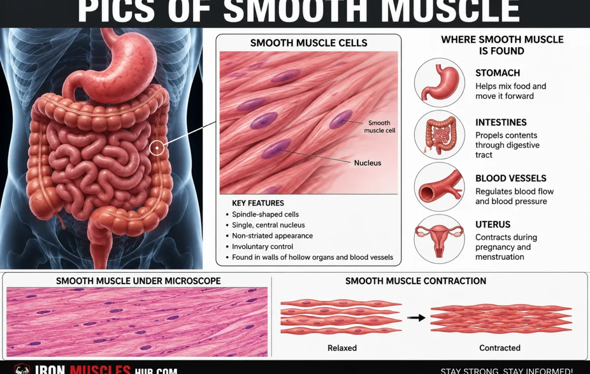
Pics of smooth muscle usually show long, narrow, spindle-shaped cells with a single centrally located nucleus and no visible striations. Smooth muscle is an involuntary type of muscle tissue found in places such as the digestive tract, blood vessels, airways, urinary system, reproductive organs, and parts of the eye.
- Smooth Muscle Anatomy at a Glance
- What Do Pics of Smooth Muscle Look Like?
- What a labeled smooth muscle picture should show
- Smooth Muscle Cells Under a Microscope
- Where Is Smooth Muscle Found?
- Smooth Muscle in Hollow Organs
- Why Doesn't Smooth Muscle Have Stripes?
- Smooth Muscle vs. Skeletal and Cardiac Muscle
- How Smooth Muscle Contracts
- Single-Unit vs. Multi-Unit Smooth Muscle
- Common Mistakes When Looking at Smooth Muscle Pictures
- Mistake 1: Assuming smooth muscle means "flat"
- Mistake 2: Thinking smooth muscle has no actin or myosin
- Mistake 3: Expecting every microscope image to look identical
- Mistake 4: Looking only for a nucleus
- How to Read a Smooth Muscle Diagram
- Frequently Asked Questions
- What does smooth muscle look like under a microscope?
- Where is smooth muscle found in the human body?
- Why is smooth muscle called smooth?
- Does smooth muscle have a nucleus?
- Is smooth muscle voluntary or involuntary?
- Does smooth muscle have actin and myosin?
- A Clearer Way to Understand Smooth Muscle Pictures
- A Closer Look at Smooth Muscle Cell Structure
- Main Parts You May See in a Smooth Muscle Picture
- Longitudinal vs. Cross-Section Pictures
- Longitudinal section
- Cross-section
- Smooth Muscle in the Digestive System
- Smooth Muscle in Blood Vessels
- Smooth Muscle in the Respiratory System
- Smooth Muscle in the Urinary System
- Smooth Muscle in the Reproductive System
- Why Smooth Muscle Cells Are Spindle-Shaped
- How to Identify Smooth Muscle in a Microscope Image
- Step 1: Look for striations
- Step 2: Examine the nuclei
- Step 3: Check cell shape
- Step 4: Look at the surrounding tissue
- Step 5: Compare several features
- Smooth Muscle vs. Skeletal Muscle Pictures
- Smooth Muscle vs. Cardiac Muscle Pictures
- Quick identification trick
- Why Smooth Muscle Can Contract for Long Periods
- Does Smooth Muscle Get Tired?
- Smooth Muscle Pictures: What to Look For
- A Simple Memory Trick
- Final Takeaway: Recognizing Smooth Muscle Gets Easier
- What a Good Smooth Muscle Picture Should Show
- Smooth Muscle Diagram vs. Microscope Picture
- How Smooth Muscle Looks in Different Organs
- Intestine
- Blood vessel
- Airway
- Bladder
- Uterus
- Why Smooth Muscle Pictures Sometimes Look Like Other Tissues
- The tissue may be cut at an angle
- Nuclei may be difficult to see
- Cells can overlap
- Magnification changes the appearance
- A Five-Second Smooth Muscle Identification Test
- Smooth Muscle and Involuntary Control
- Can You See Smooth Muscle With the Naked Eye?
- Is Smooth Muscle a Skeletal Muscle?
- Can Smooth Muscle Build Muscle Like Skeletal Muscle?
- What Is the Difference Between Smooth Muscle and Cardiac Muscle?
- What Is the Difference Between Smooth Muscle and Skeletal Muscle?
- Beginner's Smooth Muscle Study Checklist
- Smooth Muscle Picture Study Strategy
- Stage 1: Learn the diagram
- Stage 2: Study a real histology image
- Stage 3: Compare it with other muscle types
- Frequently Asked Questions
- What are the main features of smooth muscle?
- What does smooth muscle look like in a picture?
- Where can I find smooth muscle pictures?
- How do you identify smooth muscle under a microscope?
- Does smooth muscle have striations?
- Why is smooth muscle important?
- Final Takeaway: Learn the Pattern, Not Just the Picture
- Final Thoughts
If you’re looking at a smooth muscle picture for the first time, it can be difficult to know what you’re seeing. Unlike the obvious stripes of skeletal muscle, smooth muscle has a much simpler appearance under a light microscope. This guide breaks down its anatomy, explains what the cells look like, and shows where this important tissue works inside your body. For more insights read here :- how to grow muscle and how to gain muscle fast
Quick answer: Smooth muscle cells are typically spindle-shaped, non-striated, and contain one centrally located nucleus. They contract involuntarily to move substances through organs, regulate the diameter of blood vessels and airways, and perform other automatic functions.
Smooth Muscle Anatomy at a Glance
| Feature | Smooth muscle |
|---|---|
| Cell shape | Spindle-shaped, tapered at both ends |
| Nucleus | Usually one, centrally located |
| Striations | Absent |
| Control | Primarily involuntary |
| Main contractile proteins | Actin and myosin |
| Sarcomeres | Not arranged into sarcomeres |
| Common locations | Organs, blood vessels, airways, urinary and reproductive systems |
| Main job | Contracting and controlling internal structures |
These features help distinguish smooth muscle from skeletal and cardiac muscle. Smooth muscle still contains actin and myosin, but these proteins aren’t organized into the regular sarcomere pattern responsible for the obvious striations seen in skeletal and cardiac muscle. Power stroke pulls actinActinTropomyosinCalcium presentADPPi leavesMyosinSarcomere shortens
power stroke; Pi leaves
Current stage
3Power stroke pulls actin
CaOffOn
OffOn
Cycle
Cycle
What Do Pics of Smooth Muscle Look Like?
A typical smooth muscle picture shows cells that resemble tiny elongated spindles. The middle portion is wider, while each end gradually becomes narrower.
One of the easiest features to identify is the central nucleus. Because smooth muscle cells generally contain one nucleus, microscopic images often show elongated, darker structures running through the middle of the cells.
Another important clue is what you don’t see: regular stripes. Smooth muscle is called “smooth” because its contractile proteins aren’t arranged into the visible repeating pattern found in striated muscle.
What a labeled smooth muscle picture should show
A useful anatomy illustration may label:
- Smooth muscle cell — the individual contractile cell
- Nucleus — located near the center
- Cell membrane — the boundary surrounding the cell
- Actin and myosin — proteins responsible for contraction
- Dense bodies — anchoring structures associated with the contractile network
- Connective tissue — supports and connects the cells
At higher magnification, the internal arrangement is more complicated than a basic textbook diagram suggests. Smooth muscle contains a network of contractile and structural proteins rather than the neatly organized sarcomeres found in skeletal muscle.
Smooth Muscle Cells Under a Microscope


4
When viewing a microscope image of smooth muscle, start with the overall pattern instead of trying to identify every individual structure immediately.
Look for closely packed elongated cells. The nuclei may appear as darker oval or elongated areas, while the surrounding tissue can look relatively uniform compared with skeletal muscle.
The appearance can also change depending on how the tissue was cut. A longitudinal section may show elongated cells and nuclei, while a cross-section can make the same cells look more rounded.
That’s an important point for beginners: a smooth muscle cell doesn’t always look like the perfect spindle shown in a diagram, because microscope images depend on the direction and location of the tissue section.
Where Is Smooth Muscle Found?
Smooth muscle is widely distributed throughout the body. It is especially important in structures that need to change diameter, push contents forward, or maintain tension without conscious effort.
| Location | What smooth muscle does |
|---|---|
| Stomach and intestines | Helps move and mix digestive contents |
| Blood vessels | Changes vessel diameter and influences blood flow |
| Airways | Helps regulate airway diameter |
| Urinary bladder | Contracts during bladder emptying |
| Uterus | Produces contractions |
| Reproductive tract | Helps move contents through reproductive passages |
| Iris and eye | Helps regulate pupil size |
| Skin | Raises hairs through tiny muscles attached to hair follicles |
Smooth muscle therefore isn’t one large muscle that you can see from outside the body. It’s tissue distributed throughout many internal structures.
Smooth Muscle in Hollow Organs
One of the clearest ways to understand smooth muscle is to picture the wall of a hollow organ.
Smooth muscle cells can form layers around structures such as the intestines and blood vessels. When those cells contract, the surrounding tube or organ can change its shape or diameter.
In the digestive tract, coordinated smooth muscle activity helps move material through the gastrointestinal system. In blood vessels, smooth muscle contraction can alter the vessel’s internal diameter and therefore affect resistance to blood flow.
This arrangement explains why smooth muscle doesn’t need to look like the large muscles of your arms or legs. Its job is often to control an internal passageway rather than move a bone.
Why Doesn’t Smooth Muscle Have Stripes?
The answer comes down to organization.
Skeletal and cardiac muscle contain sarcomeres, highly organized repeating units that produce their characteristic striated appearance. Smooth muscle has actin and myosin too, but these filaments aren’t organized into sarcomeres in the same way.
Instead, the contractile machinery forms a less visibly regular network inside the cell. This gives smooth muscle its relatively uniform appearance under a standard light microscope.
So if you’re comparing pictures, remember:
Stripes visible = think skeletal or cardiac muscle.
No obvious stripes + spindle-shaped cells = think smooth muscle.
Smooth Muscle vs. Skeletal and Cardiac Muscle
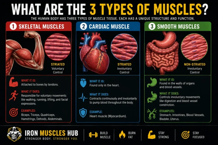
| Feature | Smooth | Skeletal | Cardiac |
|---|---|---|---|
| Visible striations | No | Yes | Yes |
| Typical control | Involuntary | Voluntary | Involuntary |
| Cell shape | Spindle-shaped | Long cylindrical fibers | Branched cells |
| Typical nuclei | One central nucleus | Multiple, usually peripheral | Usually one central nucleus |
| Sarcomeres | No | Yes | Yes |
| Main locations | Internal organs and vessels | Attached to bones | Heart |
All three types are specialized for contraction, but their structures are adapted to different jobs.
How Smooth Muscle Contracts
Smooth muscle contraction still depends on interactions between actin and myosin, but its control mechanism differs from skeletal muscle.
Calcium is an important signal. In smooth muscle, calcium activates a pathway that ultimately enables myosin to interact with actin and generate contraction.
The result is useful for organs that need slow, sustained, or automatically controlled changes. For example, smooth muscle can help maintain tension in a blood vessel or produce coordinated contractions in the digestive tract.A bandI bandH zoneZ-lineZ-lineactinmyosin
sarcomere contraction 40%
Contraction
%
Contraction
The sarcomere visualization is useful here mainly as a comparison: smooth muscle produces force without organizing its actin and myosin into the sarcomeres characteristic of striated muscle.
Single-Unit vs. Multi-Unit Smooth Muscle
Smooth muscle isn’t organized in exactly the same way everywhere.
Single-unit smooth muscle is common in the walls of visceral organs. Cells communicate with neighboring cells through connections that help groups contract together.
Multi-unit smooth muscle allows more individual control because cells can receive more independent signals. This arrangement is found in specific locations where finer control is useful.
You don’t need to memorize every example to understand the main idea: some smooth muscle behaves more like a coordinated sheet, while some allows more precise cell-by-cell control.
Common Mistakes When Looking at Smooth Muscle Pictures
Mistake 1: Assuming smooth muscle means “flat”
Smooth muscle cells are generally fusiform, meaning they are wider around the middle and taper toward the ends. “Smooth” refers primarily to their lack of visible striations, not that the cells are physically flat.
Mistake 2: Thinking smooth muscle has no actin or myosin
It does. Actin and myosin are essential contractile proteins in smooth muscle; they’re simply arranged differently from those in striated muscle.
Mistake 3: Expecting every microscope image to look identical
The appearance depends on the organ, tissue section, staining method, and viewing angle. A cross-section and longitudinal section can make the same type of tissue look surprisingly different.
Mistake 4: Looking only for a nucleus
A central nucleus is a helpful clue, but identification should use several characteristics together: cell shape, lack of striations, tissue arrangement, and anatomical location.
How to Read a Smooth Muscle Diagram
If you’re studying anatomy, use this simple four-step method:
- Find the individual cells. Look for elongated, tapered shapes.
- Locate the nuclei. They are typically central within the cells.
- Check for striations. Smooth muscle doesn’t show the regular stripes of skeletal or cardiac muscle.
- Identify the tissue location. Ask whether the tissue surrounds an organ, vessel, airway, or other internal structure.
This approach is usually more useful than trying to memorize one particular smooth muscle photograph.
Frequently Asked Questions
What does smooth muscle look like under a microscope?
Smooth muscle generally appears as closely packed, elongated cells with tapered ends and centrally located nuclei. Unlike skeletal and cardiac muscle, it doesn’t display obvious cross-striations. Its exact appearance varies depending on the organ, tissue section, staining technique, and magnification.
Where is smooth muscle found in the human body?
Smooth muscle is found in many internal structures, including the digestive tract, blood vessels, respiratory passages, urinary bladder, reproductive organs, skin, and parts of the eye. It helps these structures contract or change their diameter without requiring conscious control.
Why is smooth muscle called smooth?
It’s called smooth muscle because its cells don’t have the visible striated pattern characteristic of skeletal and cardiac muscle. The absence of organized sarcomeres produces a comparatively uniform appearance when viewed with a conventional microscope.
Does smooth muscle have a nucleus?
Yes. A typical smooth muscle cell has one centrally located nucleus. In a longitudinal tissue section, these nuclei can be particularly useful for recognizing smooth muscle under a microscope.
Is smooth muscle voluntary or involuntary?
Smooth muscle is generally involuntary, meaning you don’t consciously control most of its contractions. Its activity can be influenced by the autonomic nervous system, hormones, and local signals, depending on the tissue.
Does smooth muscle have actin and myosin?
Yes. Smooth muscle contains both actin and myosin, which generate contraction. The major structural difference is that these filaments aren’t arranged into the repeating sarcomeres found in skeletal and cardiac muscle.
A Clearer Way to Understand Smooth Muscle Pictures
The easiest way to recognize pics of smooth muscle is to combine three clues: spindle-shaped cells, centrally located nuclei, and no obvious striations. Then look at where the tissue is located. If you’re viewing a tissue surrounding an internal organ, blood vessel, airway, or similar structure, smooth muscle becomes a strong possibility.
Remember that smooth muscle is much more than a microscope-study topic. Its contractions help regulate blood vessel diameter, move digestive contents, control passages in the urinary and reproductive systems, and perform other automatic tasks throughout the body.
A Closer Look at Smooth Muscle Cell Structure
A smooth muscle cell is much smaller and simpler in appearance than the large skeletal muscle fibers used to move your limbs. Each cell is generally elongated, with a broader middle section and tapered ends.
The nucleus sits near the center of the cell. In microscope images, this central nucleus can be one of the easiest features to recognize, especially when the tissue has been cut lengthwise.
The inside of the cell contains contractile proteins, mainly actin and myosin. These proteins allow the cell to shorten and generate force, even though smooth muscle doesn’t have the highly organized sarcomeres seen in skeletal and cardiac muscle.
Main Parts You May See in a Smooth Muscle Picture
| Structure | What it means |
|---|---|
| Cell membrane | Surrounds the smooth muscle cell |
| Cytoplasm | Internal material containing the cell’s structures |
| Nucleus | Usually positioned in the center |
| Actin | Contractile protein involved in force production |
| Myosin | Contractile protein that interacts with actin |
| Dense bodies | Structures that help anchor contractile filaments |
| Connective tissue | Provides support around and between cells |
A basic educational diagram may show these structures much more clearly than a real microscope photograph. That’s normal. Real tissue contains many overlapping cells, connective tissue, blood vessels, and other structures.
Longitudinal vs. Cross-Section Pictures
One reason smooth muscle pictures can be confusing is that the same tissue can look different depending on how it was sliced.
Longitudinal section
A longitudinal section cuts along the length of the cells. Smooth muscle cells may appear elongated, and their centrally positioned nuclei can look like narrow or oval structures.
This is often the easiest view for beginners because the spindle-like cell shape is easier to recognize.
Cross-section
A cross-section cuts across the cells rather than along their length. Instead of seeing long spindle shapes, you may see many small circular or oval profiles.
The size of these profiles can vary because the cells are tapered. A slice through the wider middle of a cell won’t look the same as a slice through its narrow end.
Study tip: Don’t assume every rounded shape in a microscope image is a different type of cell. The direction of the tissue cut can dramatically change what you see.
Smooth Muscle in the Digestive System
Smooth muscle plays an important mechanical role throughout the digestive tract.
Muscle layers in the walls of the gastrointestinal tract contract in coordinated patterns. These contractions help mix digestive contents and move them through the digestive system.
The important idea is that smooth muscle doesn’t need conscious commands for every contraction. Its activity is regulated by internal control systems involving neural, hormonal, and local signals.
This is very different from deliberately contracting your biceps to lift an object.
Smooth Muscle in Blood Vessels
Blood vessel walls contain smooth muscle, particularly in the muscular layer of many arteries and other vessels.
When vascular smooth muscle contracts, the vessel’s diameter can decrease. When it relaxes, the diameter can increase.
This ability to change vessel diameter is one reason smooth muscle is so important to circulation. It allows blood vessels to adjust their size in response to signals from the body.
When studying a blood-vessel diagram, look for the muscular layer surrounding the central opening, known as the lumen.
Smooth Muscle in the Respiratory System
Smooth muscle is also present in the walls of the airways.
Airway smooth muscle can change the diameter of the passages through which air travels.
For anatomy students, this is another useful example of why smooth muscle is different from skeletal muscle. You don’t normally decide consciously to contract individual airway smooth muscle cells. Their activity is regulated automatically.
A picture of an airway may therefore show smooth muscle arranged around the passage rather than as a large standalone muscle.
Smooth Muscle in the Urinary System
The urinary bladder contains smooth muscle in its wall.
When the bladder fills, its wall stretches. During urination, coordinated contraction of the bladder’s smooth muscle helps push urine toward the outlet.
This gives smooth muscle another important job: moving material through an internal organ.
The same general principle appears in other organs. Instead of moving a bone, smooth muscle changes the shape, pressure, or diameter of internal structures.
Smooth Muscle in the Reproductive System
Smooth muscle is also found in several reproductive structures.
In the uterus, smooth muscle forms a major part of the muscular wall. Its contractions can produce powerful changes in the uterus.
Smooth muscle is also present in other parts of the reproductive tract, where contractions can help move material through internal passages.
For beginners, the main point is simple: smooth muscle is widely distributed in organs where automatic contractions are useful.
Why Smooth Muscle Cells Are Spindle-Shaped
The tapered shape of a smooth muscle cell isn’t just a random feature.
Smooth muscle cells are arranged closely together, allowing them to form sheets or layers around organs and tubes. Their elongated shape works well for this type of tissue organization.
Under a microscope, you may therefore see many cells packed together with their long axes running in similar directions.
This organization allows groups of cells to contract and change the shape or diameter of the surrounding structure.
How to Identify Smooth Muscle in a Microscope Image
If you’re given an unlabeled microscope picture, don’t immediately try to memorize what it looks like. Use a checklist.
Step 1: Look for striations
If you see strong, regular horizontal stripes across long muscle fibers, you’re probably looking at striated muscle rather than smooth muscle.
Smooth muscle generally does not show visible striations with ordinary light microscopy.
Step 2: Examine the nuclei
Look for centrally located nuclei within elongated cells.
A single central nucleus is a useful clue when distinguishing smooth muscle from typical skeletal muscle fibers.
Step 3: Check cell shape
Smooth muscle cells are generally elongated and tapered.
However, remember that a cross-section can make them appear much more rounded.
Step 4: Look at the surrounding tissue
Ask where the muscle appears to be located.
Is it surrounding an intestine, blood vessel, airway, bladder, or another internal structure? The location can provide an important clue.
Step 5: Compare several features
Never identify tissue from one feature alone.
A stronger identification comes from combining:
- Cell shape
- Nuclear position
- Lack of obvious striations
- Tissue arrangement
- Anatomical location
Smooth Muscle vs. Skeletal Muscle Pictures
The easiest visual comparison is between smooth and skeletal muscle.
Skeletal muscle usually appears as long fibers with obvious repeating striations. Individual fibers can contain multiple nuclei positioned near the edges.
Smooth muscle generally appears less striped and consists of smaller spindle-shaped cells with centrally positioned nuclei.
| Picture feature | Smooth muscle | Skeletal muscle |
| Stripes | Not visibly striated | Clearly striated |
| Cell shape | Tapered/spindle-shaped | Long cylindrical fibers |
| Nuclei | Usually central | Usually peripheral |
| Typical location | Internal organs | Attached to bones |
| Conscious control | Generally involuntary | Generally voluntary |
These visual differences make muscle tissue identification much easier once you know what clues to search for.
Smooth Muscle vs. Cardiac Muscle Pictures
Cardiac muscle can sometimes cause confusion because it is also involuntary.
The key visual difference is that cardiac muscle is striated, while smooth muscle isn’t.
Cardiac muscle cells also tend to branch and connect with neighboring cells. Specialized structures called intercalated discs can be seen in appropriately prepared cardiac tissue.
Smooth muscle instead has elongated, tapered cells without the obvious striated pattern.
Quick identification trick
If you’re looking at a microscope image:
- Striated + branching + heart tissue = cardiac muscle
- Striated + long fibers + multiple peripheral nuclei = skeletal muscle
- Non-striated + spindle-shaped + central nucleus = smooth muscle
This isn’t a substitute for formal histology training, but it’s a useful beginner’s framework.
Why Smooth Muscle Can Contract for Long Periods
Smooth muscle is particularly suited to sustained contraction.
Its contractile system can maintain tension without requiring the same rapid cycling associated with many skeletal muscle movements.
That matters in organs such as blood vessels, where maintaining an appropriate level of muscle tone can help regulate vessel diameter.
It also matters in hollow organs, where contractions may need to continue in coordinated patterns rather than producing one quick movement.
Does Smooth Muscle Get Tired?
Smooth muscle has characteristics that allow it to maintain force efficiently for relatively long periods.
Its activity is generally slower than the rapid contractions you might associate with skeletal muscle, but the exact behavior varies between different types of smooth muscle and different organs.
So it’s better to think of smooth muscle as specialized for automatic, controlled contraction rather than simply calling it “slow muscle.”
Smooth Muscle Pictures: What to Look For
When searching for or studying pictures of smooth muscle, prioritize images that clearly show the tissue and provide useful labels.
A good educational image should ideally make it possible to identify:
- Individual smooth muscle cells
- The centrally located nuclei
- The absence of obvious striations
- The direction of the cells
- The surrounding tissue or organ
- The magnification or scale when available
A labeled diagram is particularly useful when you’re learning the anatomy for the first time. A real histology photograph is valuable afterward because it shows how the tissue actually appears rather than presenting an idealized drawing.
A Simple Memory Trick
Use the phrase:
“Smooth = spindle + single central nucleus + no stripes.”
It isn’t a complete description of smooth muscle, but it’s a useful starting point for identifying it in basic anatomy and histology images.
Then add the function:
“Smooth muscle automatically moves or controls internal structures.”
Together, those two ideas cover much of what a beginner needs to recognize.
Final Takeaway: Recognizing Smooth Muscle Gets Easier
The best way to understand pics of smooth muscle is to stop treating each photograph as something you have to memorize. Instead, look for a consistent group of clues: tapered cells, central nuclei, no obvious striations, and an arrangement around internal structures.
Smooth muscle is found throughout the body and performs jobs that happen mostly without conscious effort. It helps move digestive contents, regulate blood vessel diameter, influence airway size, empty the bladder, and control contractions in other internal organs.
Once you understand its basic structure and purpose, both diagrams and microscope photographs become much easier to interpret.
How to Read and Compare Pics of Smooth Muscle
If you’re studying pics of smooth muscle, the most useful skill isn’t memorizing one photograph. It’s learning how to recognize the same structural clues across different diagrams, microscope slides, and anatomy illustrations.
Smooth muscle can look different depending on the organ, staining method, magnification, and direction of the tissue section. Once you know what to look for, those differences become much less confusing.
What a Good Smooth Muscle Picture Should Show
A useful smooth muscle image should make the important features easy to identify.
Look for:
- Spindle-shaped cells
- A single central nucleus
- No obvious cross-striations
- Closely packed cells
- Cells arranged in layers or bundles
- The surrounding organ or tissue when relevant
For beginners, a labeled illustration is often easier to understand than a high-magnification histology photograph. Once you know the basic anatomy, real microscope images become much easier to interpret.
Smooth Muscle Diagram vs. Microscope Picture
These two types of images serve different purposes.
| Image type | Best for | What you’ll notice |
|---|---|---|
| Labeled diagram | Learning anatomy | Clear cell shapes and labels |
| Histology photograph | Recognizing real tissue | Natural variation and overlapping cells |
| Organ diagram | Understanding location | Shows where smooth muscle occurs |
| Comparison image | Studying tissue types | Highlights differences between muscle types |
| High-magnification image | Detailed study | Shows cellular structures more closely |
A common mistake is expecting a real histology slide to look exactly like a textbook illustration. A diagram simplifies structures so they’re easier to learn. A microscope photograph shows the actual complexity of biological tissue.
How Smooth Muscle Looks in Different Organs
The basic characteristics remain the same, but the overall appearance can change based on where the tissue is located.
Intestine
In the intestine, smooth muscle is organized into layers in the wall of the digestive tract.
The arrangement allows coordinated contractions that help mix and move intestinal contents.
Blood vessel
In a blood vessel, smooth muscle is arranged around the lumen. Its contraction can change the vessel’s diameter.
This makes the surrounding anatomical structure particularly useful when identifying smooth muscle in a vessel photograph.
Airway
Smooth muscle is also found around parts of the respiratory tract. Its contraction can affect the diameter of an airway.
Bladder
The bladder wall contains smooth muscle that contracts during bladder emptying.
Uterus
The muscular wall of the uterus contains abundant smooth muscle. Its contractions are especially important during labor.
The key lesson is that smooth muscle is usually part of a larger structure, rather than appearing as a separate external muscle like the biceps or quadriceps.
Why Smooth Muscle Pictures Sometimes Look Like Other Tissues
Some microscope images are difficult even for beginners who understand the basic differences.
There are several reasons.
The tissue may be cut at an angle
If cells are cut diagonally, their normal spindle-like shape can become harder to recognize.
Nuclei may be difficult to see
Depending on the preparation and image quality, nuclei may not stand out clearly.
Cells can overlap
Smooth muscle is made of tightly packed cells. In a real tissue sample, one cell may partially obscure another.
Magnification changes the appearance
At low magnification, you can see the overall organization. At higher magnification, individual cells and nuclei become easier to examine.
That’s why context matters. Don’t make your identification based on a single tiny feature.
A Five-Second Smooth Muscle Identification Test
If you’re taking an anatomy quiz, try this quick method:
1. Are there obvious stripes?
If yes, consider skeletal or cardiac muscle.
2. Are the cells elongated and tapered?
If yes, smooth muscle becomes more likely.
3. Is the nucleus centrally positioned?
That’s another clue supporting smooth muscle.
4. Is the tissue part of an internal organ or vessel?
That strengthens the identification.
5. Do several clues agree?
If they do, your identification is much more reliable.
This method is faster and more useful than trying to remember a particular image from a textbook.
Smooth Muscle and Involuntary Control
Smooth muscle is generally considered involuntary muscle because you don’t consciously control most of its activity.
The autonomic nervous system can influence smooth muscle, but it isn’t the only source of regulation. Hormones, local chemical signals, stretching, and other factors can influence smooth muscle activity depending on the tissue.
That helps explain why smooth muscle can keep working while you’re sleeping, eating, exercising, or simply going about your day.
Your digestive tract doesn’t wait for you to consciously tell every muscle cell to contract.
Can You See Smooth Muscle With the Naked Eye?
Individual smooth muscle cells are microscopic, so you can’t normally see them as individual cells without magnification.
However, the smooth muscle layers made from many cells can be visible as part of dissected organs or anatomical specimens.
This is an important distinction:
You can see a smooth muscle layer as tissue, but you need microscopy to examine individual smooth muscle cells.
Is Smooth Muscle a Skeletal Muscle?
No.
Smooth muscle and skeletal muscle are two different types of muscle tissue.
Skeletal muscle is primarily associated with movement of the skeleton and is under voluntary control. Smooth muscle is mainly found in internal organs and structures such as blood vessels and is generally involuntary.
Their microscopic structures are also different.
Skeletal muscle has obvious striations and long fibers, while smooth muscle has spindle-shaped cells without visible cross-striations.
Can Smooth Muscle Build Muscle Like Skeletal Muscle?
Smooth muscle can adapt to physiological demands, including changes in cell size and function, but it isn’t something you train through ordinary resistance exercise in the same way you train skeletal muscle.
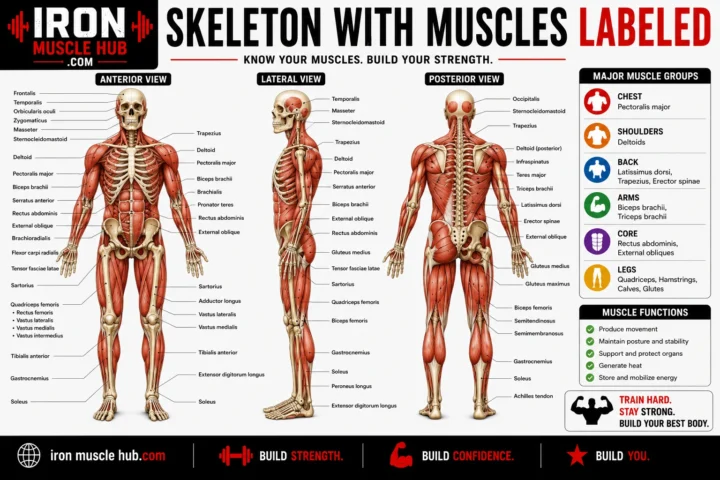
For example, lifting weights directly trains skeletal muscles such as the quadriceps, chest, shoulders, and biceps.
Smooth muscle has different jobs and is regulated differently.
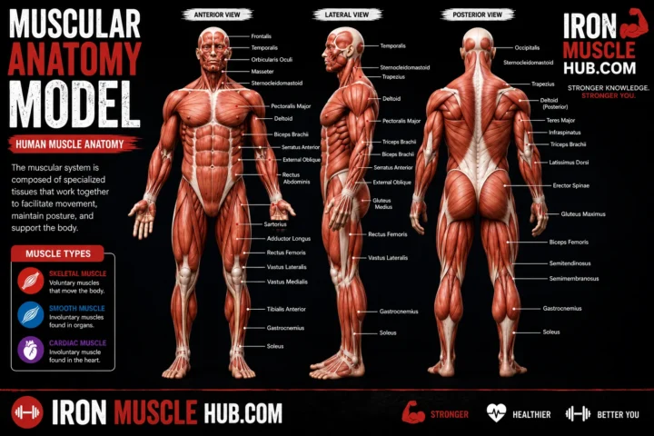
So if you’re looking at fitness-related muscle diagrams, don’t confuse the smooth muscle found inside your organs with the skeletal muscles responsible for most visible body movement.
What Is the Difference Between Smooth Muscle and Cardiac Muscle?
Both smooth and cardiac muscle are generally involuntary, but their structures are quite different.
Cardiac muscle forms the muscular wall of the heart and has visible striations. Its cells are generally branched and connected in a specialized network.
Smooth muscle is found in many internal organs and vessels. Its cells are typically tapered and don’t have visible striations.
| Characteristic | Smooth muscle | Cardiac muscle |
| Main location | Internal organs and vessels | Heart |
| Control | Involuntary | Involuntary |
| Striations | No | Yes |
| Cell shape | Spindle-shaped | Branched |
| Main role | Controls internal structures | Pumps blood |
This comparison is especially helpful when studying microscope photographs.
What Is the Difference Between Smooth Muscle and Skeletal Muscle?
The biggest visual difference is the presence of striations.
Skeletal muscle fibers have a regular striped appearance because their contractile proteins are organized into repeating sarcomeres.
Smooth muscle doesn’t have that same sarcomere organization, so it lacks the obvious striped appearance under standard microscopy.
Functionally, skeletal muscle is primarily responsible for voluntary movement, posture, and other body movements, while smooth muscle performs automatic functions inside the body.
Beginner’s Smooth Muscle Study Checklist
Before moving on from the topic, make sure you can answer these questions:
- What shape are smooth muscle cells?
- Where is the nucleus located?
- Does smooth muscle have visible striations?
- Is smooth muscle generally voluntary or involuntary?
- Where can smooth muscle be found?
- How does smooth muscle differ from skeletal muscle?
- How does it differ from cardiac muscle?
- Why can a cross-section look different from a longitudinal section?
- What are actin and myosin doing inside the cell?
- Why is the surrounding organ useful when identifying smooth muscle?
If you can answer these without looking at your notes, you have the core concept down.
Smooth Muscle Picture Study Strategy
For students, one of the best approaches is to study smooth muscle images in three stages.
Stage 1: Learn the diagram
Start with a clean labeled illustration. Learn the cell shape, nucleus, and basic contractile structures.
Stage 2: Study a real histology image
Next, look at a microscope photograph. Don’t worry if it doesn’t look as neat as the diagram.
Try to find the same features in the real tissue.
Stage 3: Compare it with other muscle types
Place smooth, skeletal, and cardiac muscle images next to each other.
Ask yourself:
- Which has stripes?
- Which cells are branched?
- Which cells are spindle-shaped?
- Where are the nuclei?
- Where is each tissue found?
This comparison-based approach can make memorization much easier.
Frequently Asked Questions
What are the main features of smooth muscle?
The main identifying features are elongated, spindle-shaped cells, a centrally located nucleus, and the absence of obvious cross-striations. Smooth muscle also contains actin and myosin for contraction, but these proteins aren’t organized into the repeating sarcomeres characteristic of skeletal and cardiac muscle.
What does smooth muscle look like in a picture?
A typical smooth muscle picture shows closely packed elongated cells with tapered ends. Their nuclei are usually located near the center of each cell. In a standard microscope image, smooth muscle generally lacks the obvious horizontal stripes seen in skeletal and cardiac muscle.
Where can I find smooth muscle pictures?
Smooth muscle images are commonly available in anatomy and histology textbooks, university educational resources, and reputable medical or biology resources. When using images online, check the licensing terms before republishing them on a website. For a commercial website, creating an original illustration or using a properly licensed image is the safest approach.
How do you identify smooth muscle under a microscope?
Start by checking for the absence of visible striations. Then look for elongated, tapered cells with centrally positioned nuclei. Finally, consider the tissue’s location and arrangement. Using several characteristics together is more reliable than relying on one visual feature.
Does smooth muscle have striations?
No. Smooth muscle doesn’t show the regular visible striations associated with skeletal and cardiac muscle under conventional light microscopy. This difference is related to how its contractile proteins are organized inside the cells.
Why is smooth muscle important?
Smooth muscle controls many automatic processes throughout the body. It helps move material through the digestive tract, changes the diameter of blood vessels and airways, contributes to bladder emptying, and produces contractions in several reproductive structures.
Final Takeaway: Learn the Pattern, Not Just the Picture
The easiest way to understand pics of smooth muscle is to learn the pattern behind them. Look for spindle-shaped cells, central nuclei, and no obvious striations, then use the surrounding tissue to confirm what you’re seeing.
Smooth muscle may look different from one microscope image to another, but its fundamental characteristics remain consistent. Understanding those characteristics is much more useful than memorizing a single textbook photograph.
For readers who want to continue learning about human muscle anatomy, related topics on Iron Muscle Hub could include what muscles are, the major muscles of the human body, and how the muscular system works with other body systems.
Final Thoughts
Understanding pics of smooth muscle becomes much easier once you know the key features to look for. Smooth muscle cells are typically spindle-shaped, have centrally located nuclei, and lack the obvious striations seen in skeletal and cardiac muscle.
These cells may look different depending on the organ, microscope magnification, staining technique, or direction of the tissue section. That’s why it’s better to learn the basic pattern rather than memorize one specific picture.
From the digestive tract and blood vessels to the bladder, airways, and reproductive organs, smooth muscle performs important automatic functions throughout the body. Once you can recognize its structure and understand what it does, smooth muscle diagrams and microscope images become much easier to read.