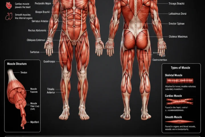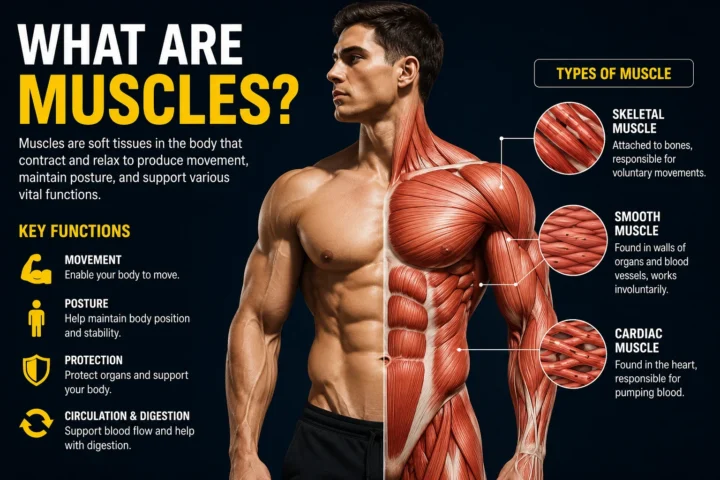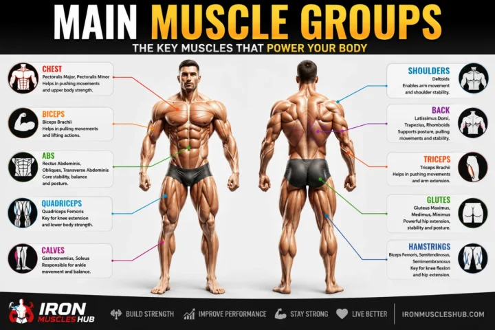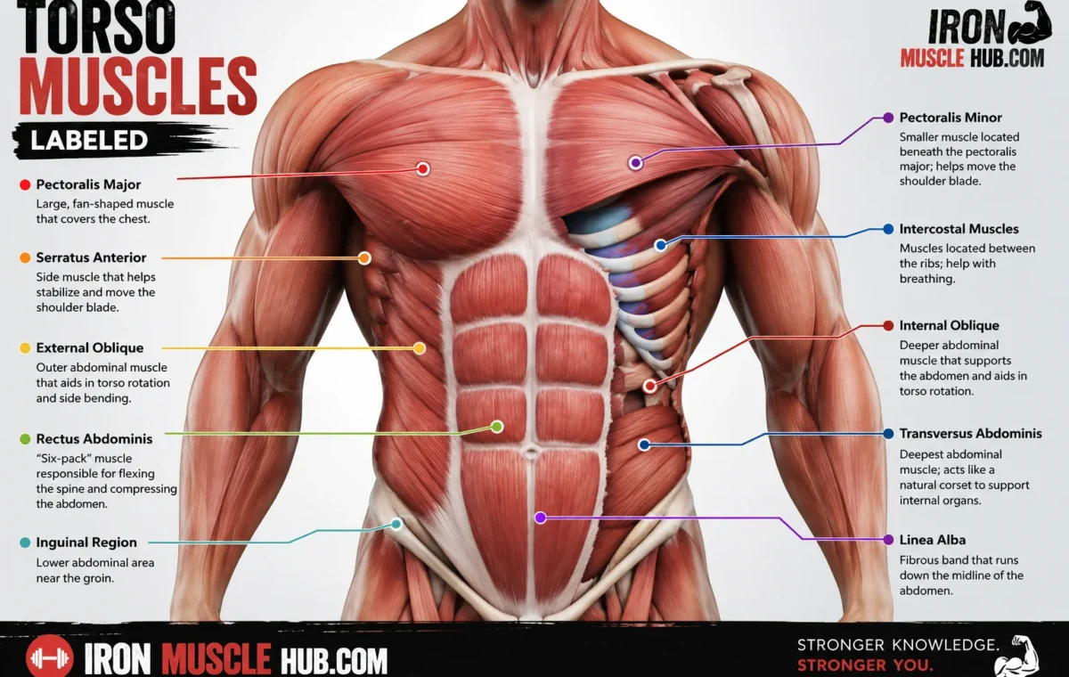
If you’re looking for torso muscles labeled, the main muscles to learn are the pectoralis major, serratus anterior, rectus abdominis, external and internal obliques, transversus abdominis, latissimus dorsi, trapezius, and erector spinae. Together, these muscles help move and stabilize the trunk, support posture, assist breathing, and protect the body’s internal structures.
- Torso Muscles Labeled: Quick Reference
- What Are the Torso Muscles?
- Anterior Torso Muscles: The Front View
- 1. Pectoralis Major
- 2. Serratus Anterior
- 3. Rectus Abdominis
- 4. External Oblique
- 5. Internal Oblique
- 6. Transversus Abdominis
- Posterior Torso Muscles: The Back View
- 1. Trapezius
- 2. Latissimus Dorsi
- 3. Erector Spinae
- 4. Rhomboids
- 5. Serratus Posterior
- Torso Muscles and Breathing
- A Simple Way to Read a Labeled Torso Diagram
- Torso Muscles by Region
- Common Mistakes When Studying Torso Anatomy
- Mistake 1: Treating the "core" as one muscle
- Mistake 2: Learning only the front
- Mistake 3: Confusing visible muscles with all muscles
- Mistake 4: Assuming every chart uses the same labels
- How Torso Muscles Work Together
- Where to Find a Copyright-Safe Torso Muscle Reference
- Suggested Internal Links for Iron Muscle Hub
- Frequently Asked Questions
- What are the main muscles of the torso?
- What is the difference between the torso and the trunk?
- What are the abdominal muscles called?
- Which muscles are on the back of the torso?
- What is the largest muscle on the torso?
- Why do some torso muscle charts look different?
- A Clearer Way to Remember Torso Anatomy
- The Torso Has Multiple Muscle Layers
- A Simple Layering Model
- Internal Oblique: The Middle Abdominal Layer
- Transversus Abdominis: The Deep Abdominal Muscle
- How to Recognize It
- Intercostal Muscles
- The Diaphragm
- Deeper Back Muscles
- Rhomboid Major and Rhomboid Minor
- Levator Scapulae
- Deeper Erector Spinae Region
- Why the Back Looks Different From the Front
- Torso Muscles by Basic Function
- Anterior vs. Posterior Torso: Easy Comparison
- How to Memorize a Torso Muscles Labeled Diagram
- Step 1: Learn the landmarks
- Step 2: Add the side muscles
- Step 3: Add deeper structures
- Step 4: Test yourself without labels
- Step 5: Learn function last
- A Common Diagram Mistake: Confusing Muscle Groups
- Another Common Mistake: Thinking "Core" Means "Abs"
- How to Choose a Torso Muscle Chart
- For beginners
- For fitness study
- For school or anatomy courses
- For medical study
- A Practical Torso Anatomy Checklist
- Final Takeaway
- The Torso at a Glance
- Pectoralis Major
- Where to find it on a diagram
- Serratus Anterior
- Diagram clue
- Rectus Abdominis
- Easy memory trick
- External Oblique
- Internal Oblique
- Transversus Abdominis
- Quick visual comparison
- Trapezius
- How to recognize it
- Latissimus Dorsi
- Diagram clue
- Rhomboids
- Erector Spinae
- Why beginners often miss it
- Intercostals and the Rib Cage
- The Diaphragm
- Front vs. Back: A Fast Study Method
- Front view
- Back view
- Torso Muscle Functions: Beginner Reference
- What Muscles Are Used During Common Exercises?
- Push-up
- Row
- Pull-up
- Crunch
- Back extension
- Why Muscle Diagrams Don't Always Match
- Different views
- Different layers
- Different educational goals
- Different terminology
- How to Study a Torso Muscle Diagram More Efficiently
- Use the cover-and-test method
- Use location + function together
- A 10-Muscle Torso Quiz
- Answers
- The Most Important Labels to Learn First
- A Note About Fitness and Anatomy
- More Torso Anatomy Topics to Explore
- Final Thoughts on Torso Muscles Labeled
Learning the names can feel confusing at first because some muscles sit on top of others. A simple front-and-back approach makes the anatomy much easier to remember. For more insights read here :-muscular system and muscle anatomy .
Quick answer: The torso, also called the trunk, includes the chest, abdominal wall, and back. A useful labeled study chart should show both the anterior (front) and posterior (back) views.
Torso Muscles Labeled: Quick Reference
| Muscle | Where it is | Main role |
|---|---|---|
| Pectoralis major | Front of upper chest | Moves the upper arm |
| Serratus anterior | Side of chest near ribs | Helps move and stabilize the shoulder blade |
| External oblique | Outer side of abdomen | Helps rotate and bend the trunk |
| Internal oblique | Under external oblique | Helps rotate and stabilize the trunk |
| Rectus abdominis | Center front of abdomen | Helps flex the trunk |
| Transversus abdominis | Deep abdominal wall | Supports abdominal and trunk stability |
| Trapezius | Upper back and neck | Moves and stabilizes the shoulder blades |
| Latissimus dorsi | Broad middle and lower back | Helps move the upper arm |
| Erector spinae | Along the spine | Helps extend and stabilize the vertebral column |
| Intercostals | Between the ribs | Assist movements involved in breathing |
The exact muscle shown on a diagram depends on how deep the illustration goes. A basic fitness chart normally emphasizes large, visible muscles, while a medical anatomy diagram may include deeper layers and smaller muscles.
What Are the Torso Muscles?
The torso, or trunk, is the central portion of the body connecting the head and limbs. Its muscles include structures associated with the thoracic wall, abdominal wall, vertebral column, and back.
For beginners, it’s easiest to divide torso anatomy into three areas:
- Front: chest and abdominal muscles
- Sides: muscles around the ribs and abdominal wall
- Back: muscles surrounding the shoulder blades and spine
Some muscles also cross between regions. For example, the latissimus dorsi is a back muscle that attaches to the upper limb, so anatomy classifications don’t always match the simple way we divide muscles for exercise.
Anterior Torso Muscles: The Front View
The anterior view means you’re looking at the body from the front.
1. Pectoralis Major
The pectoralis major is the large, superficial muscle covering much of the front of the upper chest. It connects the chest region with the upper arm and contributes to movements of the arm.
On a labeled torso diagram, it’s usually one of the easiest muscles to identify because it covers a broad area across the upper chest.
2. Serratus Anterior
The serratus anterior sits along the side of the chest, with portions attaching to the ribs. It plays an important role in moving and stabilizing the scapula, or shoulder blade.
You may recognize it as the muscle with a distinctive “saw-tooth” appearance along the ribs.
3. Rectus Abdominis
The rectus abdominis runs vertically along the front of the abdomen. Its segmented appearance is associated with the familiar “six-pack” look when the muscle is developed and visible.
Its important anatomical role includes helping flex the trunk.
4. External Oblique
The external oblique is located on the outer sides of the abdomen. Its fibers run diagonally, giving the muscle a different appearance from the vertically oriented rectus abdominis.
It contributes to trunk movement, including rotation and side bending.
5. Internal Oblique
The internal oblique lies beneath the external oblique. Because it’s deeper, you may not see it clearly on a basic superficial-muscle diagram.
It contributes to trunk movement and stabilization.
6. Transversus Abdominis
The transversus abdominis is a deeper abdominal muscle whose fibers run horizontally around the abdomen. OpenStax describes its arrangement as similar to a belt around the body.
This is one reason a simple “abs chart” doesn’t show every muscle involved in core function.
Posterior Torso Muscles: The Back View
The posterior view means you’re looking at the torso from behind.
1. Trapezius
The trapezius is a large muscle extending across the upper back and into the neck region. It helps control movements and positioning of the shoulder blades.
On a back-muscle diagram, it’s usually the broad, roughly diamond-shaped muscle occupying much of the upper back.
2. Latissimus Dorsi
The latissimus dorsi, often called the “lats,” is a broad muscle covering much of the middle and lower back.
It also connects the trunk with the upper limb and contributes to movements of the arm.
For fitness beginners, this is the muscle commonly associated with exercises such as pulldowns and rows.
3. Erector Spinae
The erector spinae is a group of muscles running along the vertebral column. These muscles help extend the spine and contribute to maintaining an upright posture.
A common mistake is to think of the back as one large muscle. In reality, the back contains several overlapping muscle groups and layers.
4. Rhomboids
The rhomboid major and rhomboid minor are located between the spine and shoulder blades. They help position and stabilize the scapulae.
They’re often easier to understand when studying a posterior diagram because their location becomes clearer from the back.
5. Serratus Posterior
The serratus posterior muscles are located deeper toward the back of the rib cage. They are less prominent on beginner fitness charts but can appear on more detailed anatomy diagrams.
This is a good example of why two labeled torso charts can look different even though both are anatomically useful.
Torso Muscles and Breathing
Not every torso muscle exists primarily to create visible movement.
The intercostal muscles occupy the spaces between the ribs and contribute to movements of the thoracic cage during breathing. The diaphragm is a dome-shaped muscle separating the thoracic and abdominal cavities and plays a major role in respiration.
This is one of the most useful anatomy facts to remember: the torso isn’t just an “abs and back” area. Its muscles also help with breathing and stabilization.
A Simple Way to Read a Labeled Torso Diagram
If you’re studying a torso muscle chart for the first time, don’t try to memorize every label at once.
Use this sequence:
- Find the chest. Identify the pectoralis major.
- Move to the sides of the ribs. Look for the serratus anterior.
- Find the center of the abdomen. That’s the rectus abdominis.
- Identify the diagonal abdominal muscles. These are the external obliques.
- Turn the diagram around. Find the large trapezius at the upper back.
- Look below the trapezius. Identify the broad latissimus dorsi.
- Trace the muscles beside the spine. These are associated with the erector spinae group.
- Only then study deeper muscles.
This approach gives you a basic mental map before you add smaller anatomical structures.
Torso Muscles by Region
| Region | Major muscles to learn first | What to remember |
| Upper chest | Pectoralis major | Large front-chest muscle |
| Side of chest | Serratus anterior | Rib-side muscle associated with scapular movement |
| Front abdomen | Rectus abdominis | Vertical abdominal muscle |
| Abdominal sides | External and internal obliques | Diagonal layers |
| Deep abdomen | Transversus abdominis | Horizontal fibers around the abdomen |
| Upper back | Trapezius | Large upper-back muscle |
| Middle/lower back | Latissimus dorsi | Broad back muscle |
| Along spine | Erector spinae | Helps extend and stabilize the spine |
| Rib spaces | Intercostals | Assist thoracic movement during breathing |
The U.S. National Cancer Institute’s SEER Training material groups trunk muscles according to their roles in moving the vertebral column, forming the thoracic and abdominal walls, and supporting other functions such as breathing.
Common Mistakes When Studying Torso Anatomy
Mistake 1: Treating the “core” as one muscle
The core isn’t simply another name for the rectus abdominis. The abdominal wall contains several muscle layers, including the external oblique, internal oblique, and transversus abdominis.
Mistake 2: Learning only the front
A front-only diagram leaves out important muscles of the back. If your goal is to understand torso anatomy, compare anterior and posterior views.
Mistake 3: Confusing visible muscles with all muscles
A basic fitness chart usually emphasizes superficial muscles. More detailed anatomy resources may show muscles that are underneath or between other muscles.
Mistake 4: Assuming every chart uses the same labels
Anatomical diagrams can differ in how much detail they provide. One chart might label a large muscle group, while another separates it into individual muscles or deeper layers.
How Torso Muscles Work Together
Torso muscles rarely work completely alone.
For example, movement of the trunk can involve the abdominal muscles and back muscles working together to control the spine. The chest and back muscles can also interact with the shoulder girdle because several muscles connect the trunk with the upper limb.
That matters for exercise, too. A movement that looks like a “chest exercise” may involve the shoulders, arms, and trunk muscles for positioning and stability.
Where to Find a Copyright-Safe Torso Muscle Reference
For studying, reputable educational anatomy resources are preferable to grabbing a random image from Google Images.
The U.S. National Cancer Institute’s SEER Training Modules provides educational material on the muscles of the trunk, while OpenStax Anatomy & Physiology provides openly available educational anatomy content covering the abdominal wall and thorax.
For your own website’s featured image, the safest option is an original illustration created specifically for your site. If you use a third-party anatomy image, check its exact license, attribution requirements, and whether commercial website use is permitted before publishing.
Suggested Internal Links for Iron Muscle Hub
Consider linking this article to related beginner-friendly guides such as:
- Body Muscle Chart: A Beginner’s Guide to Major Muscle Groups
- Muscle Anatomy Chart: Names, Locations, and Functions
- Body Diagram of Muscles: A Simple Visual Guide
These links can help readers move from a labeled torso diagram to a broader understanding of the muscular system.
Frequently Asked Questions
What are the main muscles of the torso?
The major torso muscles include the pectoralis major, serratus anterior, abdominal muscles such as the rectus abdominis and obliques, trapezius, latissimus dorsi, and erector spinae. The torso also contains deeper muscles and muscles involved in breathing, including the intercostals and diaphragm.
What is the difference between the torso and the trunk?
In everyday anatomy language, torso and trunk are often used to describe the central part of the body between the neck and limbs. The trunk includes the thorax, abdomen, and back, although exact anatomical boundaries can vary depending on the context.
What are the abdominal muscles called?
The major layers of the abdominal wall include the rectus abdominis, external oblique, internal oblique, and transversus abdominis. These muscles have different fiber directions and contribute to trunk movement and stabilization.
Which muscles are on the back of the torso?
Major muscles visible from the back include the trapezius, latissimus dorsi, and erector spinae. Other muscles, including the rhomboids and deeper back muscles, may also appear depending on how detailed the anatomical diagram is.
What is the largest muscle on the torso?
There isn’t a single universally useful answer based simply on visual size because the torso contains broad muscles, muscle groups, and muscles that cross different regions. The latissimus dorsi is one of the broadest muscles of the back, while the trapezius covers a substantial area of the upper back.
Why do some torso muscle charts look different?
Different charts have different purposes. A beginner chart may show only major superficial muscles, while a medical or university anatomy resource may reveal deeper layers and smaller structures. The difference doesn’t necessarily mean one chart is wrong.
A Clearer Way to Remember Torso Anatomy
A torso muscles labeled chart becomes much easier to understand when you stop trying to memorize dozens of names simultaneously. Start with the big landmarks: pectoralis major on the chest, rectus abdominis and obliques on the abdomen, trapezius and latissimus dorsi on the back, and erector spinae along the spine.
Once those locations are familiar, add the deeper muscles and smaller structures. That approach gives you a useful working map of the torso instead of a page full of unfamiliar labels. For deeper study, compare an anterior diagram with a posterior diagram and use a reputable anatomy textbook or educational reference alongside it.
Understanding a torso muscles labeled diagram becomes much easier once you move beyond simply memorizing names. The next step is learning where each muscle sits, what it does, and how different muscles work together.
This second part focuses on the deeper abdominal and back muscles, the muscles around the ribs, and practical ways to read anatomy diagrams. It also explains why different labeled charts can look surprisingly different.
The Torso Has Multiple Muscle Layers
One of the biggest reasons anatomy diagrams can be confusing is that muscles aren’t arranged in a single flat layer.
Some muscles are superficial, meaning they’re closer to the skin. Others sit underneath them. A detailed anatomy illustration may therefore remove or “peel back” superficial muscles to reveal deeper structures.
For example, the rectus abdominis is visible on a typical front-view muscle chart, but the transversus abdominis sits deeper within the abdominal wall. OpenStax identifies four major muscles forming much of the abdominal wall: rectus abdominis, external oblique, internal oblique, and transversus abdominis.
A Simple Layering Model
| Layer | Examples | Why it matters |
|---|---|---|
| More superficial | Rectus abdominis, external oblique | Easy to see on basic charts |
| Middle/deeper | Internal oblique | Often hidden by superficial muscles |
| Deep | Transversus abdominis | Important part of the abdominal wall |
| Posterior layers | Trapezius, latissimus dorsi, deeper back muscles | Different structures appear as layers are removed |
The exact appearance depends on the diagram and the anatomical viewpoint.
Internal Oblique: The Middle Abdominal Layer
The internal oblique lies underneath the external oblique. Because it is deeper, you may not see it on a standard front-facing torso illustration.
Its fibers run in a different direction from those of the external oblique. Together with other abdominal muscles, it contributes to movements and stabilization of the trunk.
A useful study trick is to remember:
External oblique = outer layer.
Internal oblique = underneath it.
That simple distinction prevents a common beginner mistake.
Transversus Abdominis: The Deep Abdominal Muscle
The transversus abdominis is the deepest of the four major muscles of the abdominal wall. Its fibers run horizontally around the trunk, unlike the vertical direction of the rectus abdominis and diagonal directions of the obliques.
Think of it as a broad muscular layer wrapping around the abdomen.
It isn’t usually the muscle you notice first in a basic torso picture, but it is important for understanding the structure of the abdominal wall.
How to Recognize It
If you’re looking at a layered anatomy diagram:
- Find the rectus abdominis.
- Identify the external oblique toward the side.
- Look underneath the external oblique for the internal oblique.
- The deeper horizontal abdominal layer is the transversus abdominis.
This is much easier than trying to memorize its position from a list of names.
Intercostal Muscles
The intercostal muscles are located between the ribs. They’re part of the thoracic wall and contribute to movements of the rib cage during breathing.
A simplified diagram may not label every intercostal muscle individually.
A more detailed anatomy chart can show different layers, including external and internal intercostal muscles.
This illustrates an important point: a missing label doesn’t automatically mean the structure doesn’t exist. The illustrator may simply have chosen a different level of detail.
The Diaphragm
The diaphragm is a large, dome-shaped muscle located beneath the lungs. It separates the thoracic cavity from the abdominal cavity and plays a central role in breathing.
Because of its position, the diaphragm can be included in torso anatomy even though it doesn’t look like the typical muscles shown on a bodybuilding chart.
If your goal is basic anatomy, remember its main location:
The diaphragm sits below the thoracic cavity and above much of the abdominal cavity.
Deeper Back Muscles
The back contains several layers rather than one giant “back muscle.”
The superficial muscles are easier to see on a basic posterior diagram. Deeper layers become visible when superficial muscles are removed in an anatomical illustration.
Rhomboid Major and Rhomboid Minor
The rhomboid major and rhomboid minor are positioned between the spine and scapulae.
They are associated with movement and stabilization of the shoulder blades.
On a posterior torso diagram, look around the area between the spine and the medial border of each shoulder blade.
Levator Scapulae
The levator scapulae runs from the upper part of the shoulder blade toward the neck.
Although it’s commonly discussed with shoulder and neck muscles, it can appear on a detailed posterior torso diagram because it connects the upper back and neck region.
Deeper Erector Spinae Region
The erector spinae refers to a group of muscles running longitudinally along the vertebral column. These muscles help extend and stabilize the spine.
On a simple back chart, they may appear as two long muscular columns on either side of the spine.
Why the Back Looks Different From the Front
The front of the torso has recognizable landmarks such as the chest and abdominal wall.
The back has a more complicated layered arrangement.
For example, the trapezius covers a large portion of the upper back, while the latissimus dorsi occupies a broad region farther down. Other muscles sit underneath these structures.
That’s why an anatomy textbook may show several posterior diagrams rather than attempting to place every muscle on one crowded image.
Torso Muscles by Basic Function
Instead of memorizing muscles alphabetically, group them by what they help accomplish.
| Function | Examples |
| Trunk flexion | Rectus abdominis |
| Trunk rotation | Obliques |
| Abdominal wall support | Transversus abdominis and other abdominal muscles |
| Spinal extension | Erector spinae |
| Scapular movement | Trapezius, rhomboids, serratus anterior |
| Arm movement | Pectoralis major, latissimus dorsi |
| Rib-cage movement | Intercostal muscles |
This isn’t a complete list of every action performed by these muscles. It’s a beginner’s study map designed to connect each muscle with a memorable function.
Anterior vs. Posterior Torso: Easy Comparison
| Anterior — front | Posterior — back |
| Pectoralis major | Trapezius |
| Serratus anterior | Rhomboids |
| Rectus abdominis | Latissimus dorsi |
| External oblique | Erector spinae |
| Internal oblique | Deeper back muscles |
| Transversus abdominis* | — |
*The transversus abdominis is a deep muscle, so it generally requires a layered diagram rather than a simple superficial view.
How to Memorize a Torso Muscles Labeled Diagram
You don’t need to memorize everything in one sitting.
Try this five-minute method.
Step 1: Learn the landmarks
Start with only five:
- Pectoralis major
- Rectus abdominis
- External oblique
- Trapezius
- Latissimus dorsi
These give you a basic map of the front and back.
Step 2: Add the side muscles
Next, learn the serratus anterior and the abdominal obliques.
Step 3: Add deeper structures
Once the superficial muscles are familiar, learn:
- Internal oblique
- Transversus abdominis
- Rhomboids
- Deeper back muscles
Step 4: Test yourself without labels
Look at a blank torso diagram and point to each muscle.
If you can identify its location without reading the name, you’ve moved beyond simple recognition.
Step 5: Learn function last
After you know where a muscle is, attach one simple action to it.
For example:
Rectus abdominis → trunk flexion.
Erector spinae → spinal extension.
Latissimus dorsi → upper-arm movement.
This creates stronger associations than memorizing isolated vocabulary.
A Common Diagram Mistake: Confusing Muscle Groups
Some anatomy terms describe individual muscles, while others describe groups.
For instance, erector spinae is a muscle group rather than one single muscle. A detailed anatomy resource may therefore break it into separate components.
The same issue appears elsewhere in anatomy. A beginner chart might use one broad label, while an advanced chart provides several individual labels.
Before comparing two diagrams, check their level of anatomical detail.
Another Common Mistake: Thinking “Core” Means “Abs”
The word core is commonly used in fitness, but it isn’t simply a synonym for the visible abdominal muscles.
Core training can involve muscles around the abdomen, back, pelvis, and trunk that contribute to stability and controlled movement.
For anatomy study, it’s better to learn the actual names and locations rather than treating the entire area as one muscle group.
How to Choose a Torso Muscle Chart
The best chart depends on what you’re trying to learn.
For beginners
Choose a diagram that labels around 10–20 major structures rather than one containing dozens of tiny labels.
For fitness study
Look for front and back views with major superficial muscles clearly identified.
For school or anatomy courses
Use a detailed educational anatomy resource and learn the terminology required by your course.
For medical study
Use your assigned textbook, course materials, or a recognized anatomy reference. A general fitness infographic shouldn’t replace formal educational material.
A Practical Torso Anatomy Checklist
Use this as a self-test after studying a labeled diagram:
- Can you identify the pectoralis major?
- Can you locate the serratus anterior?
- Can you identify the rectus abdominis?
- Can you distinguish the external and internal obliques?
- Can you locate the transversus abdominis on a deep diagram?
- Can you identify the trapezius from the back?
- Can you locate the latissimus dorsi?
- Can you find the rhomboid region?
- Can you identify the erector spinae?
- Can you explain the basic purpose of the intercostal muscles?
If you can answer those questions, you have a solid beginner-level map of torso musculature.
Final Takeaway
A torso muscles labeled diagram is most useful when you treat it as a map rather than a vocabulary test. Start with the large superficial muscles, learn the front and back separately, and then add deeper layers such as the internal oblique and transversus abdominis.
The most important names to recognize first are the pectoralis major, serratus anterior, rectus abdominis, obliques, trapezius, latissimus dorsi, and erector spinae. Once those landmarks are familiar, the smaller structures become much easier to place.
For accurate study, compare your diagram with a reputable anatomy reference rather than relying on an unlabeled fitness graphic or an image whose source is unclear.
Exercise Reference
If you’ve already learned the major front and back muscles, the next step is connecting torso muscles labeled diagrams with movement, exercise, and anatomy. A good muscle chart isn’t just a collection of names. It should help you understand where a muscle sits, what structures surround it, and why that location matters.
This guide brings the major torso muscles together into one beginner-friendly reference. It also explains how to read muscle diagrams, distinguish superficial from deeper structures, and avoid common anatomy mistakes.
The Torso at a Glance
The torso contains muscles associated with the chest, abdominal wall, ribs, spine, shoulder blades, and back.
For basic study, divide it into four areas:
| Area | Major structures to learn |
|---|---|
| Upper front | Pectoralis major |
| Front and sides | Rectus abdominis, external oblique, internal oblique, transversus abdominis |
| Upper and middle back | Trapezius, rhomboids |
| Middle and lower back | Latissimus dorsi, erector spinae |
This isn’t a complete list of every muscle in the trunk. It’s a practical starting point for beginners.
Pectoralis Major
The pectoralis major is the large superficial muscle covering much of the upper front chest.
It has broad attachments across the chest region and connects with the upper limb. Its actions include movements of the arm at the shoulder.
Where to find it on a diagram
Look at the front of the torso, immediately below the shoulder region. The large muscle covering each side of the upper chest is the pectoralis major.
A common mistake is to label the entire chest as “pecs.” For basic fitness language that’s understandable, but anatomy diagrams use the specific name pectoralis major.
Serratus Anterior
The serratus anterior is found along the side of the rib cage.
Its name comes from the characteristic appearance of its muscle slips along the ribs. It plays an important role in movement and stabilization of the scapula.
Diagram clue
On a front or side-view illustration, look for the finger-like portions between the chest and side of the abdomen.
The serratus anterior can be partly covered by other muscles, so its appearance changes depending on the diagram.
Rectus Abdominis
The rectus abdominis is the long vertical muscle along the front of the abdominal wall.
It’s commonly associated with the “six-pack” appearance, although the visibility of abdominal definition depends on factors beyond simply having developed abdominal muscles.
Anatomically, the rectus abdominis contributes to flexion of the vertebral column.
Easy memory trick
Think:
Rectus = straight.
Its fibers run primarily vertically along the front of the abdomen.
External Oblique
The external oblique forms the outer layer of the lateral abdominal wall.
Its fibers run diagonally, which makes it easy to distinguish from the vertically oriented rectus abdominis.
It contributes to trunk movement, including rotation and lateral flexion.
On a labeled torso diagram, you’ll find it on either side of the rectus abdominis.
Internal Oblique
The internal oblique lies beneath the external oblique.
Because it is deeper, you may only see it on a diagram where the external oblique has been removed or made transparent.
Its position is an important example of how anatomy works in layers rather than as a flat picture.
Transversus Abdominis
The transversus abdominis forms the deepest of the four major abdominal wall muscle layers.
Its fibers run horizontally across the abdomen. This is very different from the vertical fibers of the rectus abdominis and diagonal fibers of the obliques.
Quick visual comparison
| Muscle | General fiber direction |
| Rectus abdominis | Vertical |
| External oblique | Diagonal |
| Internal oblique | Diagonal in a different orientation |
| Transversus abdominis | Horizontal |
Fiber direction is one of the quickest ways to distinguish these muscles in a detailed diagram.
Trapezius
Turn your torso diagram around and you’ll find the trapezius covering a large part of the upper back.
It’s a broad muscle extending from the neck region across the upper back. It contributes to movements and stabilization of the scapula.
How to recognize it
The trapezius has a broad, roughly diamond-shaped outline.
On a superficial posterior diagram, it’s usually one of the first muscles you’ll identify.
Latissimus Dorsi
The latissimus dorsi is a broad muscle covering much of the middle and lower back.
Its name is often shortened to lat in exercise discussions.
It contributes to movement of the upper limb and connects the trunk with the arm.
Diagram clue
Look below and to the sides of the trapezius. The latissimus dorsi spreads across a broad area of the lower portion of the back.
Its large surface area is why it stands out on many posterior muscle charts.
Rhomboids
The rhomboid major and rhomboid minor are located between the spine and shoulder blades.
They help control the position of the scapulae.
Because the trapezius can cover them in a superficial view, a basic chart may not display them clearly.
A deeper posterior diagram is much more useful for learning their exact location.
Erector Spinae
The erector spinae is a group of muscles running along the vertebral column.
These muscles contribute to extension and stabilization of the spine.
On a back diagram, look for the long muscular columns on either side of the spine.
Why beginners often miss it
People sometimes expect the erector spinae to look like one narrow muscle.
It’s actually a group of muscles, which is why a detailed anatomy chart can show several separate structures in this region.
Intercostals and the Rib Cage
The intercostal muscles occupy spaces between the ribs.
They contribute to movement of the thoracic cage during respiration.
They aren’t usually emphasized on bodybuilding-style torso charts because they’re small compared with muscles such as the pectoralis major and latissimus dorsi.
However, they’re important if your goal is anatomy rather than simply identifying visible muscles.
The Diaphragm
The diaphragm is a major muscle involved in breathing.
It sits beneath the lungs and separates the thoracic cavity from the abdominal cavity. When the diaphragm contracts, it changes the dimensions of the thoracic cavity and contributes to inhalation.
It may not appear on a basic external muscle chart because it’s located internally.
Front vs. Back: A Fast Study Method
If you’re given an unlabeled diagram in class or while studying, use landmarks rather than guessing individual names.
Front view
Start at the top:
- Find the chest → pectoralis major
- Move toward the ribs → serratus anterior
- Find the center abdomen → rectus abdominis
- Move sideways → external oblique
- Think beneath it → internal oblique
- Go deeper → transversus abdominis
Back view
Start at the top:
- Upper back → trapezius
- Between the shoulder blades → rhomboids
- Broad middle/lower back → latissimus dorsi
- Along the spine → erector spinae
This sequence gives you a reliable visual map.
Torso Muscle Functions: Beginner Reference
| Muscle | Basic function to remember |
| Pectoralis major | Moves the arm at the shoulder |
| Serratus anterior | Moves/stabilizes the scapula |
| Rectus abdominis | Helps flex the trunk |
| External oblique | Helps rotate and bend the trunk |
| Internal oblique | Helps rotate and stabilize the trunk |
| Transversus abdominis | Supports abdominal-wall function and trunk stability |
| Trapezius | Controls and stabilizes the scapula |
| Rhomboids | Help position the scapula |
| Latissimus dorsi | Helps move the upper arm |
| Erector spinae | Extends and stabilizes the spine |
| Intercostals | Help move the rib cage during breathing |
These are simplified learning descriptions, not a complete list of every action performed by each muscle.
What Muscles Are Used During Common Exercises?
Exercise movements usually involve several muscles rather than isolating one structure completely.
Push-up
A push-up primarily challenges pushing muscles, including the chest, triceps, and anterior shoulder muscles. The torso also has to remain controlled during the movement.
Row
A rowing movement involves muscles of the back and shoulder region, including the latissimus dorsi, trapezius, and rhomboid region, along with muscles of the arms and trunk.
Pull-up
Pulling movements heavily involve the latissimus dorsi and other muscles around the shoulder and upper back.
Crunch
A crunch involves trunk flexion, with the rectus abdominis playing an important role.
Back extension
Back-extension movements involve muscles that extend the spine, including the erector spinae.
The exact muscles involved can change according to technique, body position, resistance, range of motion, and the specific exercise.
Why Muscle Diagrams Don’t Always Match
Search for several “torso muscles labeled” diagrams and you may notice that the labels don’t always agree.
There are several normal reasons.
Different views
An anterior diagram shows the front. A posterior diagram shows the back. A lateral diagram shows the side.
Different layers
A superficial diagram shows muscles close to the skin. A deep diagram can remove superficial structures to reveal muscles underneath.
Different educational goals
A beginner chart may label only major muscles. A university anatomy atlas may label much smaller structures.
Different terminology
Some resources use a broad group name while another resource separates the individual muscles inside that group.
So before deciding that two diagrams conflict, check whether they’re showing the same view and anatomical layer.
How to Study a Torso Muscle Diagram More Efficiently
If you’re learning anatomy for the first time, try active recall rather than repeatedly looking at a labeled picture.
Use the cover-and-test method
Look at a labeled chart for a minute or two.
Then cover the names.
Point to each structure and say its name aloud.
After that, uncover the chart and check your answers.
Repeat the process until you can identify the major muscles without looking at the labels.
Use location + function together
Instead of memorizing:
“Latissimus dorsi.”
Build a stronger association:
“Broad muscle of the back that helps move the upper arm.”
The second version gives your brain both a location and a purpose.
A 10-Muscle Torso Quiz
Test yourself before checking the answers.
- Which large muscle covers much of the upper front chest?
- Which muscle has a serrated appearance along the ribs?
- What is the vertical muscle on the front of the abdomen called?
- Which abdominal muscle forms the outer lateral layer?
- Which abdominal muscle lies beneath the external oblique?
- Which deep abdominal muscle has mostly horizontal fibers?
- What large muscle covers much of the upper back?
- What broad muscle occupies much of the middle and lower back?
- What muscles lie along the spine and help extend it?
- What muscles occupy the spaces between the ribs?
Answers
- Pectoralis major
- Serratus anterior
- Rectus abdominis
- External oblique
- Internal oblique
- Transversus abdominis
- Trapezius
- Latissimus dorsi
- Erector spinae
- Intercostal muscles
The Most Important Labels to Learn First
If you only have time to learn a small number of muscles, prioritize these:
Front:
- Pectoralis major
- Serratus anterior
- Rectus abdominis
- External oblique
Back:
- Trapezius
- Latissimus dorsi
- Rhomboids
- Erector spinae
Then add the deeper abdominal muscles: internal oblique and transversus abdominis.
This gives you a useful foundation without overwhelming yourself with every small structure at once.
A Note About Fitness and Anatomy
An anatomy diagram tells you where a muscle is, but that doesn’t automatically tell you everything about how to train it.
Exercise selection depends on the movement being performed, your technique, training experience, equipment, and individual circumstances.
If you’re studying anatomy for fitness, use muscle charts as a learning tool rather than assuming that one exercise works only one muscle.
More Torso Anatomy Topics to Explore
For a larger anatomy section on Iron Muscle Hub, useful follow-up articles could include:
- Body Muscle Chart: Major Muscles From Head to Toe
- Muscle Anatomy Chart: Beginner’s Guide to Muscle Names
- Body Diagram of Muscles: Front and Back Views
- Chest Muscles Labeled: Pectoral Anatomy Explained
- Back Muscles Labeled: A Simple Beginner’s Guide
These topics can form a useful internal-link cluster around muscle anatomy.
Final Thoughts on Torso Muscles Labeled
The easiest way to learn torso muscles labeled diagrams is to build your knowledge in layers. First identify the major muscles on the front and back. Next learn the deeper abdominal and back structures. Finally, connect each muscle with its basic function.
You don’t need to memorize an entire anatomy atlas to understand the torso. Start with the pectoralis major, serratus anterior, rectus abdominis, obliques, trapezius, latissimus dorsi, rhomboids, and erector spinae. Once those landmarks become familiar, deeper structures are much easier to place.
For detailed anatomy study, always compare fitness diagrams with a reputable anatomy textbook or educational source. A clear, accurately labeled diagram is far more useful than a crowded image containing dozens of unfamiliar names.