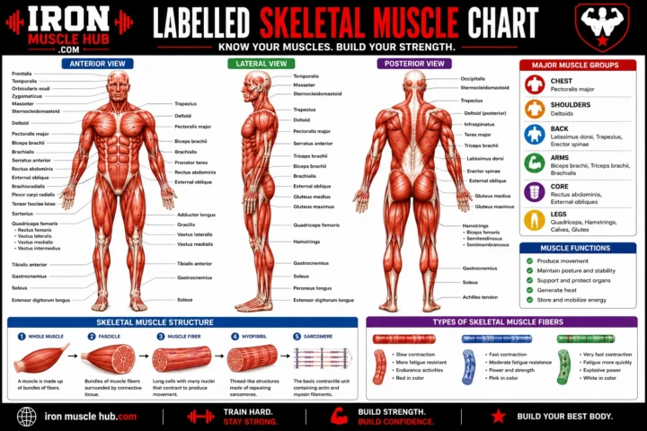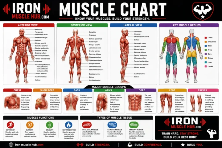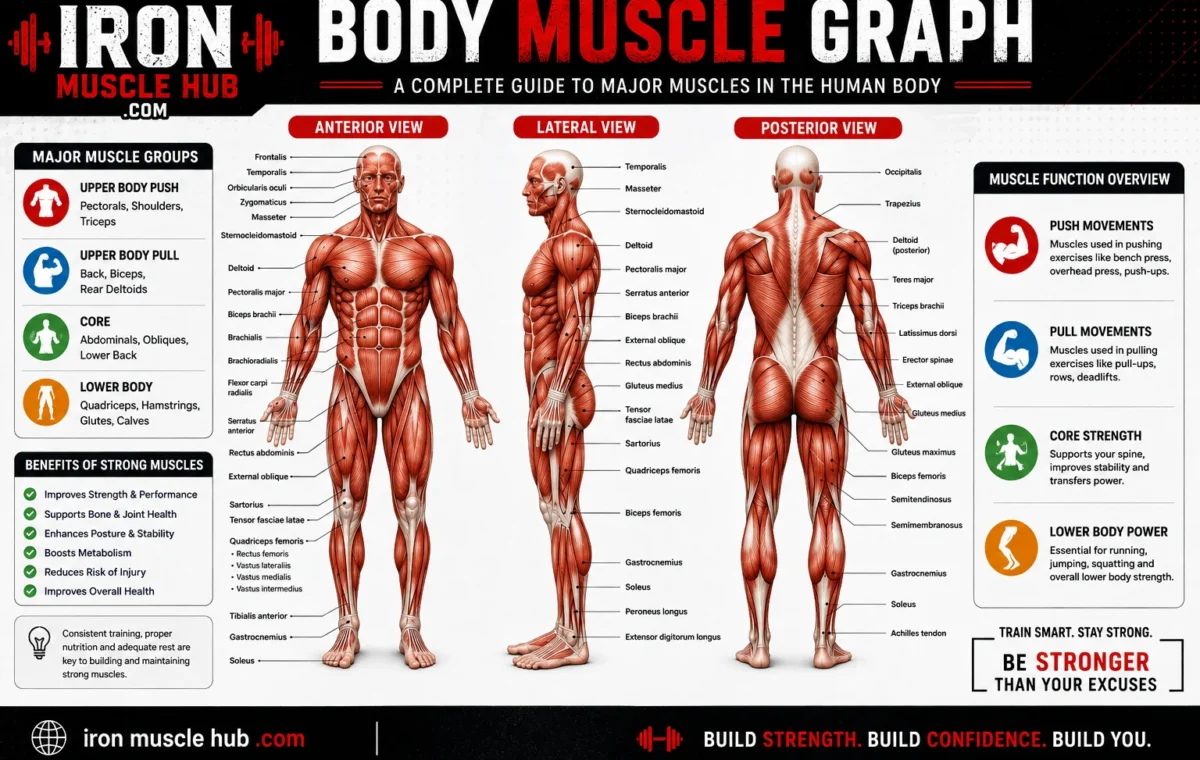
A body muscle graph is essentially a visual chart that shows where major muscles are located and, in many cases, what they do. It can help beginners identify muscles such as the biceps, triceps, chest, abdominal muscles, quadriceps, hamstrings, glutes and calf muscles without trying to memorize an entire anatomy textbook.
- Quick Answer: What Does a Body Muscle Graph Show?
- How to Read a Body Muscle Graph
- 1. Check the viewing angle
- 2. Start with large muscle groups
- 3. Follow the label carefully
- 4. Learn location before function
- Body Muscle Graph: Major Muscles to Know
- Chest: Pectoralis Major
- Shoulders: Deltoid
- Upper Back: Trapezius
- Back: Latissimus Dorsi
- Upper Arm: Biceps and Triceps
- Abdomen: Rectus Abdominis and Obliques
- Glutes: Gluteus Maximus
- Front Thigh: Quadriceps
- Back Thigh: Hamstrings
- Calf: Gastrocnemius and Soleus
- Front vs. Back Muscle Graph
- Why Muscle Charts Are Useful
- For beginners
- For exercise learners
- For students
- For general body awareness
- Three Types of Muscle Tissue
- A Simple Way to Memorize the Muscle Chart
- Day 1: Upper body
- Day 2: Core and hips
- Day 3: Legs
- A Worked Example: How to Use a Muscle Graph
- Common Mistakes When Using a Muscle Chart
- Mistake 1: Treating the chart as a complete anatomy textbook
- Mistake 2: Confusing a muscle with a muscle group
- Mistake 3: Assuming every muscle is visible
- Mistake 4: Using an anatomy graph to diagnose pain
- What Makes a Good Body Muscle Graph?
- Frequently Asked Questions
- What is a body muscle graph?
- How many muscles are in the human body?
- What are the major muscles shown on a body muscle chart?
- What is the difference between a muscle and a muscle group?
- Are all muscles shown on a body muscle graph?
- Can I use a muscle graph to learn exercise anatomy?
- Related Iron Muscle Hub Reading Ideas
- Final Takeaway: Use the Graph as Your Muscle Map
- Body Muscle Graph by Region
- Front View: What Should You Learn First?
- Pectoralis Major
- Deltoids
- Biceps Brachii
- Rectus Abdominis
- Quadriceps
- Back View: The Muscles You'll See
- Trapezius
- Latissimus Dorsi
- Triceps Brachii
- Gluteus Maximus
- Hamstrings
- Gastrocnemius
- Body Muscle Graph: A Simple Memory System
- Upper body
- Middle body
- Lower body
- How Muscle Groups Work Together
- Agonists and Antagonists: A Beginner Explanation
- Why Some Muscle Graphs Look Different
- Superficial vs. Deep Muscles
- Superficial muscles
- Deep muscles
- How to Study a Body Muscle Graph in 10 Minutes
- Minute 1–2: Scan the whole body
- Minute 3–4: Learn the upper body
- Minute 5–6: Study the trunk
- Minute 7–8: Study the legs
- Minute 9–10: Test yourself
- A Beginner's Muscle Chart Quiz
- Body Muscle Graph vs. Muscle Anatomy Diagram
- What a Muscle Chart Cannot Tell You
- Best Way to Build Your Own Muscle Reference
- The Most Important Muscle-Chart Lesson
- Frequently Asked Questions
- What is the easiest way to learn a body muscle graph?
- What are the five major muscle groups beginners should learn first?
- Why does my muscle chart have different labels from another chart?
- Is the biceps the biggest muscle in the arm?
- Should I memorize every muscle on a body muscle graph?
- Related Articles
- Final Takeaway
- Body Muscle Graph: The Complete Beginner Map
- 1. Head and Neck Muscles
- Sternocleidomastoid
- Muscles of Facial Expression
- 2. Chest and Shoulder Muscles
- Pectoralis Major
- Pectoralis Minor
- Deltoid
- 3. Back Muscle Graph
- Trapezius
- Latissimus Dorsi
- Erector Spinae
- 4. Arm Muscle Graph
- Biceps Brachii
- Triceps Brachii
- Forearm Muscles
- 5. Abdominal and Trunk Muscles
- Rectus Abdominis
- External Oblique
- Internal Oblique
- Transversus Abdominis
- 6. Hip and Thigh Muscle Graph
- Gluteus Maximus
- Gluteus Medius
- Quadriceps
- Hamstrings
- 7. Lower-Leg Muscle Graph
- Gastrocnemius
- Soleus
- Tibialis Anterior
- Example: A Push-Up
- Example: A Squat
- Location clues
- Number clues
- Shape or direction clues
- Biceps vs. Triceps
- Quadriceps vs. Hamstrings
- Gastrocnemius vs. Soleus
- Pectoralis vs. Deltoid
- What is a body muscle graph used for?
- What is the easiest muscle group to learn first?
- What are the major muscles on the front of the body?
- What are the major muscles on the back of the body?
- Can a body muscle graph show muscle function?
- Is a muscle graph the same as a muscular system diagram?
If you’ve searched for a body muscle graph because a diagram looked confusing, you’re not alone. Human muscle anatomy includes many structures, but you can make it much easier by learning the body region by region. This guide explains how to read a muscle chart, which major muscles to learn first, and how to use a diagram for studying, exercise or general body awareness. For more insights read here :- how to get bigger and Lean and Muscular.
Quick Answer: What Does a Body Muscle Graph Show?
A body muscle graph normally presents the major muscles of the human body visually, often using front (anterior) and back (posterior) views. Labels identify individual muscles or muscle groups and may include information about their location or function.
| Body region | Major muscles commonly shown | Basic role |
|---|---|---|
| Chest | Pectoralis major | Helps move the arm and shoulder |
| Shoulders | Deltoids | Help raise and move the arms |
| Upper back | Trapezius, latissimus dorsi | Assist shoulder and arm movement |
| Upper arm | Biceps, triceps | Help move the elbow and forearm |
| Abdomen | Rectus abdominis, obliques | Help stabilize and move the trunk |
| Buttocks | Gluteus maximus | Important for hip movement |
| Front thigh | Quadriceps | Extend the knee |
| Back thigh | Hamstrings | Help bend the knee and move the hip |
| Lower leg | Gastrocnemius, soleus | Help produce ankle movement |
These are major muscles and groups, not a complete list of every muscle in the body. The National Institute of Arthritis and Musculoskeletal and Skin Diseases says there are more than 650 muscles in the human body.
How to Read a Body Muscle Graph
A detailed anatomy chart can look complicated at first. The easiest approach is to read it in sections instead of attempting to learn every label at once.
1. Check the viewing angle
Most muscle diagrams use one or more of these perspectives:
- Anterior: the front of the body
- Posterior: the back of the body
- Lateral: the side of the body
A muscle that isn’t visible from the front may be much easier to identify from the back.
2. Start with large muscle groups
Don’t begin by memorizing small muscles.
Learn the major regions first:
- Shoulders
- Chest
- Back
- Arms
- Abdomen
- Glutes
- Thighs
- Calves
Once those areas make sense, individual muscle names become easier to remember.
3. Follow the label carefully
An anatomy diagram may place a line from a muscle to its name. Follow the line all the way to the structure rather than assuming that the nearest label belongs to the nearest muscle.
This is especially useful around the shoulder, forearm and thigh, where several muscles sit close together.
4. Learn location before function
For beginners, remembering where a muscle is located is often easier than immediately memorizing its full anatomical function.
For example, first recognize the biceps as a muscle on the front of the upper arm. Then learn that the biceps brachii contributes to elbow flexion and forearm rotation.
Body Muscle Graph: Major Muscles to Know
A useful beginner’s chart doesn’t need hundreds of labels. Start with the muscles that appear frequently in basic anatomy and movement discussions.
Chest: Pectoralis Major
The pectoralis major is the large muscle covering much of the front of the chest. It contributes to movements of the shoulder and upper arm.
Shoulders: Deltoid
The deltoid forms much of the rounded contour of the shoulder. It helps move the arm in several directions.
Upper Back: Trapezius
The trapezius covers a large area across the upper back and neck region. It participates in movements of the shoulder blades and shoulder girdle.
Back: Latissimus Dorsi
The latissimus dorsi, often called the “lats,” is a broad back muscle involved in movements of the upper arm.
Upper Arm: Biceps and Triceps
The biceps brachii is located on the front of the upper arm, while the triceps brachii is on the back.
They contribute to different elbow movements and are therefore commonly paired when studying arm anatomy.
Abdomen: Rectus Abdominis and Obliques
The rectus abdominis runs vertically along the front of the abdomen. The oblique muscles are positioned along the sides of the trunk.
Together with other muscles of the trunk, these structures contribute to movements and stabilization of the torso.
Glutes: Gluteus Maximus
The gluteus maximus is the large superficial muscle of the buttock region. It plays an important role in hip movement.
Front Thigh: Quadriceps
The quadriceps are a group of muscles on the front of the thigh. They are important for extending the knee.
Back Thigh: Hamstrings
The hamstrings are a group of muscles at the back of the thigh. They contribute to knee flexion and hip movement.
Calf: Gastrocnemius and Soleus
The gastrocnemius is the prominent superficial calf muscle. The soleus lies deeper and also contributes to movement at the ankle.
NCBI’s educational material similarly identifies structures such as the biceps brachii, deltoid, gastrocnemius, gluteus maximus, latissimus dorsi, pectoralis major, quadriceps and rectus abdominis among the major skeletal muscles.
Front vs. Back Muscle Graph
One of the most useful tricks is to divide your study into two views.
| Front view | Back view |
| Pectoralis major | Trapezius |
| Deltoids | Posterior deltoid |
| Biceps | Latissimus dorsi |
| Rectus abdominis | Erector spinae region |
| Obliques | Gluteus maximus |
| Quadriceps | Hamstrings |
| Tibialis anterior | Gastrocnemius |
This isn’t an exhaustive anatomy chart. It’s a beginner’s map designed to help you recognize the larger structures before moving into more detailed anatomy.
Why Muscle Charts Are Useful
A muscle graph isn’t only useful for medical students.
For beginners
A labeled chart gives you a visual reference when you hear names such as “deltoid” or “quadriceps.”
For exercise learners
Understanding which muscles are involved in a movement can make workout instructions easier to understand. It can also help you communicate more clearly with a qualified fitness or healthcare professional.
For students
Charts can turn a long list of anatomical names into a visual study aid.
For general body awareness
Learning basic anatomy can help you understand terms used in health and fitness information. It shouldn’t, however, be used to diagnose an injury or medical condition.
Cleveland Clinic explains that skeletal muscles attach to bones through tendons and contribute to movement, posture, joint stability and other functions.
Three Types of Muscle Tissue
A body muscle graph usually focuses on skeletal muscles, but the human body has three major types of muscle tissue:
- Skeletal muscle: generally under voluntary control and responsible for much of the body’s movement.
- Cardiac muscle: specialized muscle tissue found in the heart.
- Smooth muscle: found in organs and other internal structures and generally operates involuntarily.
The National Institute of Arthritis and Musculoskeletal and Skin Diseases identifies skeletal, smooth and cardiac muscle as the three main muscle types.
So, if you’re looking at a typical front-and-back body muscle chart, you’re usually looking primarily at the skeletal muscular system, not every type of muscle tissue in the body.
A Simple Way to Memorize the Muscle Chart
Instead of trying to memorize dozens of names in one sitting, use a regional approach.
Day 1: Upper body
Learn:
- Deltoid
- Pectoralis major
- Trapezius
- Latissimus dorsi
- Biceps
- Triceps
Day 2: Core and hips
Learn:
- Rectus abdominis
- Obliques
- Gluteus maximus
Day 3: Legs
Learn:
- Quadriceps
- Hamstrings
- Gastrocnemius
- Soleus
Then test yourself by looking at an unlabeled diagram. Point to a muscle and try to name it before checking the answer.
This active-recall approach is more useful than repeatedly staring at the same labeled picture.
A Worked Example: How to Use a Muscle Graph
Suppose you’re studying the movement of a basic squat.
Rather than trying to identify every muscle involved, start with the major groups shown on a beginner chart:
- Find the quadriceps on the front of the thigh.
- Find the gluteus maximus around the buttocks.
- Find the hamstrings on the back of the thigh.
- Look at the lower-leg muscles.
- Then learn how these muscles work together with the joints, bones and nervous system.
This gives you a useful mental map without pretending that one simple chart explains every detail of human movement.
Skeletal muscles attach to bones through tendons, and their contraction produces movement. They also contribute to posture, joint stability and heat production.
Common Mistakes When Using a Muscle Chart
Mistake 1: Treating the chart as a complete anatomy textbook
A simplified graph highlights selected structures. It may leave out smaller muscles, deeper muscles, nerves, blood vessels and connective tissues.
Better approach: Use the chart as your starting map, then move to a detailed anatomy reference when you need more precision.
Mistake 2: Confusing a muscle with a muscle group
“Quadriceps” refers to a group of muscles rather than one single muscle. “Hamstrings” also describes a group.
Better approach: Learn the group name first, then study the individual muscles inside it.
Mistake 3: Assuming every muscle is visible
Some muscles are deeper beneath other tissues.
Better approach: Check whether your diagram is showing superficial muscles, deeper layers or both.
Mistake 4: Using an anatomy graph to diagnose pain
A chart can show where a structure is located, but location alone doesn’t establish the cause of pain.
Better approach: For persistent, severe or concerning symptoms, seek advice from an appropriately qualified healthcare professional.
What Makes a Good Body Muscle Graph?
If you’re choosing a diagram for studying, look for these features:
- Clear front and back views
- Correct anatomical terminology
- Readable labels
- Logical grouping by body region
- High enough resolution to read small text
- A reputable educational or medical source
- Clear indication of whether the illustration shows superficial or deeper muscles
NCBI’s anatomy resources, for example, include detailed illustrations showing relationships among skeletal muscle, muscle fibers, connective-tissue layers, tendons and related structures.
For a website such as Iron Muscle Hub, an originally commissioned or created illustration is preferable to copying a commercial anatomy poster. It can also be designed specifically for beginner readers.
Frequently Asked Questions
What is a body muscle graph?
A body muscle graph is a visual anatomy reference that identifies muscles on the human body. Most beginner charts focus on skeletal muscles and may show front, back or side views. Labels usually identify major muscles such as the biceps, triceps, deltoids, pectorals, quadriceps, hamstrings and calf muscles.
How many muscles are in the human body?
There isn’t one universally useful number for every way of counting muscles, but the National Institute of Arthritis and Musculoskeletal and Skin Diseases states that humans have more than 650 muscles. Different sources can use different counting conventions, so a simple muscle chart will normally display only selected major muscles.
What are the major muscles shown on a body muscle chart?
Commonly labeled muscles include the pectoralis major, deltoid, trapezius, latissimus dorsi, biceps brachii, triceps brachii, rectus abdominis, gluteus maximus, quadriceps, hamstrings and gastrocnemius. The exact selection depends on how detailed the chart is.
What is the difference between a muscle and a muscle group?
A muscle is an individual anatomical structure, while a muscle group is a collection of muscles that are commonly discussed together because of their location or related actions. For example, the quadriceps is a group of muscles on the front of the thigh, not one single muscle.
Are all muscles shown on a body muscle graph?
No. A typical educational graph shows selected muscles rather than every muscle in the human body. Smaller muscles, deeper structures and detailed connective tissues may be omitted to keep the illustration readable.
Can I use a muscle graph to learn exercise anatomy?
Yes. A muscle graph can be a useful starting point for learning which major muscles are located in different body regions. However, a basic chart doesn’t explain every movement or muscle interaction, so more detailed anatomy and exercise-science resources may be needed for advanced study.
Related Iron Muscle Hub Reading Ideas
For readers building their basic anatomy knowledge, related articles could include:
- Muscles of the Body Labeled: A Beginner’s Visual Guide
- Muscle Anatomy Chart: Major Muscles and Their Functions
- Muscular System Diagram: How Muscles Work With Bones
These topics can be connected with descriptive internal links rather than repeating the same keyword on every page.
Final Takeaway: Use the Graph as Your Muscle Map
A body muscle graph is best treated as a visual starting point. Instead of trying to memorize every anatomical name, learn the large regions first: chest, shoulders, back, arms, abdomen, hips, thighs and calves. Then add individual muscles as your knowledge improves.
For beginners, the most useful chart is usually one that is clear, accurately labeled and easy to compare between front and back views. Medical and educational references such as NCBI and Cleveland Clinic can help you move beyond a simple diagram when you need more detailed information.
Body Muscle Graph: Part 2 — Major Muscle Groups Explained
A body muscle graph becomes much easier to understand when you stop treating it as one giant list of names. Instead, divide the body into regions and learn the major muscles in each area. This approach helps beginners recognize anatomy diagrams faster and understand basic movement without memorizing unnecessary detail.
Quick takeaway: Start with the chest, shoulders, back, arms, core, hips, thighs and lower legs. Learn the location first, then learn the basic function.
Body Muscle Graph by Region
| Region | Muscle or group | Where you’ll find it | Main movement role |
|---|---|---|---|
| Chest | Pectoralis major | Front of chest | Moves the upper arm |
| Shoulder | Deltoid | Around shoulder | Raises and moves the arm |
| Upper back | Trapezius | Upper back and neck | Helps move and position the shoulder blades |
| Back | Latissimus dorsi | Broad middle/lower back | Helps move the upper arm |
| Upper arm | Biceps brachii | Front of upper arm | Helps flex the elbow |
| Upper arm | Triceps brachii | Back of upper arm | Helps extend the elbow |
| Abdomen | Rectus abdominis | Front of abdomen | Helps flex the trunk |
| Side abdomen | Obliques | Sides of abdomen | Help rotate and bend the trunk |
| Hip | Gluteus maximus | Buttocks | Powerful hip extension |
| Thigh | Quadriceps | Front of thigh | Extends the knee |
| Thigh | Hamstrings | Back of thigh | Help flex the knee and extend the hip |
| Lower leg | Gastrocnemius | Back of calf | Helps move the ankle |
| Lower leg | Soleus | Deep calf | Helps produce ankle movement |
The table is intentionally simplified. Human movement involves many muscles working together, and individual muscles can perform more than one action.
Front View: What Should You Learn First?
A front-facing muscle graph typically makes several large structures easy to identify.
Pectoralis Major
The pectoralis major occupies much of the front of the chest. It contributes to movements of the upper arm at the shoulder.
When looking at a chart, find the broad muscle on each side of the upper chest. That’s usually the easiest starting point for identifying the chest region.
Deltoids
The deltoid sits over the shoulder joint and gives the shoulder much of its rounded appearance.
A useful memory trick is simple: deltoid = shoulder cap.
Different portions of the deltoid contribute to different arm movements, so a detailed anatomy resource may divide the muscle into sections.
Biceps Brachii
The biceps brachii is on the front of the upper arm.
It contributes to bending the elbow and also participates in rotating the forearm.
On a basic body muscle graph, it’s one of the easiest arm muscles to recognize.
Rectus Abdominis
The rectus abdominis runs vertically along the front of the abdomen.
It’s commonly associated with trunk flexion and is part of the abdominal wall.
Don’t assume that visible abdominal definition tells you everything about the underlying anatomy. A diagram is showing structures, not judging someone’s fitness level.
Quadriceps
The quadriceps occupy the front of the thigh and consist of four muscles.
For beginner study, remember:
Quadriceps → front of thigh → knee extension.
You can learn the individual muscles later if your course or training requires more detail.
Back View: The Muscles You’ll See
Turn the body muscle graph around and the picture changes considerably.
Trapezius
The trapezius covers a large area of the upper back and extends toward the neck and shoulders.
Its different portions have different functions involving the shoulder blades and neck.
Latissimus Dorsi
The latissimus dorsi is a broad muscle across much of the back.
It’s commonly called the lats in fitness settings.
It contributes to movements of the upper arm, including pulling actions.
Triceps Brachii
The triceps occupy the back of the upper arm.
The name gives you a useful clue: the muscle has three heads.
For basic learning, the important association is:
Triceps → back of upper arm → elbow extension.
Gluteus Maximus
The gluteus maximus is the large superficial muscle of the buttock.
It’s an important hip extensor and contributes to powerful movements involving the hip.
Hamstrings
The hamstrings are a group of muscles located on the back of the thigh.
They contribute to knee flexion and hip extension.
A simple comparison helps:
- Quadriceps: front of thigh
- Hamstrings: back of thigh
Gastrocnemius
The gastrocnemius is the prominent superficial muscle of the calf.
It crosses the knee and ankle joints and contributes to movement at the ankle.
Body Muscle Graph: A Simple Memory System
Trying to memorize anatomy alphabetically isn’t particularly useful.
Instead, create a mental map.
Upper body
Think:
Chest → shoulders → upper back → arms
Then attach the names:
- Chest → pectoralis major
- Shoulder → deltoid
- Upper back → trapezius
- Front arm → biceps
- Back arm → triceps
Middle body
Think:
Front abdomen → sides → lower back
Then identify:
- Rectus abdominis
- Obliques
- Back muscles
Lower body
Think:
Hip → front thigh → back thigh → calf
Then identify:
- Gluteus maximus
- Quadriceps
- Hamstrings
- Gastrocnemius
- Soleus
This gives you a simple mental filing system.
How Muscle Groups Work Together
One important limitation of a body muscle graph is that it can make muscles appear isolated.
Your body doesn’t work that way.
During a movement, multiple muscles can contribute to the action. Some muscles create movement, some stabilize joints, and others assist or oppose the movement.
For example, a squat isn’t simply a “quadriceps exercise.” The movement involves the hips, knees, ankles and trunk, with multiple muscles contributing to control and force production.
Skeletal muscles produce movement by contracting and transmitting force through connective tissues to the skeleton. They also contribute to posture and joint stability. (Cleveland Clinic; NCBI Bookshelf)
Agonists and Antagonists: A Beginner Explanation
You may eventually encounter the terms agonist and antagonist.
An agonist is generally the muscle primarily responsible for producing a particular movement, while an antagonist performs an opposing action.
A common beginner example is the relationship between the biceps and triceps around the elbow.
But don’t think of this as a permanent “one muscle works, the other switches off” arrangement. Real movement is more coordinated, and muscles can have different roles depending on the movement.
Why Some Muscle Graphs Look Different
You may find two anatomy charts that don’t look identical.
That doesn’t automatically mean one is wrong.
Differences can occur because:
- One chart shows only superficial muscles.
- Another includes deeper muscles.
- One combines muscles into groups.
- Another labels individual muscles.
- Different viewing angles are used.
- Different educational purposes require different levels of detail.
Before comparing two charts, check what each illustration is designed to teach.
Superficial vs. Deep Muscles
This is one of the most important concepts for reading anatomy diagrams.
Superficial muscles
These are closer to the surface and are generally easier to see in a basic muscular-system illustration.
Examples include:
- Deltoid
- Pectoralis major
- Biceps brachii
- Triceps brachii
- Rectus abdominis
- Gluteus maximus
- Gastrocnemius
Deep muscles
These lie beneath other structures.
A simplified graph may not show them at all.
This is why a basic internet muscle chart shouldn’t be treated as a complete anatomical reference.
How to Study a Body Muscle Graph in 10 Minutes
If you’re learning anatomy for the first time, try this routine.
Minute 1–2: Scan the whole body
Don’t memorize anything yet.
Identify the major regions.
Minute 3–4: Learn the upper body
Point to:
- Chest
- Shoulder
- Upper back
- Front upper arm
- Back upper arm
Name each structure.
Minute 5–6: Study the trunk
Find:
- Rectus abdominis
- Obliques
- Major back structures
Minute 7–8: Study the legs
Find:
- Quadriceps
- Hamstrings
- Gastrocnemius
- Gluteus maximus
Minute 9–10: Test yourself
Cover the labels.
Point to each muscle and name it without looking.
If you miss one, reveal the answer and try again.
This is more productive than simply rereading the labels.
A Beginner’s Muscle Chart Quiz
Try answering these without looking back.
- Which major muscle group is on the front of the thigh?
- Which muscle is commonly associated with the front of the upper arm?
- Which muscle group is found on the back of the thigh?
- What is the large superficial muscle of the buttock?
- Which major muscle covers much of the shoulder?
- Where is the gastrocnemius located?
- Which large muscle lies across much of the upper back?
Answers:
- Quadriceps
- Biceps brachii
- Hamstrings
- Gluteus maximus
- Deltoid
- Back of the lower leg/calf
- Trapezius
Body Muscle Graph vs. Muscle Anatomy Diagram
These phrases are sometimes used interchangeably, but they can describe slightly different resources.
A body muscle graph usually emphasizes location and labels.
A muscle anatomy diagram may go further by showing:
- Muscle attachments
- Tendons
- Muscle fibers
- Layers
- Origins and insertions
- Detailed anatomical relationships
If you’re simply trying to learn the names and locations of major muscles, a clear labeled chart is usually enough.
If you’re studying anatomy formally, you’ll probably need a much more detailed reference.
What a Muscle Chart Cannot Tell You
A picture is useful, but it has limits.
It generally cannot tell you:
- Why a particular area hurts
- Whether an injury has occurred
- How much weight you should lift
- Whether a specific exercise is appropriate for you
- Whether a muscle is “weak” based only on appearance
- How your individual anatomy differs from another person’s
Those questions require additional context and, where appropriate, guidance from qualified professionals.
Best Way to Build Your Own Muscle Reference
If you’re creating notes, don’t copy an entire anatomy poster.
Build a compact reference with four columns:
| Muscle | Location | Basic role | Easy memory clue |
| Deltoid | Shoulder | Arm movement | Shoulder cap |
| Biceps | Front upper arm | Elbow flexion | Front arm |
| Triceps | Back upper arm | Elbow extension | Back arm |
| Pectoralis major | Chest | Upper-arm movement | Chest |
| Quadriceps | Front thigh | Knee extension | Front thigh |
| Hamstrings | Back thigh | Knee flexion/hip movement | Back thigh |
| Gastrocnemius | Calf | Ankle movement | Calf |
This format forces you to connect name + location + function, rather than memorizing words separately.
The Most Important Muscle-Chart Lesson
Don’t judge your progress by how many muscle names you can recite.
The real goal is being able to look at a diagram and quickly answer three questions:
Where is the muscle?
What is it called?
What does it generally help the body do?
Once you can answer those questions for the major muscle groups, more advanced anatomy becomes much easier.
Frequently Asked Questions
What is the easiest way to learn a body muscle graph?
Start with eight regions: chest, shoulders, back, arms, abdomen, hips, thighs and lower legs. Learn the largest and most commonly labeled muscles in each region before studying smaller structures. Then practice with an unlabeled diagram so you’re recalling the information rather than simply recognizing it.
What are the five major muscle groups beginners should learn first?
There isn’t one official five-group system for every purpose. For a simple study framework, beginners can start with the chest, back, shoulders, arms and legs, then add the abdominal and hip regions. This makes it easier to organize individual muscles without trying to memorize the entire muscular system at once.
Why does my muscle chart have different labels from another chart?
Charts can differ because they show different anatomical layers, levels of detail or educational purposes. One diagram might label muscle groups while another identifies individual muscles. Check whether each image represents superficial or deeper anatomy before deciding that the labels conflict.
Is the biceps the biggest muscle in the arm?
No. The biceps brachii is a prominent muscle on the front of the upper arm, but the upper arm contains several muscles. The triceps brachii is located on the back of the upper arm and has three heads. A basic body muscle graph often labels both because they’re important landmarks.
Should I memorize every muscle on a body muscle graph?
Not if you’re a beginner. Start with the major muscles and groups that appear most often in basic anatomy resources. Once you know their locations, you can gradually add smaller or deeper muscles if your studies or professional training require them.
Related Articles
For a complete beginner-friendly anatomy content cluster, consider linking this article to:
- [Muscle Anatomy Chart: Major Muscles Explained]
- [Body Diagram of Muscles: Front and Back Views]
- [Muscular System Diagram: A Beginner’s Guide]
These internal links can help readers move from a simple visual chart to more detailed anatomy explanations.
Final Takeaway
A body muscle graph works best as a map, not as a giant memorization exercise. Learn the large regions first, connect each major muscle to its location, and then add its basic movement role. Front and back views are especially helpful because many muscles are easier to identify from one side than the other.
Most importantly, remember that a chart is a simplified representation of a complex biological system. Use reputable anatomy resources when you need greater detail, and don’t use a diagram alone to diagnose pain or injury.
Body Muscle Graph: Part 3 — Complete Beginner Reference
A body muscle graph is most useful when you can turn a visual label into a simple mental picture: where the muscle sits, what it generally does, and whether it’s on the front, back, or side of the body. You don’t need to memorize every anatomical structure in one sitting.
This final part brings the major concepts together into a practical reference you can use for revision, anatomy study, or understanding basic fitness terminology.
Body Muscle Graph: The Complete Beginner Map
Think of the body as seven connected regions:
- Head and neck
- Shoulders and chest
- Back
- Arms and forearms
- Abdomen and trunk
- Hips and thighs
- Lower legs
This regional approach is easier than memorizing a long alphabetical list.
1. Head and Neck Muscles
Most basic body muscle charts don’t label every facial or neck muscle. They usually focus on a few larger or easily identifiable structures.
Sternocleidomastoid
The sternocleidomastoid, often shortened to SCM, is a prominent muscle on each side of the neck.
It helps with movements of the head and neck.
Because the name is long, beginners often find it easier to learn the location first and the full name second.
Muscles of Facial Expression
The face contains several muscles that help create expressions such as smiling, frowning and closing the eyes.
A basic whole-body muscle graph usually leaves most of these out to avoid making the diagram too crowded.
2. Chest and Shoulder Muscles
The upper torso contains several muscles that appear frequently on anatomy charts.
Pectoralis Major
The pectoralis major is the large superficial muscle on the front of the chest.
It contributes to movement of the upper arm at the shoulder.
Pectoralis Minor
The pectoralis minor lies deeper than the pectoralis major.
Because it’s beneath a more superficial muscle, it may not appear on a simple front-view chart.
Deltoid
The deltoid covers the shoulder.
Its three portions are commonly described as anterior, middle and posterior parts. Together, they contribute to different movements of the arm.
Easy memory clue:
Deltoid = shoulder cap.
3. Back Muscle Graph
The back contains several large muscles that are easy to recognize on a posterior anatomy diagram.
Trapezius
The trapezius occupies a broad region across the upper back and neck.
It helps move and stabilize the shoulder blades and contributes to neck and shoulder movement.
Latissimus Dorsi
The latissimus dorsi is a broad muscle covering much of the middle and lower back.
It’s often called the lats.
It contributes to movements of the upper arm, particularly movements that pull the arm toward the body.
Erector Spinae
The erector spinae refers to a group of muscles running along the back of the spine.
They play important roles in extending and stabilizing the spine.
A simplified muscle graph may represent this area differently depending on how detailed the illustration is.
4. Arm Muscle Graph
The arm is one of the easiest regions to learn because the major muscles have clear positions.
Biceps Brachii
The biceps brachii is located on the front of the upper arm.
It helps flex the elbow and contributes to forearm supination.
Memory clue:
Biceps = front of upper arm.
Triceps Brachii
The triceps brachii is located on the back of the upper arm.
It contributes to elbow extension.
Memory clue:
Triceps = back of upper arm.
Forearm Muscles
The forearm contains numerous muscles rather than one large muscle.
Some control movements of the wrist and fingers, while others help rotate the forearm.
This is why a detailed forearm anatomy diagram can look considerably more complicated than a basic body muscle graph.
5. Abdominal and Trunk Muscles
The trunk provides another useful group of landmarks.
Rectus Abdominis
The rectus abdominis runs vertically along the front of the abdomen.
It contributes to flexion of the trunk.
External Oblique
The external obliques are positioned on the sides of the abdomen.
They contribute to trunk rotation and side-bending movements, along with other abdominal muscles.
Internal Oblique
The internal obliques lie deeper than the external obliques.
They also contribute to trunk movement and stabilization.
Transversus Abdominis
The transversus abdominis is a deeper abdominal muscle with fibers running generally horizontally.
It’s involved in abdominal compression and trunk stability.
A basic graph often doesn’t show this muscle clearly because it lies beneath more superficial layers.
6. Hip and Thigh Muscle Graph
The hips and thighs contain some of the largest muscle groups in the body.
Gluteus Maximus
The gluteus maximus is the large superficial muscle of the buttock.
It is an important muscle for hip extension.
Gluteus Medius
The gluteus medius sits more toward the outer side of the hip.
It contributes to hip movement and helps stabilize the pelvis during activities such as walking.
Quadriceps
The quadriceps are a four-muscle group at the front of the thigh.
The group includes:
- Rectus femoris
- Vastus lateralis
- Vastus medialis
- Vastus intermedius
The quadriceps collectively play a major role in extending the knee.
Hamstrings
The hamstrings are located at the back of the thigh.
They include several muscles that contribute to movements at the knee and hip.
For beginners, the easiest association is:
Quadriceps = front thigh
Hamstrings = back thigh
7. Lower-Leg Muscle Graph
The lower leg has several important muscles, but two are particularly familiar to beginners.
Gastrocnemius
The gastrocnemius is the large superficial calf muscle.
It crosses the knee and ankle and contributes to plantarflexion of the ankle.
Soleus
The soleus sits deep to the gastrocnemius.
It also contributes to plantarflexion at the ankle.
Because the soleus lies underneath the gastrocnemius, a basic external muscle chart may not show its complete shape.
Tibialis Anterior
The tibialis anterior is located along the front of the lower leg.
It contributes to dorsiflexion of the ankle.
Memory clue:
Tibialis anterior = front of lower leg.
Front and Back Muscle Chart
Here’s a compact reference you can save for revision.
| Body area | Front view | Back view |
|---|---|---|
| Shoulder | Deltoid | Deltoid |
| Chest | Pectoralis major | — |
| Upper back | — | Trapezius |
| Middle/lower back | — | Latissimus dorsi |
| Upper arm | Biceps | Triceps |
| Abdomen | Rectus abdominis | — |
| Side trunk | Obliques | — |
| Hip | — | Gluteus maximus |
| Thigh | Quadriceps | Hamstrings |
| Lower leg | Tibialis anterior | Gastrocnemius |
The dash doesn’t mean there are no muscles in that area. It simply means the listed muscle isn’t normally the main structure highlighted from that particular view.
How to Use a Body Muscle Graph for Exercise
A muscle graph can make fitness terminology easier to understand.
For example, you might see an exercise described as targeting the “lats.”
If you know that the lats are the latissimus dorsi, you can locate the muscle on a back-view chart.
But don’t interpret “targeting” to mean that only one muscle is working.
Most movements involve several muscles and joints.
Example: A Push-Up
A push-up involves more than the chest.
A simplified anatomy review might identify contributions from:
- Pectoralis major
- Triceps brachii
- Deltoids
- Trunk and shoulder stabilizing muscles
The exact contribution of each muscle changes with technique, body position and other factors.
Example: A Squat
A squat also involves multiple structures.
A beginner’s graph can help you identify:
- Quadriceps
- Gluteus maximus
- Hamstrings
- Calf muscles
- Trunk musculature
The exercise can’t be reduced to one muscle simply because a fitness article gives it a particular label.
Muscle Graph vs. Muscle Group Chart
These terms can sound interchangeable, but they can serve different purposes.
| Resource | Main purpose | Best for |
| Body muscle graph | Shows muscles on the body | Beginners |
| Muscle group chart | Organizes muscles by region/function | Fitness learners |
| Detailed anatomy diagram | Shows structures and relationships | Anatomy students |
| Muscle origin/insertion chart | Shows attachment points | Advanced study |
| Movement diagram | Connects muscles with actions | Exercise/anatomy study |
If you’re just beginning, start with the body muscle graph.
Don’t jump immediately into highly detailed origin-and-insertion charts unless you actually need that level of information.
A Better Way to Remember Muscle Names
Muscle names often contain useful clues.
Location clues
Some names tell you where a muscle is found.
For example:
- Rectus abdominis → abdomen
- Tibialis anterior → front of lower leg
- Pectoralis major → chest
Number clues
Some names describe the number of parts.
- Biceps → two heads
- Triceps → three heads
- Quadriceps → four muscles in the group
Shape or direction clues
Anatomical terminology may also describe shape, direction, size or position.
Once you recognize these naming patterns, unfamiliar terms become less intimidating.
Body Muscle Graph Study Checklist
Use this as a quick revision checklist.
- Identify the front and back views.
- Find the deltoids.
- Find the pectoralis major.
- Find the trapezius.
- Find the latissimus dorsi.
- Find the biceps.
- Find the triceps.
- Find the rectus abdominis.
- Find the obliques.
- Find the gluteus maximus.
- Find the quadriceps.
- Find the hamstrings.
- Find the gastrocnemius.
- Find the tibialis anterior.
- Test yourself using an unlabeled diagram.
Common Anatomy Confusions
Biceps vs. Triceps
Biceps: front of upper arm.
Triceps: back of upper arm.
Don’t rely only on the word “bi” or “tri.” Learn their locations and basic actions together.
Quadriceps vs. Hamstrings
Quadriceps: front of thigh.
Hamstrings: back of thigh.
This front/back pairing is one of the simplest landmarks on a muscle graph.
Gastrocnemius vs. Soleus
Both are major calf muscles.
The gastrocnemius is more superficial, while the soleus lies deeper.
Pectoralis vs. Deltoid
The pectoralis major is primarily associated with the chest.
The deltoid surrounds the shoulder.
On a front-view diagram, the two structures sit close together, so following the label line carefully matters.
Why Accurate Muscle Diagrams Matter
A muscle chart isn’t just decoration.
A poorly labeled diagram can teach incorrect associations that become harder to correct later.
When selecting a reference, check whether the source identifies the anatomy clearly and whether the diagram is intended for beginners, fitness education or formal anatomical study.
Reputable educational resources such as NCBI and Cleveland Clinic provide anatomy information that can be used alongside visual diagrams. The National Institute of Arthritis and Musculoskeletal and Skin Diseases also explains the basic roles and types of muscle tissue. (NCBI; Cleveland Clinic; NIAMS)
How to Make a High-Quality Body Muscle Graph
If you’re designing a muscle chart for a website, classroom or study notes, clarity should come before decoration.
A useful chart should have:
- Front and back views
- Readable labels
- Consistent terminology
- Enough space between labels
- Clear leader lines
- A logical order
- A note explaining what is and isn’t included
Avoid putting dozens of labels into a tiny image. A crowded diagram may technically contain more information but can be harder for beginners to use.
A better option is sometimes to create two diagrams: one for major superficial muscles and another for deeper structures.
Frequently Asked Questions
What is a body muscle graph used for?
A body muscle graph is primarily used as a visual reference for identifying muscles and understanding their general locations. Students, fitness beginners and readers learning basic anatomy can use one to connect anatomical names with body regions. A detailed medical anatomy reference is more appropriate for advanced study.
What is the easiest muscle group to learn first?
The upper arm is often a convenient starting point because the biceps and triceps have clear front-and-back locations. From there, you can move to the chest, shoulders, back, abdomen and legs. Learning muscles by body region is generally easier than trying to memorize a long list in alphabetical order.
What are the major muscles on the front of the body?
A beginner chart commonly identifies the pectoralis major, deltoids, biceps, rectus abdominis, obliques, quadriceps and tibialis anterior. There are many additional muscles, including deeper structures that may not appear on a simplified diagram.
What are the major muscles on the back of the body?
Commonly labeled posterior muscles include the trapezius, latissimus dorsi, triceps, gluteus maximus, hamstrings and gastrocnemius. A detailed chart may also show muscles such as the erector spinae and smaller structures.
Can a body muscle graph show muscle function?
Some charts include basic functions, while others only provide names and locations. For learning movement, it’s better to combine a labeled anatomy diagram with a reliable explanation of muscle actions. A picture alone usually doesn’t provide enough information to understand complex movement.
Is a muscle graph the same as a muscular system diagram?
Not necessarily. The terms are sometimes used loosely, but a muscle graph may be a simple labeled picture, while a muscular-system diagram can be much more detailed. Always check what structures and anatomical layers the particular diagram includes.
Final Takeaway: Turn the Chart Into a Mental Map
The best way to use a body muscle graph isn’t to stare at it until the names look familiar. Use it actively. Identify a region, name the muscle, recall its general role, then test yourself without the labels.
Start with the big landmarks: pectoralis major, deltoid, trapezius, latissimus dorsi, biceps, triceps, rectus abdominis, gluteus maximus, quadriceps, hamstrings and gastrocnemius. Once those become familiar, smaller muscles and deeper anatomical structures are much easier to learn.
A good chart should make anatomy simpler, not more confusing. Use reputable references for detailed study, and remember that an educational diagram isn’t a substitute for professional assessment of pain, injury or other medical concerns.