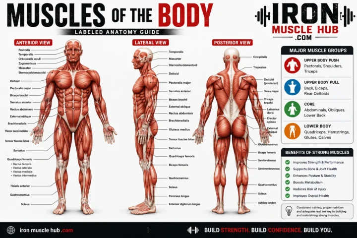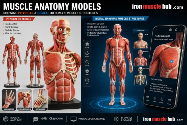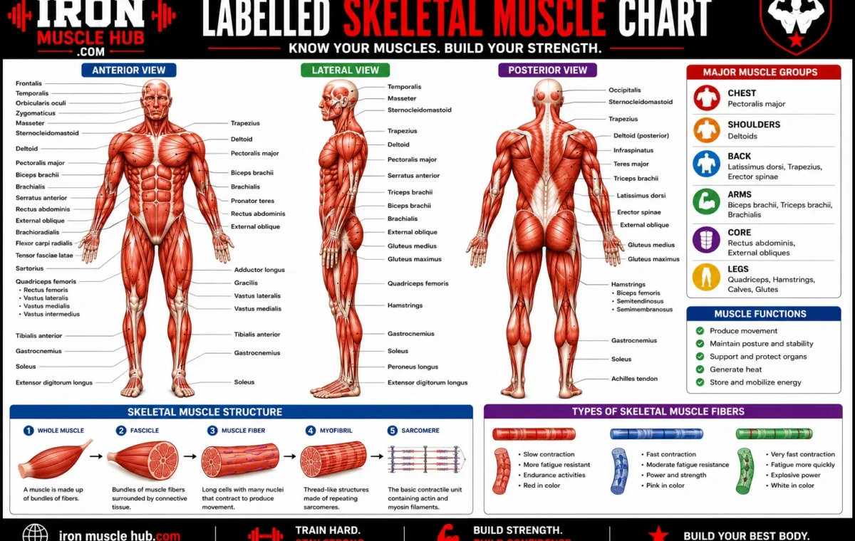
To label skeletal muscle correctly, start with the large structures and work inward: bone, tendon, whole muscle, fascicle, muscle fiber, myofibril and sarcomere. The connective-tissue coverings are epimysium, perimysium and endomysium, while the microscopic contractile proteins include actin and myosin. Skeletal muscles attach to bones through tendons and contract in response to signals from the nervous system, producing movement and helping maintain posture.
- Quick Answer: Skeletal Muscle Labels
- What Is Skeletal Muscle?
- How to Label a Skeletal Muscle Diagram
- 1. Bone
- 2. Tendon
- 3. Muscle Belly
- 4. Epimysium
- 5. Fascicle
- 6. Perimysium
- 7. Muscle Fiber
- 8. Endomysium
- The Smaller Parts: Sarcolemma, Myofibrils and Sarcomeres
- Sarcolemma
- Myofibrils
- Sarcomere
- How Skeletal Muscle Produces Movement
- A Simple Worked Example
- The Most Common Labeling Mistakes
- Mixing up epimysium, perimysium and endomysium
- Calling a fascicle a muscle fiber
- Confusing a tendon with a ligament
- Forgetting the microscopic labels
- A Fast Memory System for Exams
- Why Skeletal Muscle Matters
- Internal-Link Ideas for Iron Muscle Hub
- Frequently Asked Questions
- What are the main parts of skeletal muscle?
- What are the three layers of skeletal muscle connective tissue?
- What is a fascicle in skeletal muscle?
- Why is skeletal muscle called striated muscle?
- Is skeletal muscle voluntary or involuntary?
- What is the difference between a tendon and a skeletal muscle?
- A Simple Way to Remember the Whole Diagram
- Understanding the Structure Makes Anatomy Easier
- How the Skeletal Muscle Hierarchy Fits Together
- Whole Muscle
- Fascicles
- Muscle Fibers
- Myofibrils
- Sarcomeres
- The Three Connective-Tissue Layers Made Easy
- Epi = Entire Muscle
- Peri = Pack or Fascicle
- Endo = Each Fiber
- What Is the Sarcolemma?
- What Is the Sarcoplasm?
- Actin and Myosin: The Proteins Behind Contraction
- What Are Z Discs?
- What Are the A Band and I Band?
- How Nerves Connect With Skeletal Muscle
- Why Blood Vessels Appear on Muscle Diagrams
- A Better Way to Read a Labeling Diagram
- Quick Study Chart
- One-Line Memory Trick
- Final Part 2 Takeaway
- A Step-by-Step Labeling Method
- Step 1: Find the Bone
- Step 2: Find the Tendon
- Step 3: Identify the Muscle Belly
- Step 4: Locate the Epimysium
- Step 5: Identify the Fascicle
- Step 6: Find the Perimysium
- Step 7: Identify the Muscle Fiber
- Step 8: Find the Endomysium
- Step 9: Look Inside the Muscle Fiber
- Step 10: Find the Myofibrils
- Step 11: Identify the Sarcomere
- Step 12: Find Actin and Myosin
- The Complete Skeletal Muscle Label Sequence
- How to Recognize a Skeletal Muscle Under a Microscope
- Striations
- Long Fibers
- Multiple Nuclei
- Skeletal Muscle vs. Cardiac Muscle vs. Smooth Muscle
- What Does Skeletal Muscle Do?
- Movement
- Posture
- Joint Stability
- Heat Production
- Common Exam Questions
- “What surrounds a fascicle?”
- “What surrounds the entire muscle?”
- “What surrounds an individual muscle fiber?”
- “What is the functional unit of skeletal muscle?”
- “What proteins are responsible for muscle contraction?”
- “What connects muscle to bone?”
- A 30-Second Revision Test
- Answers
- The Biggest Mistake to Avoid
- Final Study Summary
- Final Thoughts
If you’re looking at a muscle diagram for a class, exam or anatomy assignment, the hardest part usually isn’t memorizing every term. It’s understanding how the pieces fit together. Once you see the structure from the whole muscle down to the tiny sarcomere, the labels become much easier to remember. For more insights read here :- how to build muscle and leaner physique .
Quick Answer: Skeletal Muscle Labels
| Label | What it means |
|---|---|
| Bone | The skeletal structure that the muscle helps move |
| Tendon | Strong connective tissue that attaches muscle to bone |
| Muscle belly | The main fleshy portion of a muscle |
| Epimysium | Connective-tissue layer surrounding the whole muscle |
| Fascicle | A bundle of muscle fibers |
| Perimysium | Connective tissue surrounding each fascicle |
| Muscle fiber | An individual skeletal muscle cell |
| Endomysium | Connective tissue surrounding an individual muscle fiber |
| Sarcolemma | Cell membrane surrounding a muscle fiber |
| Myofibril | Contractile structure running inside a muscle fiber |
| Sarcomere | Repeating functional unit responsible for muscle contraction |
| Actin and myosin | Protein filaments involved in contraction |
These labels describe a hierarchy: whole muscle → fascicle → muscle fiber → myofibril → sarcomere → actin and myosin.
What Is Skeletal Muscle?
Skeletal muscle is the type of muscle tissue primarily responsible for moving your skeleton. It is called striated muscle because its fibers have a striped appearance under a microscope, and most skeletal muscle is under voluntary control through signals from the nervous system.
Skeletal muscle doesn’t work alone. It connects with bones through tendons, and its contractions create forces that move bones around joints. It also contributes to posture, joint stability and heat production.
There are three major types of muscle tissue:
- Skeletal muscle — generally voluntary and striated.
- Cardiac muscle — found in the heart and involuntary.
- Smooth muscle — found in many internal organs and involuntary.
How to Label a Skeletal Muscle Diagram
The easiest method is to label the diagram from outside to inside.
1. Bone
Bone provides the rigid framework that skeletal muscles act upon. A muscle contraction can pull on a bone through its tendon, producing movement around a joint.
If your diagram shows a long bone at either end of a muscle, that structure should normally be labeled bone.
2. Tendon
A tendon is strong connective tissue that connects skeletal muscle to bone. It helps transfer the force produced by a contracting muscle to the skeleton.
On a typical anatomy diagram, the tendon appears at the end of the muscle and may look lighter and more fibrous than the muscle belly.
3. Muscle Belly
The muscle belly is the main fleshy portion of the muscle between its attachment areas.
It’s often the easiest part to recognize because it forms the thick, central portion of a simplified skeletal-muscle illustration.
4. Epimysium
The epimysium is the connective-tissue covering around the entire skeletal muscle.
Think of it as the outer wrapping around the whole muscle. OpenStax and NCBI describe this connective-tissue organization as one of the main structural layers that packages skeletal muscle.
5. Fascicle
A fascicle is a bundle of individual muscle fibers grouped together inside a skeletal muscle.
If a diagram shows the muscle being cut open to reveal smaller bundles inside it, those bundles are likely fascicles.
6. Perimysium
The perimysium surrounds each fascicle.
A useful memory trick is:
- Epi = entire muscle
- Peri = around a fascicle
- Endo = around an individual fiber
That simple sequence can make three commonly confused labels much easier to remember.
7. Muscle Fiber
A muscle fiber is an individual skeletal muscle cell.
Each fiber contains many smaller structures called myofibrils. Skeletal muscle fibers have the characteristic striated appearance associated with the organized arrangement of their contractile proteins.
8. Endomysium
The endomysium is the connective tissue surrounding an individual muscle fiber.
So, if you’re labeling from outside to inside, remember:
Epimysium → muscle → perimysium → fascicle → endomysium → muscle fiber.
This is one of the most important sequences to learn for a skeletal muscle labeling exercise.
The Smaller Parts: Sarcolemma, Myofibrils and Sarcomeres
Once you’ve labeled the major structures, some diagrams go deeper into the muscle fiber.
Sarcolemma
The sarcolemma is the cell membrane of a skeletal muscle fiber. It surrounds the muscle cell and helps separate its internal contents from the surrounding environment.
Myofibrils
Inside a muscle fiber are many myofibrils. These elongated structures contain the proteins and repeating units involved in contraction.
A single muscle fiber can therefore be thought of as a cell containing many myofibrils.
Sarcomere
The sarcomere is the repeating functional unit of a skeletal muscle fiber.
Sarcomeres contain organized actin and myosin filaments. Their arrangement creates the microscopic stripes associated with skeletal muscle and allows contraction to occur.
A useful hierarchy is:
Muscle → fascicle → muscle fiber → myofibril → sarcomere → actin and myosin
That’s the key structure to remember if you’re studying a detailed skeletal muscle diagram.
How Skeletal Muscle Produces Movement
The basic idea is simpler than the terminology makes it sound.
First, the nervous system sends a signal to a skeletal muscle fiber. The signal initiates processes inside the fiber that allow actin and myosin to interact. The sarcomeres then generate force, causing the muscle fiber to contract.
The force produced by the muscle is transferred through connective tissues and the tendon to the bone. Because the muscle pulls on the bone, the skeleton can move.
Skeletal muscles also make small adjustments that help keep the body upright and stabilize joints.
A Simple Worked Example
Imagine you’re labeling a diagram of the upper arm.
You see a large muscle connected to bones at either end. The thick central portion is the muscle belly, while the tougher structures connecting it toward the bones are tendons.
Now imagine the diagram zooms into the muscle. The outer covering is the epimysium. Inside, smaller bundles are fascicles, each surrounded by perimysium.
Zoom in again and you’ll see individual muscle fibers, each surrounded by endomysium. Inside those fibers are myofibrils, which contain repeating sarcomeres made with actin and myosin.
So the labels aren’t random vocabulary. Each smaller structure sits inside the larger one.
The Most Common Labeling Mistakes
Mixing up epimysium, perimysium and endomysium
This is probably the most common problem.
Remember the order:
Epimysium = whole muscle
Perimysium = fascicle
Endomysium = muscle fiber
Calling a fascicle a muscle fiber
A fascicle is a bundle of muscle fibers. A muscle fiber is one individual muscle cell.
If the diagram shows a group of many fibers surrounded by a layer of connective tissue, you’re probably looking at a fascicle.
Confusing a tendon with a ligament
A tendon connects muscle to bone. Ligaments connect bone to bone.
For a basic skeletal-muscle diagram, a fibrous structure connecting the muscle to a bone is the tendon.
Forgetting the microscopic labels
Some diagrams stop at the muscle fiber. Others continue into the cell and ask for the sarcolemma, myofibril, sarcomere, actin or myosin.
Before labeling, check how far the diagram has been magnified. That tells you which level of anatomy you’re expected to identify.
A Fast Memory System for Exams
Instead of memorizing a long list, learn the structure in layers.
Layer 1: Attachments
- Bone
- Tendon
Layer 2: Whole muscle
- Muscle belly
- Epimysium
Layer 3: Bundles
- Fascicle
- Perimysium
Layer 4: Individual cell
- Muscle fiber
- Endomysium
- Sarcolemma
Layer 5: Inside the cell
- Myofibril
- Sarcomere
- Actin
- Myosin
This approach gives you a mental map rather than a collection of disconnected terms.
Why Skeletal Muscle Matters
Skeletal muscle does much more than make movement possible. It helps maintain posture, contributes to joint stability and produces heat when muscles contract.
Muscle activity is also part of everyday life. Walking, climbing stairs, lifting objects and many forms of exercise involve skeletal muscle. Muscle-strengthening activities include exercises such as push-ups and weight lifting.
Understanding the anatomy can make fitness information easier to understand because you can connect an exercise with the muscles and structures involved.
Internal-Link Ideas for Iron Muscle Hub
Consider adding links to related articles such as:
- Skeletal Muscle vs Cardiac Muscle: What’s the Difference?
- Body Muscle Chart: Major Muscles Explained
- Muscle Anatomy Chart: A Beginner’s Guide
These would give readers natural next steps after learning how to label skeletal muscle.
Frequently Asked Questions
What are the main parts of skeletal muscle?
The major structural levels are the whole muscle, fascicles, muscle fibers, myofibrils and sarcomeres. Connective-tissue layers help organize them: epimysium surrounds the whole muscle, perimysium surrounds fascicles and endomysium surrounds individual muscle fibers. Tendons connect skeletal muscle with bones.
What are the three layers of skeletal muscle connective tissue?
The three main connective-tissue layers are epimysium, perimysium and endomysium. Epimysium surrounds the whole muscle, perimysium surrounds bundles called fascicles, and endomysium surrounds individual muscle fibers.
What is a fascicle in skeletal muscle?
A fascicle is a bundle of individual skeletal muscle fibers grouped together within a muscle. The fascicle is surrounded by connective tissue called the perimysium. Several fascicles can be organized together to form a complete skeletal muscle.
Why is skeletal muscle called striated muscle?
Skeletal muscle is called striated because its muscle fibers have a regular striped appearance under a microscope. This appearance comes from the organized arrangement of contractile proteins, particularly actin and myosin, within repeating sarcomeres.
Is skeletal muscle voluntary or involuntary?
Skeletal muscle is generally considered voluntary muscle, meaning you can consciously control many of its movements. It differs from cardiac and smooth muscle, which are primarily involuntary. Some skeletal-muscle functions, such as those involved in breathing, can also be regulated automatically by the nervous system.
What is the difference between a tendon and a skeletal muscle?
A skeletal muscle is contractile tissue that produces force. A tendon is strong connective tissue that transfers force from the muscle to a bone. In simple terms, the muscle pulls and the tendon helps transmit that pull to the skeleton.
A Simple Way to Remember the Whole Diagram
If you only have a minute before a quiz, remember this sequence:
Bone → tendon → muscle → epimysium → fascicle → perimysium → muscle fiber → endomysium → myofibril → sarcomere → actin + myosin.
Once that sequence makes sense, a labeled skeletal muscle diagram becomes much easier to read. You’re simply moving from the large structure toward progressively smaller structures.
For a reliable visual reference, the Wikimedia Commons skeletal-muscle diagram is particularly useful because its labels cover several of the major structures discussed above. For a deeper anatomy explanation, OpenStax Anatomy & Physiology: Skeletal Muscle provides an openly available educational reference.
Understanding the Structure Makes Anatomy Easier
Learning to label skeletal muscle isn’t just about memorizing unfamiliar words. The terms describe a clear organization, from the whole muscle down to the microscopic structures that actually produce contraction.
Start with the big picture: muscle connects to bone through a tendon. Then work inward through the epimysium, fascicles, perimysium, muscle fibers, endomysium and myofibrils. Finally, remember that sarcomeres contain the actin and myosin system responsible for contraction.
Once you understand that order, most basic skeletal-muscle labeling diagrams become much less intimidating.
How the Skeletal Muscle Hierarchy Fits Together
One of the easiest ways to understand skeletal muscle anatomy is to think of it like a structure made of smaller structures.
A complete skeletal muscle contains groups of fascicles. Each fascicle contains many muscle fibers, and each muscle fiber contains numerous myofibrils. Myofibrils are organized into repeating sections called sarcomeres, where the main contractile proteins actin and myosin are arranged.
The hierarchy looks like this:
Whole muscle → fascicle → muscle fiber → myofibril → sarcomere → actin and myosin
This order is useful because many diagrams ask you to identify structures at different magnifications.
Whole Muscle
The whole muscle is the largest structure in the hierarchy.
A skeletal muscle is made from muscle fibers, connective tissue, blood vessels and nerves. The connective tissue helps organize the muscle and provides pathways for structures that supply and communicate with the muscle.
The outer connective-tissue covering is called the epimysium.
Fascicles
Inside a skeletal muscle are bundles called fascicles.
Each fascicle contains multiple muscle fibers. The connective tissue surrounding a fascicle is the perimysium.
If you’re looking at a diagram and see several smaller bundles inside the larger muscle, those bundles are usually the fascicles.
A useful visual rule is:
Large muscle = many fascicles; one fascicle = many muscle fibers.
Muscle Fibers
A muscle fiber is an individual skeletal muscle cell.
Muscle fibers are elongated cells containing specialized structures that allow them to contract. They contain many myofibrils, which extend along much of the length of the cell.
The connective tissue surrounding an individual muscle fiber is the endomysium.
This gives you the three connective-tissue layers:
| Structure | Covering |
|---|---|
| Entire muscle | Epimysium |
| Fascicle | Perimysium |
| Individual muscle fiber | Endomysium |
Myofibrils
Zoom inside a muscle fiber and you’ll find myofibrils.
Myofibrils are long structures containing repeating contractile units. They are arranged in an organized pattern that gives skeletal muscle its characteristic microscopic appearance.
This is where the anatomy starts becoming more detailed.
A diagram might show a single muscle fiber opened up to reveal several long, thread-like myofibrils.
Sarcomeres
Myofibrils are divided into repeating units called sarcomeres.
The sarcomere is the basic functional unit associated with skeletal-muscle contraction. It contains overlapping protein filaments, including actin and myosin.
When a skeletal muscle contracts, changes in the interaction between these proteins cause the sarcomeres to shorten, generating force.
So if your diagram zooms in even further, you may see a sarcomere with lines and protein filaments rather than a recognizable muscle cell.
The Three Connective-Tissue Layers Made Easy
Students often confuse the three muscle coverings because the names sound similar.
Here’s a simple memory trick:
Epi = Entire Muscle
Epimysium surrounds the entire skeletal muscle.
Think:
EPI → Entire
Peri = Pack or Fascicle
Perimysium surrounds each fascicle.
Think:
PERI → around the bundle
Endo = Each Fiber
Endomysium surrounds individual muscle fibers.
Think:
ENDO → individual
So the progression is:
Epimysium → whole muscle
Perimysium → fascicle
Endomysium → muscle fiber
That three-part pattern is worth memorizing because it appears frequently in basic muscle anatomy.
What Is the Sarcolemma?
The sarcolemma is the cell membrane surrounding a skeletal muscle fiber.
It’s easy to confuse the sarcolemma with endomysium because both are associated with the individual muscle fiber.
The difference is important:
- Sarcolemma: the muscle fiber’s cell membrane.
- Endomysium: connective tissue surrounding the muscle fiber.
If a detailed diagram points directly to the boundary of the muscle cell, the label may be sarcolemma rather than endomysium.
What Is the Sarcoplasm?
The inside of a muscle fiber has a specialized name: sarcoplasm.
In ordinary cells, you’d generally call the internal fluid and cellular material cytoplasm. In muscle cells, the term sarcoplasm is commonly used.
A detailed diagram may therefore show:
Sarcolemma → membrane
Sarcoplasm → interior of the muscle fiber
This distinction becomes more useful as you move from basic anatomy toward more advanced physiology.
Actin and Myosin: The Proteins Behind Contraction
Two important proteins in skeletal muscle are actin and myosin.
They are arranged within the sarcomere and play central roles in muscle contraction.
Myosin contains structures that interact with actin. During contraction, these interactions generate movement between the filaments, allowing the sarcomere to shorten.
You don’t need to memorize every molecular step to label a basic diagram. At beginner level, remember:
Actin + myosin → interaction within the sarcomere → muscle contraction
What Are Z Discs?
A detailed sarcomere diagram may include Z discs, sometimes called Z lines.
Z discs mark the boundaries of a sarcomere. The arrangement of actin and myosin between these boundaries helps create the organized pattern seen in skeletal muscle.
If your assignment includes a sarcomere diagram, Z discs are therefore an important label to recognize.
What Are the A Band and I Band?
Advanced skeletal-muscle diagrams may also identify regions called the A band and I band.
The A band corresponds to the region associated with the length of the thick myosin filaments, including areas where they overlap with actin.
The I band contains actin but does not contain the thick myosin filaments.
These bands contribute to the striped appearance of skeletal muscle under a microscope.
You may not need these labels for a basic “label skeletal muscle” worksheet, but recognizing them can help if your diagram goes down to the sarcomere level.
How Nerves Connect With Skeletal Muscle
Skeletal muscle movement depends on communication between the nervous system and muscle fibers.
A motor neuron communicates with a skeletal muscle fiber at a specialized connection called the neuromuscular junction.
At this junction, chemical signaling helps initiate the electrical changes that lead to muscle contraction.
This is why skeletal muscle isn’t simply a collection of fibers pulling on bones. It is an organized tissue supplied by nerves and blood vessels and supported by connective tissue.
Why Blood Vessels Appear on Muscle Diagrams
You may notice small blood vessels included in detailed skeletal-muscle illustrations.
Muscle tissue needs a blood supply. Blood vessels provide oxygen and nutrients and help transport metabolic waste away from tissues.
The exact amount and arrangement of blood supply varies depending on the muscle and its activity, but blood vessels are an important part of muscle anatomy.
A Better Way to Read a Labeling Diagram
Before writing any labels, identify the magnification level of the image.
Ask yourself:
- Am I looking at the entire muscle?
- Has the muscle been divided into fascicles?
- Has a fascicle been enlarged to show individual muscle fibers?
- Has a muscle fiber been opened to show myofibrils?
- Has a myofibril been enlarged to show sarcomeres?
- Is the diagram showing actin, myosin or sarcomere bands?
This prevents one of the most common mistakes: trying to identify a microscopic structure before figuring out which part of the muscle the diagram is actually showing.
Quick Study Chart
| Level | Structure | What to Remember |
|---|---|---|
| 1 | Whole muscle | Complete skeletal muscle |
| 2 | Fascicle | Bundle of muscle fibers |
| 3 | Muscle fiber | Individual muscle cell |
| 4 | Myofibril | Contractile structure inside a fiber |
| 5 | Sarcomere | Repeating contractile unit |
| 6 | Actin | Thin contractile protein filament |
| 7 | Myosin | Thick contractile protein filament |
One-Line Memory Trick
Muscle contains fascicles; fascicles contain fibers; fibers contain myofibrils; myofibrils contain sarcomeres; sarcomeres contain actin and myosin.
That sentence captures the basic organizational structure you need for many introductory skeletal-muscle diagrams.
Final Part 2 Takeaway
If you’re studying for an anatomy quiz, don’t try to memorize skeletal muscle labels as unrelated vocabulary.
Learn the structure from large to small:
Muscle → fascicle → muscle fiber → myofibril → sarcomere → actin and myosin.
Then attach the connective-tissue layers to the correct level:
Epimysium → muscle
Perimysium → fascicle
Endomysium → muscle fiber
Once those relationships are clear, even a complicated skeletal muscle diagram becomes much easier to understand and label correctly.
How to Label Skeletal Muscle Diagrams Correctly
By the time you reach a detailed label skeletal muscle worksheet, you may see far more than just a muscle attached to a bone. Some diagrams move from the entire muscle down to a fascicle, then to a muscle fiber, myofibrils and finally a sarcomere.
The trick is to recognize which level you’re looking at before naming the structure.
A Step-by-Step Labeling Method
Step 1: Find the Bone
Start with the largest obvious structure.
A bone provides the rigid structure that skeletal muscle acts upon. When a muscle contracts, its force can be transmitted through a tendon to the bone.
If your diagram shows a long hard structure attached to the end of a muscle, it’s generally the bone.
Step 2: Find the Tendon
Next, look for the structure connecting muscle and bone.
A tendon is connective tissue that attaches skeletal muscle to bone and helps transfer muscular force to the skeleton.
A simple way to remember it:
Tendon = muscle to bone
Don’t confuse it with a ligament:
Ligament = bone to bone
Step 3: Identify the Muscle Belly
The thick, fleshy middle portion is the muscle belly.
This is the part most people picture when they hear the word muscle. It contains the organized bundles of muscle fibers responsible for producing force.
Step 4: Locate the Epimysium
If the diagram cuts through the muscle, look for the outer connective-tissue covering.
That is the epimysium.
Its job is to surround and organize the entire muscle.
Remember:
Epi = entire muscle
Step 5: Identify the Fascicle
Inside the muscle, you’ll see smaller bundles.
These are fascicles.
Each fascicle contains many individual muscle fibers.
If you see something that looks like a smaller bundle inside the main muscle, fascicle is usually the label you’re looking for.
Step 6: Find the Perimysium
The connective tissue surrounding a fascicle is the perimysium.
This is where many students lose marks because epimysium and perimysium sound almost identical.
Use the size of the structure to tell them apart:
- Around the whole muscle = epimysium
- Around a fascicle = perimysium
Step 7: Identify the Muscle Fiber
Zoom in on one fascicle.
You’ll find many individual cells called muscle fibers.
A skeletal muscle fiber is an individual muscle cell. It contains numerous myofibrils.
Step 8: Find the Endomysium
The connective tissue around each individual muscle fiber is the endomysium.
The three layers can now be remembered as:
Epimysium → whole muscle
Perimysium → fascicle
Endomysium → muscle fiber
Step 9: Look Inside the Muscle Fiber
A more detailed diagram may show structures inside the muscle fiber.
These include:
- Sarcolemma
- Sarcoplasm
- Myofibrils
- Nuclei
- Mitochondria
- Sarcoplasmic reticulum
Not every worksheet will ask for all of these.
Step 10: Find the Myofibrils
Myofibrils are long contractile structures inside a muscle fiber.
If the diagram shows several thin structures running lengthwise inside one muscle fiber, they may represent myofibrils.
Step 11: Identify the Sarcomere
If the diagram zooms in even further, you may see repeating sections along a myofibril.
These are sarcomeres.
The sarcomere is the repeating functional unit associated with skeletal-muscle contraction.
Step 12: Find Actin and Myosin
At the smallest level, a diagram may show protein filaments.
The two major ones are:
- Actin: thin filament
- Myosin: thick filament
Their organized interaction within the sarcomere is central to skeletal-muscle contraction.
The Complete Skeletal Muscle Label Sequence
Here’s the entire pathway in one place:
Bone
↓
Tendon
↓
Whole muscle
↓
Epimysium
↓
Fascicle
↓
Perimysium
↓
Muscle fiber
↓
Endomysium
↓
Myofibril
↓
Sarcomere
↓
Actin + myosin
You don’t necessarily need every label on every diagram. The exact labels depend on how detailed the illustration is.
How to Recognize a Skeletal Muscle Under a Microscope
Skeletal muscle has several characteristics that distinguish it from other muscle tissues.
Striations
Skeletal muscle fibers have a recognizable striped or striated appearance because of the organized arrangement of their contractile proteins.
Long Fibers
Skeletal muscle fibers are generally long cells with their contractile machinery arranged along the length of the fiber.
Multiple Nuclei
Skeletal muscle fibers are multinucleated, meaning a single fiber contains multiple nuclei.
These characteristics can help you identify skeletal muscle in histology images.
Skeletal Muscle vs. Cardiac Muscle vs. Smooth Muscle
If your worksheet includes several tissue samples, knowing the differences can help.
| Feature | Skeletal Muscle | Cardiac Muscle | Smooth Muscle |
|---|---|---|---|
| Striated | Yes | Yes | No |
| Main control | Voluntary | Involuntary | Involuntary |
| Typical location | Attached to bones | Heart | Walls of many organs |
| Cell appearance | Long fibers | Branched cells | Spindle-shaped cells |
| Nuclei | Multiple per fiber | Usually one per cell | Usually one per cell |
The table is a study aid rather than a substitute for a detailed histology reference, because tissue appearance can vary depending on how a specimen is prepared and viewed.
What Does Skeletal Muscle Do?
Skeletal muscle has several important roles.
Movement
Its most obvious function is producing movement of the skeleton.
Walking, running, jumping and lifting an object all require skeletal muscle activity.
Posture
Skeletal muscles also contribute to maintaining body position.
Even when you’re standing relatively still, muscles can remain active to help maintain posture.
Joint Stability
Muscles and their associated connective tissues can contribute to joint stability by controlling movement around joints.
Heat Production
Muscle contraction also produces heat. This contributes to the body’s ability to maintain its internal temperature.
Common Exam Questions
“What surrounds a fascicle?”
Perimysium.
“What surrounds the entire muscle?”
Epimysium.
“What surrounds an individual muscle fiber?”
Endomysium.
“What is the functional unit of skeletal muscle?”
Sarcomere.
“What proteins are responsible for muscle contraction?”
Actin and myosin are the primary contractile proteins involved.
“What connects muscle to bone?”
Tendon.
These short questions are worth practicing because they test whether you understand the relationship between structures rather than simply recognizing their names.
A 30-Second Revision Test
Cover the answers and try to complete these:
- A muscle contains bundles called ______.
- A fascicle contains individual ______.
- A muscle fiber contains many ______.
- Myofibrils contain repeating ______.
- The connective tissue around the entire muscle is ______.
- The connective tissue around a fascicle is ______.
- The connective tissue around a muscle fiber is ______.
- A tendon connects muscle to ______.
- The two major contractile proteins are ______ and ______.
Answers
- Fascicles
- Muscle fibers
- Myofibrils
- Sarcomeres
- Epimysium
- Perimysium
- Endomysium
- Bone
- Actin and myosin
The Biggest Mistake to Avoid
Don’t memorize epimysium, perimysium and endomysium as three unrelated words.
Instead, associate each word with its target:
Epi = whole muscle
Peri = fascicle
Endo = muscle fiber
Then connect the structural hierarchy:
Muscle → fascicle → fiber → myofibril → sarcomere
This makes the terminology much easier to retrieve during a test.
Final Study Summary
A skeletal muscle diagram is basically a series of nested structures.
The whole muscle contains fascicles. Fascicles contain muscle fibers. Muscle fibers contain myofibrils. Myofibrils are organized into sarcomeres, which contain the actin and myosin filaments involved in contraction.
The connective-tissue layers follow the same hierarchy:
Epimysium → whole muscle
Perimysium → fascicle
Endomysium → muscle fiber
Once you understand those two patterns, you can approach most basic skeletal muscle labeling diagrams logically instead of relying on pure memorization.
Final Thoughts
Learning to label skeletal muscle becomes much easier once you stop treating the terminology as a list of random anatomy words. Each structure has a specific place and relationship to the others.
Start with the big picture: a skeletal muscle attaches to bone through a tendon. Inside the muscle are fascicles, which contain individual muscle fibers. Those fibers contain myofibrils, which are organized into sarcomeres containing actin and myosin.
The three connective-tissue layers are especially important to remember:
- Epimysium surrounds the whole muscle.
- Perimysium surrounds each fascicle.
- Endomysium surrounds each muscle fiber.
If you’re preparing for an exam, practice identifying the structures from largest to smallest rather than trying to memorize them in isolation. Once you understand how the pieces fit together, even a detailed skeletal muscle diagram becomes much easier to read.
The goal isn’t simply to remember the labels. It’s to understand what each label represents and how the structures work together to produce movement.