A muscular anatomy chart is a visual guide that helps you identify the major muscles of the human body, where they are located, and what movements they help produce. For beginners, a chart is much easier to study than trying to memorize hundreds of individual muscles at once. The human body contains three types of muscle tissue: skeletal, cardiac, and smooth muscle, but most anatomy charts focus on skeletal muscles, which are responsible for voluntary movement.
- Muscular Anatomy Chart at a Glance
- What Is a Muscular Anatomy Chart?
- Why Are Front and Back Views Important?
- Major Muscles of the Human Body
- 1. Chest Muscles
- 2. Shoulder Muscles
- 3. Back Muscles
- 4. Arm Muscles
- 5. Abdominal Muscles
- 6. Gluteal Muscles
- 7. Thigh Muscles
- 8. Calf Muscles
- How to Read a Muscular Anatomy Chart
- Step 1: Start with body regions
- Step 2: Learn the large superficial muscles
- Step 3: Connect each muscle with an action
- Step 4: Test yourself without looking
- Step 5: Add deeper anatomy later
- A Simple Muscle-Chart Study Example
- Common Mistakes When Using a Muscle Anatomy Chart
- Trying to memorize everything at once
- Confusing a muscle with a muscle group
- Studying only the front view
- Memorizing names without actions
- Muscular Anatomy Chart vs. a Muscle Diagram
- Where to Find Reliable Anatomy References
- Internal Link Ideas
- Frequently Asked Questions
- What is a muscular anatomy chart?
- What are the major muscle groups in the human body?
- How many muscles are in the human body?
- What is the difference between skeletal, cardiac, and smooth muscle?
- What should I learn first from a muscle anatomy chart?
- How can I memorize muscles more easily?
- A Clear Way to Use This Chart
- Understanding Muscle Groups Beyond the Basic Chart
- Key Muscle Groups to Know
- Neck Muscles
- The Rotator Cuff
- Easy Memory Trick
- Forearm Muscles
- Understanding the Abdominal Wall
- Rectus Abdominis
- External Oblique
- Internal Oblique
- Transversus Abdominis
- Hip Muscles
- Quadriceps: Four Muscles, Not One
- How to Read Them on a Chart
- Hamstrings
- Lower-Leg Anatomy
- Gastrocnemius
- Soleus
- Tibialis Anterior
- Superficial vs. Deep Muscles
- How Muscles Work Together
- A Better Way to Memorize Muscle Anatomy
- 1. Identify the location
- 2. Learn the name
- 3. Learn one major action
- 4. Test yourself
- Muscular Anatomy Chart Study Order
- Quick Self-Test
- Final Takeaway
If you’re learning anatomy, exercising, teaching, or simply trying to understand how your body moves, start with the large muscle groups. Once those locations become familiar, you can gradually learn smaller and deeper muscles. For more insights read here :- how do you build muscle mass fast and Lean and Muscular .
Quick answer: The major muscle groups commonly shown on a basic muscular anatomy chart include the chest, shoulders, back, arms, abdominals, glutes, thighs, and calves. A useful chart should show both front and back views and clearly distinguish major superficial muscles.
Muscular Anatomy Chart at a Glance
| Body region | Major muscles to learn first | Basic role |
|---|---|---|
| Chest | Pectoralis major | Moves the arm and helps stabilize the shoulder |
| Shoulders | Deltoid | Moves the arm at the shoulder |
| Upper back | Trapezius, rhomboids | Helps position and stabilize the shoulder blades |
| Mid/lower back | Latissimus dorsi | Helps move the upper arm |
| Front of arm | Biceps brachii | Helps bend the elbow |
| Back of arm | Triceps brachii | Helps straighten the elbow |
| Abdomen | Rectus abdominis, obliques | Helps move and stabilize the trunk |
| Buttocks | Gluteus maximus and other gluteal muscles | Help move and stabilize the hip |
| Front thigh | Quadriceps | Primarily extends the knee |
| Back thigh | Hamstrings | Help flex the knee and move the hip |
| Calf | Gastrocnemius, soleus | Help move the foot and ankle |
These are simplified learning categories rather than a complete list of every muscle. Anatomy references divide the body into much more detailed regional and functional groups.
What Is a Muscular Anatomy Chart?
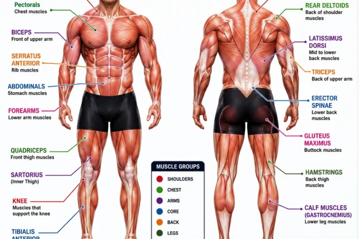
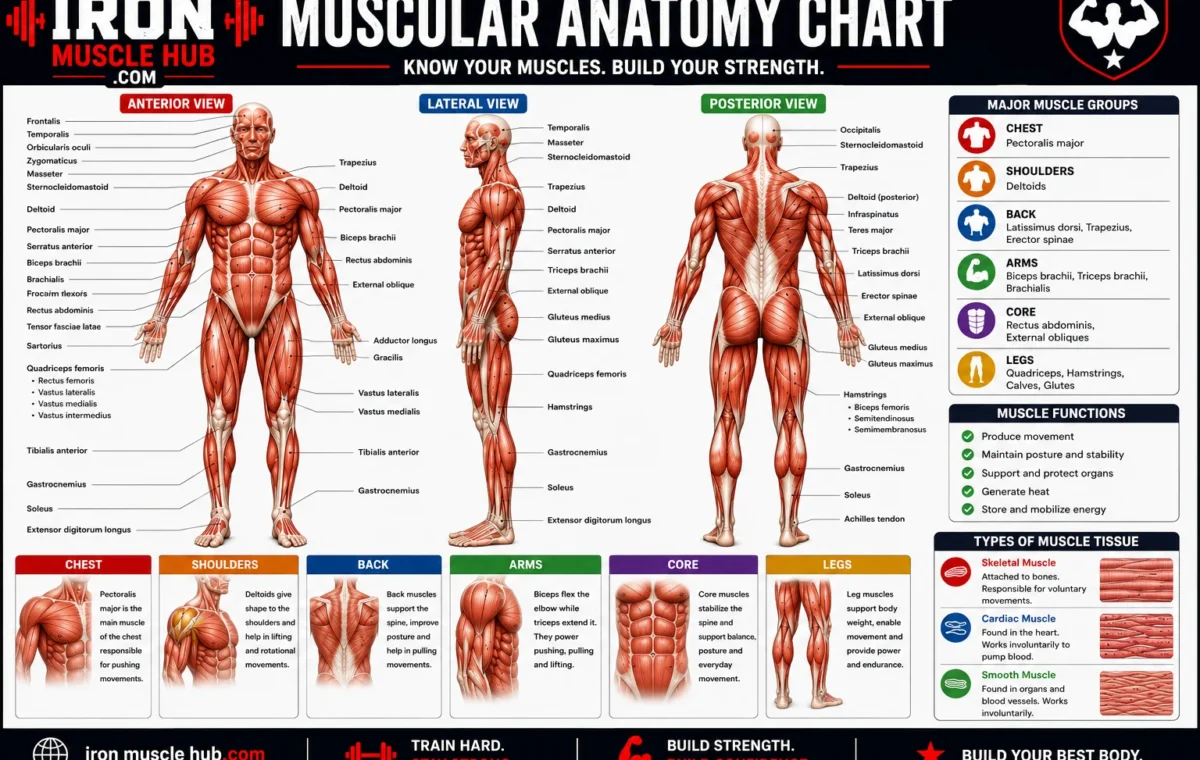
A muscular anatomy chart is a labeled visual representation of muscles and their locations. Depending on its level of detail, it may show only the large muscles visible near the surface or include deeper structures, muscle attachments, nerves, actions, and anatomical terminology.
For beginners, a simple chart is usually the best starting point. More advanced charts can include information such as a muscle’s origin, insertion, nerve supply, blood supply, and specific action. TeachMeAnatomy recommends studying muscles using several consistent features, including location, shape, attachments, action, nerve supply, and blood supply.
Why Are Front and Back Views Important?
A single front-view picture can’t show every major muscle clearly.
For example, the pectoralis major is prominent on the front of the chest, while the latissimus dorsi is mainly associated with the back. The hamstrings are on the posterior thigh, and the gluteal muscles are located around the buttocks and hip.
A useful anatomy chart therefore uses at least two views:
- Anterior view: the front of the body
- Posterior view: the back of the body
OpenStax’s muscular-system overview similarly uses anterior and posterior views to distinguish superficial and deeper muscles.
Major Muscles of the Human Body
1. Chest Muscles
The pectoralis major is the large, fan-shaped muscle covering much of the upper front of the chest. It contributes to movements of the upper limb and is one of the easiest muscles to identify on a basic muscular anatomy chart.
A more detailed chart may also show the pectoralis minor and muscles associated with the rib cage.
Beginner tip: Find the collarbone and breastbone first, then identify the large chest muscle spanning the front of the upper thorax.
2. Shoulder Muscles
The deltoid forms the rounded contour of the shoulder. It is a triangular muscle that covers the shoulder joint and contributes to movement of the arm.
The shoulder region also contains several deeper muscles, including the supraspinatus, infraspinatus, teres minor, and subscapularis. Together, these are commonly known as the rotator cuff muscles.
For a beginner chart, learn the deltoid first. Add the rotator cuff later.
3. Back Muscles
The back contains many muscles, but three useful names to recognize early are:
- Trapezius: a large upper-back muscle involved in positioning the shoulder blade.
- Latissimus dorsi: a broad muscle of the back that helps move the upper arm.
- Rhomboids: muscles between the spine and shoulder blades that help position the scapulae.
A detailed anatomy chart can reveal several layers beneath these superficial muscles. The trunk contains both superficial and deep back muscle groups.
4. Arm Muscles
The two names most beginners encounter first are biceps brachii and triceps brachii.
The biceps is located toward the front of the upper arm and helps flex the forearm at the elbow. The triceps is on the back of the upper arm and helps extend the forearm.
They provide a useful example of opposing muscle actions: the biceps and triceps work in opposite directions at the elbow during flexion and extension.
5. Abdominal Muscles
The abdominal wall contains several muscles rather than one single “abs” muscle.
Important names include:
- Rectus abdominis
- External oblique
- Internal oblique
- Transversus abdominis
The rectus abdominis runs vertically along the front of the abdomen. The oblique muscles are positioned more toward the sides, while the transversus abdominis is deeper within the abdominal wall.
This is one reason a detailed muscular anatomy chart can be more useful than a generic fitness poster: it shows that visible muscle groups can contain several separate muscles.
6. Gluteal Muscles
The gluteal region contains several muscles. The gluteus maximus is the large superficial muscle most people associate with the buttocks.
Deeper structures include the gluteus medius, gluteus minimus, and smaller muscles around the hip.
A beginner doesn’t need to memorize every small hip muscle immediately. Learning the major gluteal muscles first gives you a useful foundation.
7. Thigh Muscles
The thigh is easier to understand if you divide it into three broad compartments: anterior, medial, and posterior.
The quadriceps femoris is the major muscle group on the front of the thigh. It consists of four muscles: rectus femoris, vastus lateralis, vastus medialis, and vastus intermedius. Collectively, these muscles extend the knee.
The hamstrings are on the back of the thigh. The group includes the biceps femoris, semitendinosus, and semimembranosus, which contribute to knee flexion.
8. Calf Muscles
The gastrocnemius is the prominent superficial muscle at the back of the lower leg. The soleus lies underneath it.
A more detailed muscular anatomy chart also shows muscles in the anterior, lateral, and deep posterior compartments of the lower leg.
How to Read a Muscular Anatomy Chart
A crowded anatomy chart can look intimidating at first. Use this simple method instead.
Step 1: Start with body regions
Learn the broad areas first:
head → neck → chest → back → shoulder → arm → abdomen → hip → thigh → lower leg → foot
Don’t attempt to memorize every label on your first study session.
Step 2: Learn the large superficial muscles
Start with recognizable muscles such as:
- Pectoralis major
- Deltoid
- Biceps brachii
- Triceps brachii
- Rectus abdominis
- Latissimus dorsi
- Gluteus maximus
- Quadriceps
- Hamstrings
- Gastrocnemius
Once these are familiar, smaller muscles become easier to place.
Step 3: Connect each muscle with an action
Don’t memorize names without understanding what the muscles do.
For example:
Biceps → elbow flexion
Triceps → elbow extension
Quadriceps → knee extension
Hamstrings → knee flexion
This creates a useful mental connection between the muscle’s location and its function. OpenStax specifically describes muscles in terms of their roles in movement and opposing actions.
Step 4: Test yourself without looking
This is one of the easiest ways to find what you actually remember.
Look at a labeled chart for a few minutes. Then switch to an unlabeled diagram and try to identify the muscles yourself.
You can also cover the labels on a printed chart and point to each muscle before checking the answer.
Step 5: Add deeper anatomy later
Once the major superficial muscles are familiar, move on to smaller or deeper structures.
This regional approach is also recommended by anatomy-learning resources because trying to learn every muscle simultaneously can quickly become overwhelming.
A Simple Muscle-Chart Study Example
Suppose you’re learning the upper body for the first time.
Instead of memorizing dozens of names, begin with these eight:
- Pectoralis major
- Deltoid
- Trapezius
- Latissimus dorsi
- Biceps brachii
- Triceps brachii
- Rectus abdominis
- External oblique
Now identify each one on a front-and-back diagram.
Next, connect each name to its approximate location and a basic movement. After that, switch to an unlabeled chart and test yourself.
This approach turns a complicated picture into a small series of manageable learning tasks.
Common Mistakes When Using a Muscle Anatomy Chart
Trying to memorize everything at once
The human body has a large number of individual muscles, so a comprehensive chart can contain far more information than a beginner needs initially.
Better approach: Learn by region and gradually add detail.
Confusing a muscle with a muscle group
“Quadriceps” refers to a group of four muscles, not one individual muscle. Similarly, “hamstrings” refers to a group.
Better approach: Learn the group name first, then memorize the individual muscles underneath it.
Studying only the front view
Some of the most important muscles for understanding movement are easier to locate from the back.
Better approach: Always compare anterior and posterior views.
Memorizing names without actions
Knowing that a muscle is called the triceps isn’t as useful as knowing where it sits and what movement it contributes to.
Better approach: Pair every new muscle name with its location and a basic action.
Muscular Anatomy Chart vs. a Muscle Diagram
The terms are often used interchangeably, but there can be a practical difference.
| Resource | Best for | Typical detail |
| Basic muscle diagram | Quick identification | Major visible muscles |
| Muscular anatomy chart | Study and reference | More labels and regional detail |
| Labeled anatomy chart | Beginners and revision | Muscle names with locations |
| Unlabeled chart | Self-testing | Identification practice |
| Detailed anatomy atlas | Advanced study | Deep muscles, attachments and structures |
A good learning resource doesn’t have to show every muscle. The right level of detail depends on what you’re trying to learn.
Where to Find Reliable Anatomy References
For a free educational reference, OpenStax Anatomy and Physiology provides structured coverage of muscle tissue, skeletal muscles, muscle actions, and the muscular system. Its textbook materials are openly licensed under CC BY-NC-SA 4.0, with specific restrictions on commercial reuse.
The National Center for Biotechnology Information’s Medical Terminology reference is another useful source for anatomical terminology and major skeletal-muscle names.
If you’re publishing an anatomy image on a commercial website, check the image’s individual license rather than assuming that an educational image is automatically free for commercial use.
Internal Link Ideas
For related content on Iron Muscle Hub, consider linking this article to:
- Human Muscle Anatomy Explained for Beginners
- Body Muscle Chart: Major Muscle Groups and Their Functions
- Skeletal vs. Cardiac vs. Smooth Muscle Explained
These articles would create a useful learning path from a basic chart to more detailed muscle anatomy.
Frequently Asked Questions
What is a muscular anatomy chart?
A muscular anatomy chart is a labeled visual guide to the muscles of the human body. Basic charts identify major superficial muscles, while detailed charts can include deeper muscles, anatomical regions, attachments, and actions. They’re useful for beginners because location is easier to understand visually than from a list of muscle names alone.
What are the major muscle groups in the human body?
Common major muscle groups include the chest, shoulders, back, arms, abdomen, glutes, thighs, and calves. These broad categories contain many individual muscles. For example, the quadriceps is a four-muscle group in the front of the thigh, while the hamstrings are a group of muscles at the back of the thigh.
How many muscles are in the human body?
The exact number depends on how muscles are defined and counted, but anatomy references commonly describe the human body as having more than 600 muscles. For beginners, trying to memorize every muscle isn’t necessary. Learning major muscles region by region is a more practical starting point.
What is the difference between skeletal, cardiac, and smooth muscle?
Skeletal muscle is associated mainly with movement of the skeleton and is under voluntary nervous-system control. Cardiac muscle forms the muscular tissue of the heart, while smooth muscle is found in structures such as the digestive tract and blood vessels. These are the three major types of muscle tissue.
What should I learn first from a muscle anatomy chart?
Start with the largest and easiest-to-identify muscles: pectoralis major, deltoid, trapezius, latissimus dorsi, biceps, triceps, rectus abdominis, gluteus maximus, quadriceps, hamstrings, and gastrocnemius. Then learn their basic locations and actions before moving to smaller muscles.
How can I memorize muscles more easily?
Use a three-part system: location + name + action. Study one body region at a time, then test yourself with an unlabeled diagram. Repeatedly identifying muscles from memory is more useful than simply rereading the labels.
A Clear Way to Use This Chart
A muscular anatomy chart is most useful when it becomes a study tool rather than something you simply look at once. Start with the large muscle groups, learn their locations, and connect each one with a basic movement. Then move from superficial muscles to deeper structures.
The biggest mistake is trying to learn everything in one sitting. Anatomy becomes much easier when you break the body into regions and repeatedly test yourself. A reliable textbook or anatomy reference can then help you confirm details as your knowledge grows.
For fitness readers, anatomy can also make exercise explanations easier to understand. For students, a labeled and unlabeled chart can turn a difficult topic into manageable revision practice.
Understanding Muscle Groups Beyond the Basic Chart
A beginner muscular anatomy chart usually focuses on the large muscles you can identify easily. Part 2 goes one step further by looking at muscle groups, deeper muscles, anatomical regions, and how muscles work together.
You don’t need to memorize every small muscle immediately. A better approach is to understand how muscles are organized and then add detail gradually. This makes an anatomy chart much easier to read and helps you connect muscle names with actual movement.
Key Muscle Groups to Know
| Region | Important muscles or groups | What to remember |
|---|---|---|
| Neck | Sternocleidomastoid | Prominent muscle on the side/front of the neck |
| Chest | Pectoralis major, pectoralis minor | Major muscles of the chest |
| Upper back | Trapezius, rhomboids | Help control the shoulder blades |
| Back | Latissimus dorsi | Large superficial back muscle |
| Shoulder | Deltoid, rotator cuff muscles | Shoulder movement and stability |
| Upper arm | Biceps brachii, triceps brachii | Major elbow movers |
| Forearm | Flexor and extensor groups | Move the wrist and fingers |
| Abdomen | Rectus abdominis, obliques, transversus abdominis | Trunk movement and stability |
| Hip | Gluteal and deep hip muscles | Hip movement and stability |
| Thigh | Quadriceps, hamstrings, adductors | Knee and hip movement |
| Lower leg | Gastrocnemius, soleus, tibialis anterior | Foot and ankle movement |
Neck Muscles
One of the easiest muscles to recognize on a muscular anatomy chart is the sternocleidomastoid.
It runs diagonally along the side of the neck from the skull toward the sternum and clavicle. It contributes to movements of the head and neck.
The neck contains several other muscles, including deeper muscles that aren’t always visible on a basic chart. For beginners, learning the sternocleidomastoid first gives you a useful landmark.
The Rotator Cuff
The rotator cuff is especially important because it’s a group of four muscles, not one muscle.
The four are:
- Supraspinatus
- Infraspinatus
- Teres minor
- Subscapularis
These muscles are associated with the shoulder joint and help stabilize and move the upper arm.
On a basic front-and-back muscular anatomy chart, some of these muscles can be difficult to identify because several are positioned deep to or around larger muscles.
Easy Memory Trick
A common memory aid is SITS:
- Supraspinatus
- Infraspinatus
- Teres minor
- Subscapularis
The memory trick helps you remember the names, but you should still use a labeled anatomy diagram to learn where each muscle is located.
Forearm Muscles
The forearm contains many muscles, which is why detailed anatomy charts can suddenly become much more complicated below the elbow.
For beginner study, think in terms of two broad functional groups:
- Flexor muscles: generally associated with flexing the wrist and fingers.
- Extensor muscles: generally associated with extending the wrist and fingers.
There are several individual muscles within these groups. A detailed anatomy chart may label them separately.
You don’t need to memorize all of them during your first anatomy study session. Learn the major groups first, then add individual muscles as needed.
Understanding the Abdominal Wall
The phrase “abdominal muscles” can make it sound as if the abdomen contains one large muscle. It doesn’t.
A basic anatomy chart should help you distinguish several layers.
Rectus Abdominis
The rectus abdominis runs vertically along the front of the abdomen. It contributes to flexion of the trunk.
External Oblique
The external oblique is positioned on the sides of the abdomen. It contributes to movements and stabilization of the trunk.
Internal Oblique
The internal oblique lies deeper than the external oblique and also contributes to trunk movement.
Transversus Abdominis
The transversus abdominis is a deeper abdominal muscle whose fibers run broadly across the abdomen.
This layered arrangement is one reason an anatomy chart can be more useful than a simple fitness illustration.
Hip Muscles
The hip is surrounded by muscles that help move and stabilize the pelvis and thigh.
The gluteus maximus is the large superficial gluteal muscle. The gluteus medius and gluteus minimus are positioned more laterally and deeper.
There are also smaller muscles around the hip that are often included on advanced anatomy charts.
When studying this region, use the pelvis as your landmark. Find the pelvis first, then identify the muscles surrounding it.
Quadriceps: Four Muscles, Not One
The quadriceps femoris is one of the most useful examples of why anatomical names matter.
It consists of four muscles:
- Rectus femoris
- Vastus lateralis
- Vastus medialis
- Vastus intermedius
Three of these are generally easier to visualize on a superficial chart. The vastus intermedius is deeper and is partly covered by the rectus femoris.
Together, the quadriceps are major muscles involved in extending the knee.
How to Read Them on a Chart
From the front:
- Rectus femoris is near the center.
- Vastus lateralis is toward the outer thigh.
- Vastus medialis is toward the inner side of the front thigh.
- Vastus intermedius lies underneath rectus femoris.
This is a good example of why depth matters when reading muscle anatomy.
Hamstrings
The hamstrings are located at the back of the thigh.
The principal hamstring muscles include:
- Biceps femoris
- Semitendinosus
- Semimembranosus
A posterior-view muscular anatomy chart makes this group much easier to understand because the muscles can be viewed from behind.
The hamstrings participate in movements of both the hip and knee.
Lower-Leg Anatomy
The lower leg contains several muscle compartments.
For basic study, three muscles are especially useful to recognize:
Gastrocnemius
The gastrocnemius forms much of the visible upper calf. It crosses both the knee and ankle joints.
Soleus
The soleus sits deep to the gastrocnemius and contributes to movement at the ankle.
Tibialis Anterior
The tibialis anterior is located toward the front of the lower leg. It contributes to dorsiflexion of the ankle.
These three provide a simple starting point before you learn the many smaller muscles of the lower leg.
Superficial vs. Deep Muscles
One of the most important concepts in reading a muscular anatomy chart is depth.
A superficial muscle is closer to the body’s surface. A deep muscle is positioned underneath other structures.
For example, the soleus is underneath the gastrocnemius. The subscapularis is located on the anterior surface of the scapula and is part of the rotator cuff.
A detailed anatomy chart may therefore provide several versions of the same body region:
- Superficial muscles
- Intermediate layers
- Deep muscles
- Individual muscle attachments
If a muscle seems to “disappear” from one chart, it may simply be covered by another muscle in that particular view.
How Muscles Work Together
Muscles rarely work in complete isolation.
Consider bending your elbow. The biceps brachii contributes to elbow flexion, while the triceps brachii is the major muscle associated with elbow extension.
The same principle appears throughout the body. Some muscles act as prime movers, while others assist, stabilize joints, or oppose particular movements.
This is why studying function alongside location is more useful than memorizing names alone.
A Better Way to Memorize Muscle Anatomy
Try this four-part method.
1. Identify the location
Ask:
Where is this muscle?
Don’t worry about its detailed attachment yet.
2. Learn the name
Say the name aloud or write it down.
For example:
Front of upper arm → biceps brachii
3. Learn one major action
Connect the muscle to a basic movement.
Biceps brachii → elbow flexion
4. Test yourself
Cover the label and identify the muscle without looking at the answer.
This final step matters because recognition and recall aren’t the same thing. A chart can look familiar even when you can’t name the structures independently.
Muscular Anatomy Chart Study Order
If you’re completely new to anatomy, this sequence can keep the subject manageable:
Stage 1: Major regions
- Chest
- Back
- Shoulders
- Arms
- Abdomen
- Glutes
- Thighs
- Calves
Stage 2: Major individual muscles
- Pectoralis major
- Deltoid
- Trapezius
- Latissimus dorsi
- Biceps brachii
- Triceps brachii
- Rectus abdominis
- Gluteus maximus
- Quadriceps
- Hamstrings
- Gastrocnemius
Stage 3: Muscle groups
- Rotator cuff
- Quadriceps
- Hamstrings
- Abdominal wall
- Forearm flexors and extensors
Stage 4: Deeper anatomy
Study smaller muscles, attachments, innervation, and detailed actions only after the basic map makes sense.
Quick Self-Test
Look at an unlabeled human muscle diagram and try to answer these without checking a reference:
- What is the large muscle covering the shoulder?
- What is the major muscle on the front of the upper arm?
- Which muscle group occupies the front of the thigh?
- Which major muscles are found at the back of the thigh?
- What large muscle covers much of the upper back?
- Which prominent muscle forms much of the calf?
If you can answer most of these, you’re ready to add more detailed anatomy.
Final Takeaway
A muscular anatomy chart becomes much easier once you stop treating it as one enormous collection of names.
Start with regions, identify the large superficial muscles, connect each muscle to a basic action, and then gradually move into deeper layers. Groups such as the rotator cuff, quadriceps, hamstrings, and abdominal muscles should be learned as groups first and individual muscles second.
The goal isn’t to memorize an entire anatomy textbook in one sitting. Build a mental map of the body first. Once that map is familiar, the smaller structures have somewhere to fit.
Once you can look at an unlabeled diagram and identify the major muscles from memory, you’re no longer just memorizing a picture. You’re building a working map of human movement.