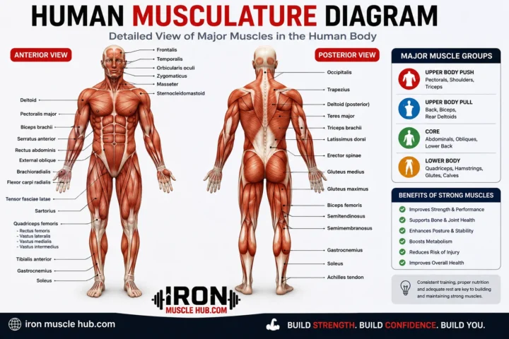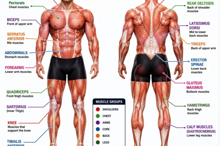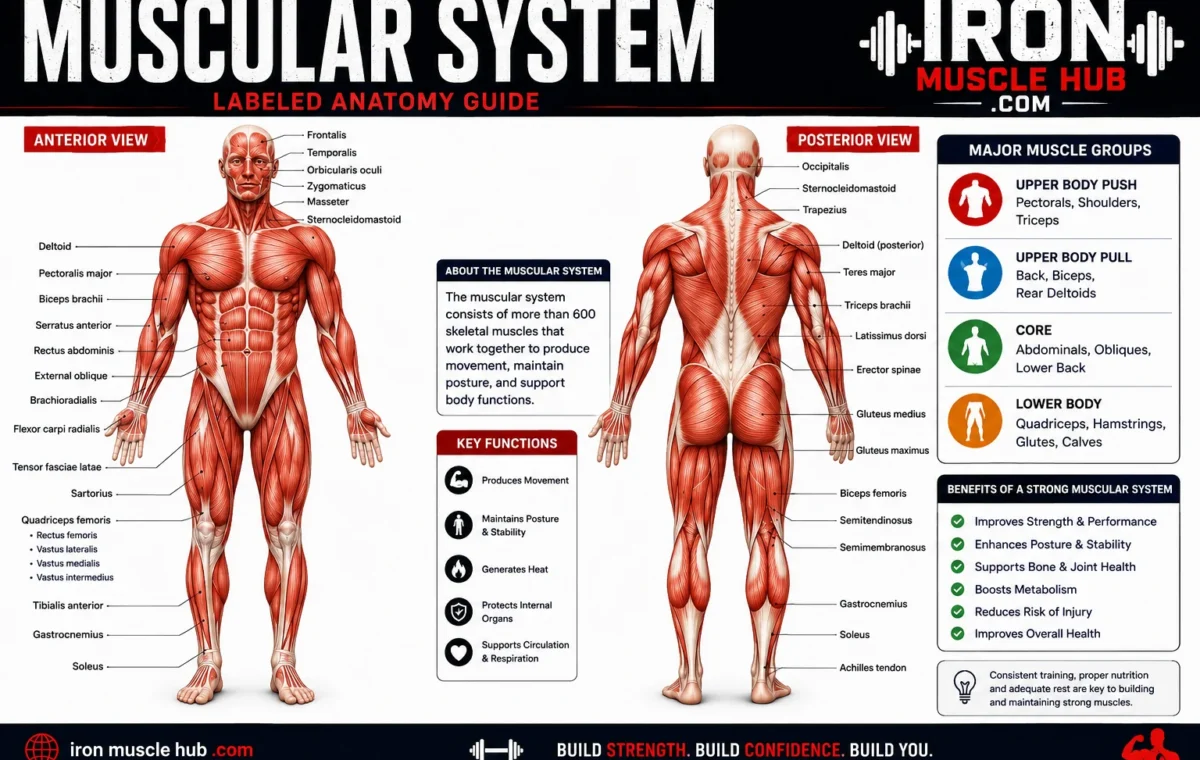
A muscular system labeled diagram helps you identify where the body’s major muscles are located and understand what they do. The muscular system includes skeletal muscles that help you move, as well as cardiac and smooth muscle tissues that perform important involuntary jobs.
- Muscular System Labeled: Quick Reference
- What Is the Muscular System?
- The Three Types of Muscle Tissue
- 1. Skeletal Muscle
- 2. Cardiac Muscle
- 3. Smooth Muscle
- Major Muscles on a Labeled Human Body Diagram
- Head and Neck
- Chest and Shoulders
- Arms
- Abdomen
- Back
- Hips and Thighs
- Lower Leg
- How Muscles Actually Produce Movement
- A Useful Way to Study a Muscular System Labeled Diagram
- A Common Mistake: Confusing Muscle Groups With Individual Muscles
- Why Labeled Muscle Diagrams Are Useful
- Muscular System vs. Skeletal System
- Internal Links to Add on Iron Muscle Hub
- Frequently Asked Questions
- What are the three types of muscle in the human body?
- What are the major muscles of the human body?
- What is the muscular system responsible for?
- Is the heart part of the muscular system?
- Are muscles connected directly to bones?
- What is the easiest way to memorize a labeled muscular system?
- A Simple Way to Remember the Major Muscle Groups
- Major Muscle Groups You Should Know
- Front View of the Muscular System
- Deltoid
- Pectoralis Major
- Biceps Brachii
- Rectus Abdominis
- Quadriceps
- Back View of the Muscular System
- Trapezius
- Latissimus Dorsi
- Triceps Brachii
- Gluteus Maximus
- Hamstrings
- Muscles That Work in Pairs
- Why Muscle Names Can Look Complicated
- Muscular System Labeled Diagram: Common Study Mistakes
- Mistake 1: Memorizing Names Without Locations
- Mistake 2: Treating Every Group as One Muscle
- Mistake 3: Only Studying the Front View
- Mistake 4: Learning Function Without Anatomy
- Mistake 5: Trying to Learn Everything in One Session
- A 7-Day Muscular Anatomy Study Plan
- Muscular System and Exercise
- The Most Important Labels to Memorize First
- Final Takeaway
- Muscle Anatomy by Body Region
- Neck
- Shoulder Region
- Chest
- Upper Back
- Lower Back
- Understanding Muscle Attachments
- Muscle Groups vs. Individual Muscles
- Quadriceps
- Hamstrings
- Gluteal Muscles
- How to Read a Detailed Muscle Diagram
- Start With the Skeleton
- Find the Large Surface Muscles
- Look for Smaller Muscles
- Follow the Muscle's Direction
- Connect the Muscle to Its Function
- Muscular System Labeled: Front vs. Back
- A Simple Muscle-Memory Trick
- Why Muscle Anatomy Matters Beyond Exercise
- Important Anatomy Terms to Know
- Flexion
- Extension
- Abduction
- Adduction
- Rotation
- Quick Quiz: Can You Identify These Muscles?
- What a Complete Muscular System Chart Should Include
- Frequently Asked Questions About Muscle Labels
- What is the most important muscle to learn first?
- Why do different muscular system diagrams have different labels?
- Are the abs one muscle?
- Are the quadriceps one muscle?
- What is the difference between a muscle and a muscle group?
- Can a muscular system diagram show every muscle?
- Final Study Checklist
- Final Takeaway
If you’re learning human anatomy for the first time, a labeled diagram can seem crowded with unfamiliar names. The easiest approach is to learn the large muscle groups first, then add smaller muscles as you become more comfortable. For more insights read here :- leaner physique and how to put on muscle fast .
Quick answer: The muscular system is made up of three main types of muscle tissue: skeletal, cardiac, and smooth. Skeletal muscles attach to bones and produce voluntary movement, cardiac muscle forms the heart, and smooth muscle works inside organs and other structures without conscious control.
Muscular System Labeled: Quick Reference
| Muscle or group | Where you’ll find it | Main job |
|---|---|---|
| Deltoids | Shoulders | Move and stabilize the shoulder |
| Pectorals | Chest | Help move the arms |
| Biceps | Front of upper arm | Bend the elbow |
| Triceps | Back of upper arm | Straighten the elbow |
| Rectus abdominis | Front of abdomen | Helps flex the trunk |
| Obliques | Sides of abdomen | Help rotate and bend the trunk |
| Trapezius | Upper back and neck | Helps move the shoulder blades and neck |
| Latissimus dorsi | Middle/lower back | Helps pull and move the upper arm |
| Gluteal muscles | Buttocks | Help extend and stabilize the hip |
| Quadriceps | Front of thigh | Straighten the knee |
| Hamstrings | Back of thigh | Bend the knee and help extend the hip |
| Gastrocnemius | Calf | Helps push the foot downward |
This table focuses on commonly identified skeletal muscles. The human body contains more than 650 muscles, so a complete anatomy chart will contain considerably more labels.
What Is the Muscular System?
The muscular system is the collection of muscle tissues that generate force and movement. Muscles also contribute to posture and other essential body functions. Skeletal muscles work with bones and joints to produce movement, while cardiac and smooth muscles perform jobs you generally don’t control consciously.
Think of a muscle as a biological engine. It receives signals, generates force, and changes tension so your body can move or maintain a position.
The term musculoskeletal system is broader. It includes muscles along with bones, joints, tendons, ligaments and related connective tissues.
The Three Types of Muscle Tissue
A labeled muscular system diagram usually focuses on visible skeletal muscles, but there are actually three major types of muscle tissue.
1. Skeletal Muscle
Skeletal muscles are attached to bones through tendons and are responsible for voluntary movement. They also help maintain posture and body position.
Examples include the biceps, quadriceps, hamstrings and deltoids.
Under a microscope, skeletal muscle has a striped or striated appearance. Its contractile proteins are organized into repeating structures called sarcomeres.
2. Cardiac Muscle
Cardiac muscle is found in the heart. It contracts involuntarily and provides the force needed for the heart to pump blood through the circulation.
Cardiac muscle is also striated, but it has specialized structural features that allow heart muscle cells to communicate and contract in a coordinated way.
3. Smooth Muscle
Smooth muscle is involuntary muscle found in places such as the walls of blood vessels and hollow organs. It helps move substances through the body and regulates the diameter of structures such as blood vessels.
Unlike skeletal and cardiac muscle, smooth muscle doesn’t have its actin and myosin arranged into sarcomeres, so it doesn’t have the same striated appearance.
Major Muscles on a Labeled Human Body Diagram
The easiest way to read a human muscular system labeled chart is to work from the head downward.
Head and Neck
Frontalis: Located across the forehead and involved in raising the eyebrows.
Masseter: One of the major muscles used for chewing.
Sternocleidomastoid: A prominent neck muscle that helps move and rotate the head.
A diagram may include several smaller facial and neck muscles, so don’t worry about memorizing every label immediately.
Chest and Shoulders
Pectoralis major: A large chest muscle involved in movements of the upper arm.
Deltoid: The rounded muscle covering much of the shoulder. It contributes to several arm movements.
Serratus anterior: Located along the side of the chest and helps position and move the shoulder blade.
These muscles are often clearly visible on front-view muscular system charts.
Arms
The two labels beginners usually recognize first are the biceps brachii and triceps brachii.
The biceps is on the front of the upper arm, while the triceps occupies the back. Their actions at the elbow are different: the biceps contributes to elbow flexion, while the triceps contributes to elbow extension.
The forearm contains numerous additional muscles responsible for movements of the wrist and fingers.
Abdomen
Rectus abdominis: Runs vertically along the front of the abdomen.
External obliques: Located on the sides of the abdominal region and contribute to trunk rotation and bending.
Internal obliques: Lie deeper than the external obliques and also contribute to trunk movement and stability.
Transversus abdominis: A deeper abdominal muscle that wraps around the trunk and contributes to abdominal wall function and trunk stability.
A common mistake is to think of the “abs” as one single muscle. A labeled anatomy chart shows that the abdominal wall contains several muscles.
Back
Trapezius: A broad muscle extending across the upper back and neck region.
Latissimus dorsi: A large back muscle that contributes to movements of the upper arm.
Erector spinae: A group of muscles running along the back that helps support and extend the spine.
Back diagrams can look more complicated than front diagrams because several layers of muscle overlap.
Hips and Thighs
Gluteus maximus: The largest of the gluteal muscles and an important muscle for hip extension.
Quadriceps: A group of muscles on the front of the thigh that works to extend the knee.
Hamstrings: A group on the back of the thigh that contributes to knee flexion and hip extension.
Remembering the front-versus-back position makes this section much easier: quads are in front; hamstrings are behind.
Lower Leg
Gastrocnemius: The large, visible calf muscle.
Soleus: A deeper calf muscle beneath the gastrocnemius.
Muscles of the lower leg work together to move the ankle and foot. A detailed muscular system diagram may therefore show many more labels than a basic beginner chart.
How Muscles Actually Produce Movement
Muscle movement begins with signals from the nervous system. In skeletal muscle, those signals cause interactions between proteins including actin and myosin, producing contraction.
Here’s a simplified example:
- You decide to bend your arm.
- Your nervous system sends a signal to the relevant skeletal muscle.
- Muscle fibers receive that signal.
- Contractile proteins interact inside the muscle fibers.
- The muscle develops tension.
- The tension is transmitted through connective tissues and the tendon.
- The bones move around a joint.
A muscle doesn’t simply “pull a bone” by itself. Movement results from coordinated work among muscles, tendons, bones, joints and the nervous system.
A Useful Way to Study a Muscular System Labeled Diagram
Trying to memorize dozens of names at once usually makes anatomy harder.
Instead, use this five-step method:
Step 1: Learn the regions.
Start with head, neck, chest, back, abdomen, arms, hips, thighs and lower legs.
Step 2: Learn the biggest muscles.
Focus first on deltoids, pectorals, biceps, triceps, trapezius, latissimus dorsi, abdominals, gluteals, quadriceps, hamstrings and gastrocnemius.
Step 3: Learn location before function.
If you can point to a muscle on a diagram, remembering its job becomes easier.
Step 4: Compare front and back views.
For example, the quadriceps are on the front of the thigh, while the hamstrings are on the back.
Step 5: Add smaller muscles gradually.
Once the major landmarks are familiar, move to more detailed anatomy.
A Common Mistake: Confusing Muscle Groups With Individual Muscles
Not every label on an anatomy chart represents one individual muscle.
The quadriceps, for example, refers to a group of four muscles in the front of the thigh. The hamstrings also describe a group rather than one single muscle.
This matters because simplified fitness diagrams sometimes use group names while more detailed anatomy diagrams identify individual muscles.
Why Labeled Muscle Diagrams Are Useful
A good labeled diagram can help you:
- Learn basic human anatomy.
- Identify major muscle groups.
- Understand where common muscles are located.
- Connect a muscle’s position with its general function.
- Study for anatomy, biology or health classes.
- Better understand exercise terminology.
For exercise beginners, anatomy diagrams can also make instructions such as “work your quadriceps” or “strengthen your upper back” easier to understand. They shouldn’t, however, be used to diagnose an injury or medical condition.
Muscular System vs. Skeletal System
These two systems work closely together but aren’t the same.
| System | Main structures | Basic role |
| Muscular system | Muscle tissues | Produces force and movement |
| Skeletal system | Bones and related structures | Supports the body and provides a framework for movement |
| Musculoskeletal system | Muscles, bones, joints and connective tissues | Works as a coordinated movement and support system |
Tendons connect muscles to bones, helping transmit muscular force to produce movement. Ligaments, by contrast, connect bones to other bones.
Internal Links to Add on Iron Muscle Hub
For a stronger anatomy content cluster, consider linking this guide to:
- Muscle Anatomy Chart: A beginner’s guide to reading common muscle diagrams.
- Three Types of Muscle Tissue: A deeper explanation of skeletal, cardiac and smooth muscle.
- Body Diagram of Muscles: A visual guide to major muscles from head to toe.
These pages can give readers a natural next step without forcing them to search for basic definitions elsewhere.
Frequently Asked Questions
What are the three types of muscle in the human body?
The three major types are skeletal, cardiac and smooth muscle. Skeletal muscle is generally under voluntary control and moves the skeleton. Cardiac muscle makes up the heart and contracts involuntarily. Smooth muscle is found in structures such as hollow organs and blood vessels and also works involuntarily.
What are the major muscles of the human body?
Commonly labeled major skeletal muscles include the deltoids, pectorals, biceps, triceps, trapezius, latissimus dorsi, abdominal muscles, gluteal muscles, quadriceps, hamstrings and gastrocnemius. A detailed anatomy chart will show many additional muscles because the human body contains more than 650 muscles.
What is the muscular system responsible for?
The muscular system generates force that allows the body to move and maintain posture. Different muscle tissues also perform involuntary functions, including pumping blood through the heart and moving substances through organs.
Is the heart part of the muscular system?
Yes. The heart contains cardiac muscle, one of the three major muscle tissue types. Cardiac muscle contracts involuntarily and provides the pumping action that moves blood through the circulatory system.
Are muscles connected directly to bones?
Skeletal muscles attach to bones through tendons. When a skeletal muscle contracts, force can be transmitted through the tendon to the bone, helping create movement around a joint.
What is the easiest way to memorize a labeled muscular system?
Start with the largest and easiest-to-locate muscle groups rather than memorizing every label at once. Learn the body region, identify the muscle, then learn its main function. Reviewing front and back diagrams repeatedly is usually more useful than trying to memorize a long list in one sitting.
A Simple Way to Remember the Major Muscle Groups
You don’t need to memorize an entire anatomy textbook to understand a muscular system labeled diagram.
Start with the major landmarks: shoulders, chest, arms, abdomen, back, glutes, thighs and calves. Then attach the key names to those regions.
Once those basics feel familiar, add smaller muscles and individual muscle names. That approach gives you a useful mental map first, instead of turning anatomy into a wall of vocabulary.
The muscular system is much more than the muscles you can see in a mirror. Skeletal, cardiac and smooth muscle tissues work together to produce movement and support essential functions throughout the body.
Major Muscle Groups You Should Know
If you’re studying a muscular system labeled diagram, don’t try to memorize every muscle immediately. Start with the major groups that appear most often on beginner anatomy charts.
| Region | Major muscles | Easy way to remember |
|---|---|---|
| Shoulders | Deltoids | Shoulder cap |
| Chest | Pectoralis major | Large front-chest muscle |
| Upper back | Trapezius | Upper-back and neck region |
| Back | Latissimus dorsi | Broad muscle along the back |
| Upper arm | Biceps, triceps | Front and back of arm |
| Abdomen | Rectus abdominis, obliques | Front and sides of trunk |
| Hips | Gluteal muscles | Hip and buttock region |
| Front thigh | Quadriceps | Four-muscle group |
| Back thigh | Hamstrings | Back of the thigh |
| Calf | Gastrocnemius, soleus | Lower-leg muscles |
This gives you a useful framework before you move into smaller and deeper muscles.
Front View of the Muscular System
A front-view muscular system diagram usually makes the chest, shoulders, arms, abdomen and thighs easiest to identify.
Deltoid
The deltoid covers the shoulder area and contributes to several movements of the upper arm. On a muscular system labeled diagram, it is usually easy to spot because it forms the rounded contour of the shoulder.
Pectoralis Major
The pectoralis major is a large muscle on the front of the chest. It contributes to movements of the upper arm and is one of the most recognizable muscles on a front-view anatomy illustration.
Biceps Brachii
The biceps brachii is located on the front of the upper arm. It crosses the shoulder and elbow and contributes to bending the elbow and rotating the forearm.
Rectus Abdominis
The rectus abdominis runs vertically along the front of the abdomen. It contributes to flexion of the trunk and is commonly associated with the “six-pack” appearance when the muscle and surrounding anatomy are clearly defined.
Quadriceps
The quadriceps femoris is a group of four muscles located at the front of the thigh. Together, these muscles are important for extending the knee.
Back View of the Muscular System
A back-view diagram reveals several large muscles that aren’t as obvious from the front.
Trapezius
The trapezius occupies a large area across the upper back and extends toward the neck and shoulders. It contributes to movement and positioning of the shoulder blades.
Latissimus Dorsi
The latissimus dorsi is a broad muscle covering much of the lower and middle back. It helps move the upper arm and is commonly labeled simply as the “lats” in fitness discussions.
Triceps Brachii
The triceps brachii occupies the back of the upper arm. Its main action at the elbow is extension, making it the counterpart to the elbow-flexing action of the biceps.
Gluteus Maximus
The gluteus maximus is the largest of the three gluteal muscles. It contributes strongly to hip extension and is important for movements such as standing up and climbing.
Hamstrings
The hamstrings are a group of muscles on the back of the thigh. They contribute to bending the knee and extending the hip.
Muscles That Work in Pairs
One useful way to understand a labeled muscular system is to stop thinking of individual muscles as isolated structures.
Many movements involve muscles working in opposing or coordinated roles.
For example, during elbow movement, the biceps and triceps have different primary actions. When one muscle group contributes to flexion, the opposing group can contribute to extension.
This doesn’t mean only one muscle is active at a time. Real movement involves coordination among multiple muscles, joints and the nervous system.
Why Muscle Names Can Look Complicated
Anatomy terminology can seem intimidating because many muscle names describe something about the muscle itself.
A name may refer to:
- Location — where the muscle is found.
- Shape — what the muscle looks like.
- Size — whether it is large or small.
- Direction — the orientation of its fibers.
- Action — what movement it helps produce.
- Attachments — where it begins or ends.
Once you understand this naming system, unfamiliar labels become easier to approach.
For example, biceps indicates a muscle with two heads, while triceps indicates three. Maximus means larger or greatest, while minimus means smaller or least.
Muscular System Labeled Diagram: Common Study Mistakes
Mistake 1: Memorizing Names Without Locations
Knowing that a muscle is called the trapezius isn’t very useful if you can’t identify it on the body.
Better approach: Learn the location first, then connect the name and function to it.
Mistake 2: Treating Every Group as One Muscle
Terms such as quadriceps and hamstrings describe groups of muscles.
Better approach: Learn the group name first. Add the individual muscle names later.
Mistake 3: Only Studying the Front View
Many large muscles are easier to understand from the back.
Better approach: Study front and back diagrams together.
Mistake 4: Learning Function Without Anatomy
Memorizing “this muscle flexes the knee” doesn’t automatically tell you where the muscle is.
Better approach: Connect three things: name → location → action.
Mistake 5: Trying to Learn Everything in One Session
A detailed anatomy chart can contain a huge amount of information.
Better approach: Learn five to ten important labels at a time and review them regularly.
A 7-Day Muscular Anatomy Study Plan
If you’re learning the muscular system for school, fitness education or general knowledge, this simple schedule can help.
Day 1: Learn the major muscles of the head, neck and shoulders.
Day 2: Study the chest, upper back and arms.
Day 3: Learn the abdominal muscles and lower back.
Day 4: Study the gluteal muscles and thighs.
Day 5: Learn the calf and other lower-leg muscles.
Day 6: Review a complete front-and-back muscular system labeled diagram.
Day 7: Test yourself without looking at the labels.
The final step is important. Recognition is easier than recall, so covering the labels and trying to name each structure yourself can reveal which areas need more review.
Muscular System and Exercise
Understanding basic muscle anatomy can make exercise terminology easier to follow.
For example, a workout program may refer to the quadriceps, hamstrings, glutes, pectorals, deltoids or latissimus dorsi. A labeled diagram lets you see where those structures are instead of treating the names as abstract terms.
However, anatomy alone doesn’t tell you how to design a safe exercise program. Exercise selection, technique, training volume, recovery and individual circumstances all matter.
If you’re dealing with persistent pain or an injury, an anatomy chart isn’t a substitute for professional medical assessment.
The Most Important Labels to Memorize First
If you only have a short time to study, focus on these:
- Deltoid — shoulder
- Pectoralis major — chest
- Biceps brachii — front upper arm
- Triceps brachii — back upper arm
- Rectus abdominis — front abdomen
- External oblique — side of abdomen
- Trapezius — upper back
- Latissimus dorsi — broad back muscle
- Gluteus maximus — buttock region
- Quadriceps — front thigh
- Hamstrings — back thigh
- Gastrocnemius — prominent calf muscle
Once these become familiar, more detailed anatomy becomes much easier to organize.
Final Takeaway
A muscular system labeled diagram is most useful when you treat it as a map rather than a list of vocabulary words. First identify the body region, then learn the major muscle, its location and its basic function.
Start with the large, recognizable structures such as the deltoids, pectorals, biceps, triceps, trapezius, abdominal muscles, glutes, quadriceps, hamstrings and calf muscles. After those become familiar, move on to smaller muscles and deeper anatomical structures.
The goal isn’t to memorize every label in one sitting. Build your understanding layer by layer, and the full muscular system will become much easier to read.
Muscle Anatomy by Body Region
Once you’ve learned the major muscles, the next step is understanding how they fit into each body region. This makes a muscular system labeled diagram much easier to read because you can identify a muscle from its position instead of memorizing names randomly.
Head and Face
The head contains several muscles responsible for facial expression, chewing and movement of the jaw.
Important examples include:
- Frontalis — helps raise the eyebrows.
- Orbicularis oculi — surrounds the eye and helps close the eyelids.
- Orbicularis oris — surrounds the mouth and contributes to movements of the lips.
- Masseter — a powerful muscle involved in chewing.
- Temporalis — located on the side of the skull and involved in jaw movement.
These muscles may be labeled on detailed anatomy charts but are often omitted from simplified muscular-system illustrations.
Neck
The neck contains muscles that help support and move the head.
The sternocleidomastoid is particularly easy to identify because it runs diagonally along the side of the neck. It contributes to rotation and movement of the head and neck.
Other deeper neck muscles may appear on advanced anatomy diagrams.
Shoulder Region
The shoulder region contains several muscles that work together to control the upper limb and stabilize the shoulder.
The deltoid is the most recognizable surface muscle. Underneath and around the shoulder are muscles of the rotator cuff, including:
- Supraspinatus
- Infraspinatus
- Teres minor
- Subscapularis
A beginner chart may simply label the deltoid, while a detailed chart can identify the individual rotator-cuff muscles.
Chest
The pectoralis major is one of the most prominent muscles on the front of the chest.
A deeper muscle called the pectoralis minor lies underneath it. The chest also works closely with muscles around the ribs and shoulder blade during breathing and upper-body movement.
This is why a detailed anatomy illustration can contain many more labels than a basic fitness chart.
Upper Back
The upper back is dominated by the trapezius, a large muscle extending across the upper back and neck region.
Beneath and around it are additional muscles that contribute to shoulder-blade and spinal movement.
Learning the trapezius first gives you a useful landmark for understanding the rest of the back.
Lower Back
The lower back contains several muscles that help support and move the trunk.
The erector spinae refers to a group of muscles running along the spine. These muscles contribute to extending and stabilizing the vertebral column.
A labeled diagram can also show deeper muscles that aren’t obvious from the surface.
Understanding Muscle Attachments
One of the most useful anatomy concepts is that muscles have attachment points.
Skeletal muscles connect to bones through tendons. When a muscle develops tension, that force can be transferred through the tendon to the attached bone.
This helps explain why muscle location matters.
For example, a muscle crossing a joint can influence movement at that joint. Its exact attachment points help determine the direction in which it can create force.
You don’t need to memorize every attachment when you’re starting. Learn the basic relationship first:
Muscle → tendon → bone → joint movement
That simple chain makes many anatomy concepts easier to understand.
Muscle Groups vs. Individual Muscles
A major source of confusion is the difference between a muscle group and an individual muscle.
Quadriceps
The quadriceps femoris is a group containing four muscles:
- Rectus femoris
- Vastus lateralis
- Vastus medialis
- Vastus intermedius
They occupy the front of the thigh and work together to extend the knee.
Hamstrings
The hamstrings are a group of muscles at the back of the thigh. Major muscles in this group include:
- Biceps femoris
- Semitendinosus
- Semimembranosus
They contribute to movements involving the knee and hip.
Gluteal Muscles
The gluteal region contains three major muscles:
- Gluteus maximus
- Gluteus medius
- Gluteus minimus
A basic muscular system labeled chart may label only the gluteal region or gluteus maximus, while a detailed chart can identify all three.
How to Read a Detailed Muscle Diagram
A crowded anatomy chart becomes much easier if you use a consistent method.
Start With the Skeleton
First identify the major bones and joints beneath the muscles.
This gives you landmarks for locating muscles.
Find the Large Surface Muscles
Next identify structures such as the deltoid, pectoralis major, trapezius, latissimus dorsi, gluteus maximus and quadriceps.
Look for Smaller Muscles
Once the large muscles are familiar, identify the smaller muscles surrounding them.
Follow the Muscle’s Direction
Muscle fibers often run in recognizable directions. Looking at their orientation can help you understand how the muscle contributes to movement.
Connect the Muscle to Its Function
Finally, ask:
Where is it? What does it cross? What movement can it help create?
That approach is much more effective than memorizing isolated names.
Muscular System Labeled: Front vs. Back
| Body area | Front view | Back view |
|---|---|---|
| Shoulder | Deltoid | Deltoid |
| Upper arm | Biceps | Triceps |
| Chest/back | Pectoralis major | Trapezius, latissimus dorsi |
| Abdomen | Rectus abdominis, obliques | Erector spinae and other back muscles |
| Hip | Hip-flexor region | Gluteal region |
| Thigh | Quadriceps | Hamstrings |
| Lower leg | Tibialis anterior region | Gastrocnemius |
The front and back views aren’t simply two copies of the same anatomy. Different muscles become visible depending on the viewing direction.
A Simple Muscle-Memory Trick
Use the location → action → name method.
For example:
Front upper arm → bends the elbow → biceps
Then:
Back upper arm → straightens the elbow → triceps
Another example:
Front thigh → extends the knee → quadriceps
Back thigh → helps flex the knee → hamstrings
This creates a mental connection between the muscle’s position and its job.
Why Muscle Anatomy Matters Beyond Exercise
Basic muscular anatomy isn’t only useful for gym terminology.
It can help students understand biology and health subjects, make anatomy diagrams easier to interpret and give beginners a clearer picture of how movement works.
It can also help you understand why the body is described in terms such as flexion, extension, rotation, abduction and adduction.
These movement terms describe what joints and body parts are doing rather than simply naming a muscle.
Important Anatomy Terms to Know
Flexion
Flexion generally describes a movement that decreases the angle between bones at a joint.
Extension
Extension generally describes a movement that increases the angle between bones at a joint.
Abduction
Abduction means moving a body part away from the body’s midline.
Adduction
Adduction means moving a body part toward the body’s midline.
Rotation
Rotation involves turning a body part around an axis.
Learning these terms can make muscle-function descriptions much easier to understand.
Quick Quiz: Can You Identify These Muscles?
Try answering without looking at a diagram.
1. Which muscle is on the front of the upper arm?
Answer: Biceps brachii
2. Which muscle group is on the front of the thigh?
Answer: Quadriceps
3. Which muscle group is on the back of the thigh?
Answer: Hamstrings
4. Which large muscle covers much of the shoulder?
Answer: Deltoid
5. Which large muscle spans much of the upper back?
Answer: Trapezius
6. Which broad muscle is found across much of the back?
Answer: Latissimus dorsi
If you can answer these comfortably, you’re ready to add more detailed labels.
What a Complete Muscular System Chart Should Include
A useful anatomy chart should ideally provide:
- Clear front and back views.
- Readable labels.
- Anatomically appropriate muscle names.
- Major muscle groups.
- Individual muscles where greater detail is needed.
- A clear distinction between surface and deeper structures.
- A legend or organization system that doesn’t overwhelm beginners.
For online educational content, the best chart isn’t necessarily the one with the most labels. A chart is more useful when readers can actually understand it.
Frequently Asked Questions About Muscle Labels
What is the most important muscle to learn first?
There isn’t one single muscle that everyone must learn first. For beginners, large and easy-to-identify muscles such as the deltoid, pectoralis major, biceps, triceps, trapezius, quadriceps and gluteus maximus provide useful landmarks.
Why do different muscular system diagrams have different labels?
Anatomy diagrams are created for different purposes. A basic educational chart may show only major surface muscles, while a medical or academic illustration may include deeper structures and individual muscles.
Are the abs one muscle?
No. The abdominal wall contains several muscles, including the rectus abdominis, external oblique, internal oblique and transversus abdominis. The word “abs” is commonly used as a casual term for the abdominal muscles collectively.
Are the quadriceps one muscle?
No. The quadriceps femoris is a group of four muscles located at the front of the thigh. They work together, particularly during knee extension.
What is the difference between a muscle and a muscle group?
An individual muscle is a specific anatomical structure. A muscle group is a collection of muscles that share a region, function or other relationship. “Biceps brachii” refers to an individual muscle, while “quadriceps” refers to a group of four muscles.
Can a muscular system diagram show every muscle?
A highly detailed anatomy reference can show a very large number of individual muscles, but beginner charts usually simplify the body by labeling the most important or recognizable structures. The amount of detail depends on the purpose of the diagram.
Final Study Checklist
Before moving on from basic muscular anatomy, make sure you can:
- Identify the major muscles on a front-view diagram.
- Identify the major muscles on a back-view diagram.
- Distinguish biceps from triceps.
- Distinguish quadriceps from hamstrings.
- Identify the deltoid and pectoralis major.
- Locate the trapezius and latissimus dorsi.
- Identify the major gluteal muscles.
- Explain the difference between skeletal, cardiac and smooth muscle.
- Explain the basic role of tendons.
- Understand common movement terms such as flexion and extension.
If you can do these things, you have a strong foundation for reading more detailed anatomy charts.
Final Takeaway
Learning a muscular system labeled diagram doesn’t have to mean memorizing hundreds of names. Start with the major regions, identify the largest muscles, and then connect each muscle with its location and basic function.
Once that foundation is clear, detailed anatomy becomes much easier. You can gradually learn muscle groups, individual muscles, attachments and movement patterns without trying to absorb everything at once.
For students, fitness beginners and anyone curious about human anatomy, a clear labeled diagram is best used as a visual map. Study it repeatedly, test yourself without the labels, and build from the major structures toward the smaller ones.