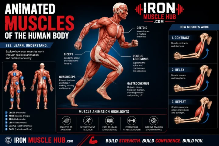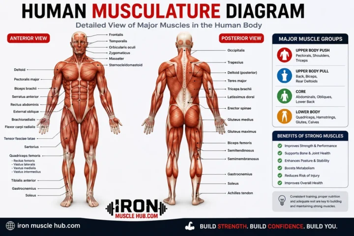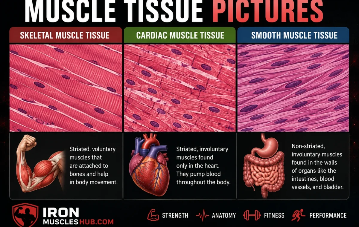
Muscle tissue pictures make it much easier to understand how the body’s three major muscle tissues differ. The three types are skeletal muscle, cardiac muscle, and smooth muscle. Skeletal muscle is striated and mainly controls voluntary movement, cardiac muscle is the specialized muscle of the heart, and smooth muscle works automatically inside organs and blood vessels.
- Muscle Tissue Pictures at a Glance
- 1. Skeletal Muscle Tissue Pictures
- What to look for in a skeletal muscle picture
- Where is skeletal muscle found?
- 2. Cardiac Muscle Tissue Pictures
- How to identify cardiac muscle in a picture
- Why does cardiac muscle look different?
- 3. Smooth Muscle Tissue Pictures
- What to look for in a smooth muscle picture
- Muscle Tissue Pictures: How to Tell Them Apart
- Step 1: Look for stripes
- Step 2: Look at the cell shape
- Step 3: Find the nuclei
- Step 4: Look for branching and connections
- Skeletal vs Cardiac vs Smooth Muscle Pictures
- Why Muscle Tissue Looks Different Under a Microscope
- A Simple Example: Identifying Three Muscle Pictures
- Common Mistakes When Reading Muscle Tissue Pictures
- Mistake 1: Assuming every striped muscle is skeletal
- Mistake 2: Thinking smooth muscle isn't contractile
- Mistake 3: Judging by color alone
- Mistake 4: Confusing a muscle organ with muscle tissue
- Where to Find Reliable Muscle Tissue Pictures
- Internal Link Ideas for Iron Muscle Hub
- Frequently Asked Questions
- What are the three types of muscle tissue?
- What does skeletal muscle tissue look like?
- What does cardiac muscle look like under a microscope?
- What does smooth muscle tissue look like?
- Which muscle tissue is voluntary?
- Why are skeletal and cardiac muscles striped?
- Seeing the Difference Makes Muscle Anatomy Easier
- How Muscle Tissue Is Organized
- Skeletal Muscle Tissue: What the Pictures Show
- Skeletal muscle in longitudinal section
- Skeletal muscle in cross-section
- Cardiac Muscle Tissue: Important Picture Features
- What should you look for?
- Smooth Muscle Tissue: Understanding the Picture
- Smooth muscle picture clues
- Muscle Tissue Pictures: Longitudinal vs Cross Section
- How to Identify Muscle Tissue in an Exam
- Question 1: Are there visible striations?
- Question 2: Are the cells long and parallel?
- Question 3: Do the cells branch?
- Question 4: Where are the nuclei?
- Question 5: Can you see intercalated discs?
- Muscle Tissue vs Muscle Fiber
- Why Striations Matter in Muscle Tissue Pictures
- A Quick Identification Chart
- What Muscle Tissue Pictures Can Tell You
- A Common Confusion: Muscle vs Tendon
- A Common Confusion: Smooth Muscle vs Dense Connective Tissue
- How to Study Muscle Tissue Pictures Faster
- Skeletal
- Cardiac
- Smooth
- Muscle Tissue Pictures and Body Function
- Key Takeaways From Muscle Tissue Pictures
- Frequently Asked Questions
- How can I identify skeletal muscle in a picture?
- How can I identify cardiac muscle in a microscope picture?
- How does smooth muscle look different?
- Why do muscle tissue pictures look different from one another?
- Which muscle tissue has striations?
- Final Thoughts on Reading Muscle Tissue Pictures
- The Three Muscle Tissues Side by Side
- How Magnification Changes Muscle Tissue Pictures
- Low magnification
- Higher magnification
- Muscle Tissue Pictures in Longitudinal Section
- Skeletal muscle
- Cardiac muscle
- Smooth muscle
- Muscle Tissue Pictures in Cross Section
- The key question
- What Are Muscle Fibers Made Of?
- Understanding Striations More Clearly
- How Nuclei Help Identify Muscle Tissue
- Skeletal muscle
- Cardiac muscle
- Smooth muscle
- Intercalated Discs in Cardiac Muscle
- Smooth Muscle and Internal Organs
- Why Muscle Tissue Is Important for the Human Body
- A Practical Muscle Tissue Identification Exercise
- Picture A
- Picture B
- Picture C
- Picture D
- Five-Second Muscle Tissue Test
- Mistakes to Avoid When Studying Muscle Tissue Pictures
- Don't rely on image color
- Don't memorize only one photograph
- Don't assume stripes automatically mean skeletal muscle
- Don't forget section orientation
- Don't confuse muscle tissue with an entire muscle
- How Muscle Tissue Pictures Connect to Fitness and Exercise
- Muscle Tissue Pictures: The Big Picture
- Skeletal muscle
- Cardiac muscle
- Smooth muscle
- Frequently Asked Questions
- What is the easiest way to identify muscle tissue pictures?
- Which muscle tissue has no striations?
- Can cardiac muscle be mistaken for skeletal muscle?
- Why does skeletal muscle look different in cross section?
- What do muscle tissue pictures teach us?
- Final Thoughts:
Looking at muscle tissue under a microscope can seem confusing at first. The good news is that each type has a few visual clues that make identification much easier. Once you know what to look for, you can usually distinguish them by their shape, stripes, nuclei, and location. For more insights read here :- lean build and fastest way to gain muscle .
Quick answer: Human muscle tissue has three main types: skeletal, cardiac, and smooth. Skeletal muscle has obvious striations and many nuclei, cardiac muscle is striated and branched with centrally located nuclei, while smooth muscle lacks visible striations and usually has one central nucleus.
Muscle Tissue Pictures at a Glance
| Muscle tissue | What it looks like | Control | Main location | Main job |
|---|---|---|---|---|
| Skeletal | Long, cylindrical, striped fibers | Voluntary | Attached to bones | Body movement and posture |
| Cardiac | Short, branched, striped cells | Involuntary | Heart | Pumps blood |
| Smooth | Spindle-shaped cells without visible stripes | Involuntary | Internal organs and blood vessels | Moves and regulates substances |
These visual differences aren’t just cosmetic. The structure of each muscle tissue is closely related to the work it performs.
1. Skeletal Muscle Tissue Pictures
Skeletal muscle is the type most people picture when they hear the word “muscle.” It attaches to bones through tendons and produces movements such as walking, lifting, jumping, and changing posture. It is also the muscle type you can generally control consciously.
Under a microscope, skeletal muscle has a distinctive striped or striated appearance. Its cells are long and cylindrical, and individual fibers contain multiple nuclei positioned toward the edges of the cell.
What to look for in a skeletal muscle picture
When identifying skeletal muscle tissue, look for:
- Long, parallel fibers
- Clear repeating stripes or striations
- Multiple nuclei
- Nuclei positioned toward the outer edge of the fibers
- An organized, relatively uniform appearance
Those stripes come from the organized arrangement of contractile proteins inside the muscle fibers. Skeletal muscle and cardiac muscle are both striated because their contractile proteins are arranged into organized structures called sarcomeres.
Where is skeletal muscle found?
Skeletal muscle is attached to bones throughout the body. It helps you move your limbs, maintain posture, produce facial expressions, and perform many other deliberate movements.
For example, when you decide to bend your elbow, your nervous system activates skeletal muscles that pull on the bones around the joint.
2. Cardiac Muscle Tissue Pictures
Cardiac muscle is found in the heart. Its primary job is to contract repeatedly and generate the pressure needed to move blood through the circulatory system. Unlike skeletal muscle, cardiac muscle operates involuntarily.
Cardiac muscle can look somewhat like skeletal muscle because both have visible striations. The major difference is the shape and organization of the cells.
Cardiac muscle cells are generally shorter and branched, with centrally located nuclei. They connect with neighboring cells through specialized structures called intercalated discs, which help the heart muscle coordinate its contractions.
How to identify cardiac muscle in a picture
Look for:
- Visible striations
- Shorter cells than skeletal muscle fibers
- Branching cells
- Usually one centrally positioned nucleus
- Connections between neighboring cells called intercalated discs
These features allow cardiac tissue to work as a coordinated muscle network rather than as isolated cells.
Why does cardiac muscle look different?
The heart needs a specialized structure because it must contract repeatedly without conscious effort. Cardiac muscle cells are physically and electrically connected, allowing contractions to spread through the heart muscle in a coordinated way.
3. Smooth Muscle Tissue Pictures
Smooth muscle is different from both skeletal and cardiac muscle. Its cells don’t have the obvious striated pattern seen in the other two types.
Smooth muscle cells are generally spindle-shaped, meaning they are wider around the middle and narrow toward the ends. Each cell typically contains a single centrally located nucleus.
Smooth muscle works automatically rather than being consciously controlled.
It is found in many internal structures, including the walls of hollow organs and blood vessels. Its contractions help move substances through the digestive system, regulate blood vessel diameter, and perform other automatic functions.
What to look for in a smooth muscle picture
The easiest clues are:
- No obvious striations
- Spindle-shaped cells
- One central nucleus per cell
- Closely packed cells
- Involuntary activity
The absence of stripes doesn’t mean smooth muscle can’t contract. It still contains actin and myosin, but these proteins aren’t arranged into the same organized sarcomeres found in skeletal and cardiac muscle.
Muscle Tissue Pictures: How to Tell Them Apart
If you’re looking at a microscope image and aren’t sure what you’re seeing, use this simple process.
Step 1: Look for stripes
Stripes present? Think skeletal or cardiac muscle.
No obvious stripes? Smooth muscle is the likely answer.
Step 2: Look at the cell shape
Long, parallel fibers suggest skeletal muscle.
Shorter, branching fibers suggest cardiac muscle.
Spindle-shaped cells suggest smooth muscle.
Step 3: Find the nuclei
Multiple nuclei toward the edges of the fibers point toward skeletal muscle.
A centrally located nucleus is more typical of cardiac or smooth muscle.
Step 4: Look for branching and connections
Branching cells and visible intercalated discs are strong clues for cardiac muscle.
This four-step approach is useful for students because you don’t have to memorize every microscopic detail at once.
Skeletal vs Cardiac vs Smooth Muscle Pictures
The biggest mistake beginners make is trying to identify muscle tissue based on just one feature.
For example, both skeletal and cardiac muscle have striations. Instead, combine several clues.
| Feature | Skeletal | Cardiac | Smooth |
| Striations | Yes | Yes | No obvious striations |
| Cell shape | Long and cylindrical | Short and branched | Spindle-shaped |
| Nuclei | Many, generally peripheral | Usually one, central | Usually one, central |
| Control | Voluntary | Involuntary | Involuntary |
| Main location | Attached to bones | Heart | Internal organs and vessels |
| Main function | Movement and posture | Pumping blood | Moving/regulating substances |
Why Muscle Tissue Looks Different Under a Microscope
Microscope images show that muscle tissue isn’t simply one uniform material.
Each muscle type has a structure suited to its job. Skeletal muscle needs to generate controlled force for movement. Cardiac muscle needs synchronized contractions inside the heart. Smooth muscle needs to perform automatic contractions in organs and passageways.
At the cellular level, all three types are specialized for contraction, but their internal organization differs.
Skeletal and cardiac muscle have organized contractile structures called sarcomeres, which contribute to their striped appearance. Smooth muscle contains actin and myosin too, but they are organized differently, so the cells don’t show the same visible banding.
A Simple Example: Identifying Three Muscle Pictures
Imagine you’re given three unlabeled microscope photographs in a biology class.
Picture A: You see long fibers with obvious stripes and several nuclei along the edges. That’s most consistent with skeletal muscle.
Picture B: You see branching striped cells with centrally located nuclei and specialized connections between cells. That’s cardiac muscle.
Picture C: You see tightly packed, tapered cells without obvious stripes and with central nuclei. That’s smooth muscle.
The trick isn’t memorizing the photographs. It’s recognizing the structural clues.
Common Mistakes When Reading Muscle Tissue Pictures
Mistake 1: Assuming every striped muscle is skeletal
Cardiac muscle is also striated. Check the cell shape, branching, and nucleus position before deciding.
Mistake 2: Thinking smooth muscle isn’t contractile
Smooth muscle contracts continuously in many parts of the body. It simply lacks the visible striations associated with skeletal and cardiac muscle.
Mistake 3: Judging by color alone
Different stains, microscopes, lighting conditions, and image-processing techniques can change how tissue appears. Color can help, but structural features are much more useful for identification.
Mistake 4: Confusing a muscle organ with muscle tissue
A whole skeletal muscle, such as the biceps, contains more than muscle fibers. It also includes connective tissue, blood vessels, nerves, and other structures.
Where to Find Reliable Muscle Tissue Pictures
For study and educational purposes, reputable medical and educational sources are preferable to random image searches.
OpenStax provides anatomy and physiology material with labeled explanations of skeletal, cardiac, and smooth muscle tissue.
The National Center for Biotechnology Information (NCBI) also provides anatomy and histology resources, including educational figures showing the three major muscle tissue types.
If you’re publishing an image on a website, always check the specific image’s license and attribution requirements rather than assuming that an educational image is automatically free to reuse.
Internal Link Ideas for Iron Muscle Hub
You could connect this article naturally to related beginner-friendly topics such as:
- What Are Muscles? A Beginner’s Guide to Muscle Anatomy
- What Are the Major Muscles in the Human Body?
- How Does the Muscular System Work With Other Body Systems?
These links can help readers move from basic tissue identification into broader muscle anatomy and physiology.
Frequently Asked Questions
What are the three types of muscle tissue?
The three major types are skeletal, cardiac, and smooth muscle. Skeletal muscle is mainly responsible for voluntary movement, cardiac muscle makes up the heart, and smooth muscle performs involuntary contractions in many internal organs and blood vessels. Their different structures allow them to perform different jobs.
What does skeletal muscle tissue look like?
Skeletal muscle tissue typically appears as long, parallel, cylindrical fibers with obvious striations. The cells contain multiple nuclei, and those nuclei are generally located toward the edges of the fibers. This combination of long fibers, stripes, and peripheral nuclei is useful when identifying skeletal muscle under a microscope.
What does cardiac muscle look like under a microscope?
Cardiac muscle has visible striations, but its cells are shorter and often branched compared with skeletal muscle fibers. The nuclei are usually centrally located, and neighboring cells connect through intercalated discs. These structural features help distinguish cardiac muscle from skeletal muscle.
What does smooth muscle tissue look like?
Smooth muscle generally consists of spindle-shaped cells that are wider in the middle and narrow at the ends. The cells usually contain one central nucleus and don’t show the obvious striations found in skeletal and cardiac muscle. Smooth muscle is common in the walls of internal organs and blood vessels.
Which muscle tissue is voluntary?
Skeletal muscle is the primary voluntary muscle tissue. You can consciously activate skeletal muscles to perform movements such as walking, lifting an object, or changing posture. Cardiac and smooth muscles generally operate involuntarily, meaning their activity isn’t normally under conscious control.
Why are skeletal and cardiac muscles striped?
Skeletal and cardiac muscle cells contain contractile proteins arranged into organized structures called sarcomeres. This regular arrangement produces the visible striated pattern seen under a microscope. Smooth muscle also contains actin and myosin, but its contractile proteins aren’t organized into sarcomeres in the same way.
Seeing the Difference Makes Muscle Anatomy Easier
Muscle tissue pictures become much easier to understand once you stop trying to memorize each photograph and start looking for a few consistent features. Skeletal muscle is long and striated, cardiac muscle is striated and branched, and smooth muscle is spindle-shaped without obvious striations.
Those differences reflect the jobs each tissue performs throughout the body. If you’re studying anatomy, focus first on stripes, cell shape, nuclei, and location. With those four clues, you’ll have a much easier time identifying muscle tissue in diagrams, microscope slides, and exam questions.
How Muscle Tissue Is Organized
Muscle tissue may look simple in a picture, but it has a highly organized structure. The cells are specialized to contract, and their arrangement differs depending on the type of muscle.
The three major types—skeletal, cardiac, and smooth muscle—all perform contraction, but they are built differently because they have different jobs.
A useful way to study muscle tissue pictures is to move from the largest structure to the smallest:
- Muscle or organ
- Muscle tissue
- Muscle cells or fibers
- Contractile structures inside the cells
- Proteins involved in contraction
This approach makes microscopic images much easier to understand.
Skeletal Muscle Tissue: What the Pictures Show
Skeletal muscle is made of long muscle fibers that are arranged in an organized pattern. In a typical microscope image, these fibers can appear as long parallel structures.
One of the easiest characteristics to recognize is striations. These alternating light and dark bands result from the organized arrangement of contractile structures within the muscle fiber.
Skeletal muscle in longitudinal section
When skeletal muscle is viewed lengthwise, the fibers usually appear long and parallel.
You may be able to see:
- Repeating striations
- Long cylindrical fibers
- Multiple nuclei
- Nuclei near the outside of the fibers
- Parallel organization
This is one of the classic appearances used to identify skeletal muscle in histology.
Skeletal muscle in cross-section
A cross-sectional picture looks very different.
Instead of seeing long fibers extending across the image, you may see many roughly circular or polygonal fiber profiles packed together.
This is an important exam tip: the same tissue can look dramatically different depending on the direction in which it was cut.
Cardiac Muscle Tissue: Important Picture Features
Cardiac muscle forms the muscular wall of the heart.
Like skeletal muscle, cardiac muscle is striated, so stripes can be visible in microscopic images. However, cardiac cells have several features that help distinguish them from skeletal muscle.
Cardiac muscle cells are generally shorter and can branch. They commonly contain a centrally located nucleus.
Another important feature is the intercalated disc. These specialized junctions connect neighboring cardiac muscle cells and help them function as a coordinated tissue.
What should you look for?
When studying cardiac muscle pictures, look for:
- Striations
- Branching cells
- Central nuclei
- Shorter fibers than typical skeletal muscle fibers
- Intercalated discs
Not every microscopic image will make every feature equally obvious. That’s why it’s better to identify cardiac muscle using several clues rather than relying on one feature.
Smooth Muscle Tissue: Understanding the Picture
Smooth muscle is often the easiest tissue to distinguish because it doesn’t have the obvious striated appearance seen in skeletal and cardiac muscle.
Its cells are generally elongated and spindle-shaped. The center of each cell is wider, while the ends become narrower.
Smooth muscle is found in many structures that need automatic contraction, including parts of the digestive tract and blood vessels.
Smooth muscle picture clues
Look for:
- No obvious stripes
- Spindle-shaped cells
- Central nuclei
- Closely packed cells
- Relatively uniform tissue organization
Because smooth muscle cells can overlap, a microscope image may sometimes look more complicated than a simple textbook illustration.
Muscle Tissue Pictures: Longitudinal vs Cross Section
One of the most useful concepts for beginners is understanding section orientation.
A longitudinal section cuts through tissue along the length of the cells. A cross section cuts across the cells.
The difference can completely change what you see.
| Section | What you usually see |
|---|---|
| Longitudinal | Cells viewed along their length |
| Cross section | Cells viewed across their width |
| Oblique section | Cells viewed at an angle |
For example, a skeletal muscle fiber looks long and striped when viewed longitudinally. In cross section, you may instead see many individual fiber profiles grouped together.
This is why comparing a microscope picture with a simplified anatomy diagram can sometimes be confusing.
How to Identify Muscle Tissue in an Exam
If you’re given an unlabeled muscle tissue image, don’t panic. Use a consistent checklist.
Question 1: Are there visible striations?
If yes, consider skeletal or cardiac muscle.
If no, smooth muscle becomes the strongest possibility.
Question 2: Are the cells long and parallel?
Long, cylindrical, parallel fibers strongly suggest skeletal muscle.
Question 3: Do the cells branch?
Branching cells point toward cardiac muscle.
Question 4: Where are the nuclei?
Multiple peripheral nuclei are characteristic of skeletal muscle.
Central nuclei are common in cardiac and smooth muscle.
Question 5: Can you see intercalated discs?
If clear intercalated discs are present, cardiac muscle is the likely identification.
Using these questions together is more reliable than memorizing the appearance of a single image.
Muscle Tissue vs Muscle Fiber
The terms muscle tissue and muscle fiber are related, but they aren’t exactly the same.
A muscle fiber generally refers to a muscle cell, particularly in skeletal muscle. Muscle tissue refers to the larger tissue made from muscle cells along with their surrounding organization.
Think of it like this:
Muscle fiber → many fibers together → muscle tissue → complete muscle structure
A whole skeletal muscle also contains connective tissue, nerves, and blood vessels. So a photograph of an entire muscle and a microscope photograph of muscle tissue are showing different levels of organization.
Why Striations Matter in Muscle Tissue Pictures
Striations are one of the most recognizable features in muscle histology.
Skeletal and cardiac muscles have contractile proteins arranged into repeating structures called sarcomeres. This regular arrangement produces the characteristic banding pattern visible under suitable microscopy.
Smooth muscle contracts too, but its contractile proteins aren’t organized into sarcomeres in the same way.
That’s why smooth muscle can contract without showing the familiar striped appearance.
A Quick Identification Chart
Here’s a simple study tool you can save for later:
| If the picture shows… | Most likely tissue |
| Long fibers + obvious stripes + many peripheral nuclei | Skeletal muscle |
| Branching fibers + stripes + central nuclei | Cardiac muscle |
| Spindle-shaped cells + no obvious stripes | Smooth muscle |
| Intercalated discs | Cardiac muscle |
| Very long parallel fibers | Skeletal muscle |
The more features you can identify, the more confident your answer will be.
What Muscle Tissue Pictures Can Tell You
A good histology picture can reveal much more than simply the name of a tissue.
It can help you understand:
- Cell shape
- Cell arrangement
- Nucleus position
- Presence or absence of striations
- Connections between cells
- How structure relates to function
For example, the branching structure of cardiac muscle and its specialized cell connections help explain how the heart coordinates its contractions.
Similarly, the elongated organization of skeletal muscle supports force production and movement.
A Common Confusion: Muscle vs Tendon
Muscle tissue and tendon tissue can sometimes be confused in anatomy images.
A tendon is primarily connective tissue, not muscle tissue. It connects skeletal muscle to bone and contains densely arranged collagen fibers.
Muscle tissue contains specialized contractile cells, while tendon tissue is specialized for transmitting force between muscle and bone.
If an image shows tightly packed, parallel collagen fibers without the characteristic muscle-cell appearance, you may be looking at tendon rather than skeletal muscle.
A Common Confusion: Smooth Muscle vs Dense Connective Tissue
Smooth muscle can also be confused with certain connective tissues in microscope images.
The key is to look for the shape and arrangement of the cells.
Smooth muscle typically has elongated cells with central nuclei. Dense connective tissue contains abundant extracellular collagen fibers and has a different overall appearance.
When the image is unclear, don’t identify the tissue based on color alone. Examine the cells, nuclei, and organization.
How to Study Muscle Tissue Pictures Faster
If you’re preparing for a biology, anatomy, or physiology test, you don’t need to memorize dozens of photographs.
Start with three words:
Stripes — Shape — Nuclei
Then add location.
Skeletal
Stripes + long fibers + peripheral nuclei
Cardiac
Stripes + branching + central nuclei
Smooth
No obvious stripes + spindle-shaped cells + central nuclei
This simple framework can make identification much faster.
Muscle Tissue Pictures and Body Function
The appearance of muscle tissue reflects what that tissue needs to accomplish.
Skeletal muscle needs to create controlled movements of the skeleton. Its long fibers and organized contractile structures support force production.
Cardiac muscle needs to contract repeatedly as part of the heart’s pumping activity. Its specialized connections help cardiac cells work together.
Smooth muscle needs to control movement and diameter within internal structures. Its cellular organization supports slow, involuntary contractions in places such as the digestive tract and blood vessels.
In other words, structure and function are closely connected.
Key Takeaways From Muscle Tissue Pictures
Before moving on, remember these essential points:
- Skeletal muscle is striated, long, and generally multinucleated.
- Cardiac muscle is striated, branched, and usually has central nuclei.
- Smooth muscle lacks obvious striations and has spindle-shaped cells.
- Skeletal muscle is mainly associated with voluntary movement.
- Cardiac muscle is found in the heart.
- Smooth muscle occurs in many internal organs and blood vessels.
- A tissue’s appearance can change depending on whether it is viewed in longitudinal or cross section.
- Use several microscopic clues together instead of relying on color or one feature.
Frequently Asked Questions
How can I identify skeletal muscle in a picture?
Look for long, parallel fibers with obvious striations. Skeletal muscle fibers also contain multiple nuclei, which are generally positioned toward the outer edges of the cells. If the image is a cross section, the fibers may instead look like many closely packed polygonal profiles.
How can I identify cardiac muscle in a microscope picture?
Look for striated cells that are shorter and more branched than typical skeletal muscle fibers. Cardiac muscle commonly has centrally located nuclei. Intercalated discs may also be visible between neighboring cells and are an important clue when they can be clearly seen.
How does smooth muscle look different?
Smooth muscle generally lacks the obvious stripes seen in skeletal and cardiac muscle. Its cells are typically spindle-shaped, with a wider middle and tapered ends. Each cell usually has a centrally positioned nucleus. The cells are commonly arranged in layers within the walls of internal organs.
Why do muscle tissue pictures look different from one another?
Several factors can change the appearance of a tissue image. The tissue may have been cut longitudinally, transversely, or at an angle. Magnification, staining, lighting, and the particular region being examined can also affect what is visible.
Which muscle tissue has striations?
Both skeletal muscle and cardiac muscle have visible striations because their contractile proteins are organized into sarcomeres. Smooth muscle also contains contractile proteins but doesn’t have the same sarcomere arrangement, so it doesn’t show the same obvious banding pattern.
Final Thoughts on Reading Muscle Tissue Pictures
Muscle tissue pictures become much easier once you know which details matter most. Start by checking for striations, then examine the cell shape, nuclei, and any signs of branching or specialized connections.
Skeletal, cardiac, and smooth muscle may all contract, but their microscopic structures are different because their jobs are different. If you’re studying anatomy, don’t try to memorize every photograph. Learn the identifying patterns first, then practice applying them to different images.
That approach will help you recognize muscle tissue even when the microscope image doesn’t look exactly like the one in your textbook.
When you look at muscle tissue pictures under a microscope, the first thing to remember is that you’re seeing cells rather than the entire muscle you see in an anatomy diagram.
A complete skeletal muscle, for example, is an organ made from many layers of tissue and supporting structures. A histology image zooms in on a much smaller part of that organization.
This is why microscope pictures can look unfamiliar even when you already know the major muscles of the body.
The Three Muscle Tissues Side by Side
A side-by-side comparison is one of the fastest ways to learn the differences.
| Feature | Skeletal Muscle | Cardiac Muscle | Smooth Muscle |
|---|---|---|---|
| Striations | Yes | Yes | No obvious striations |
| Cell shape | Long and cylindrical | Shorter and branched | Spindle-shaped |
| Nuclei | Multiple, generally peripheral | Usually central | Usually central |
| Control | Voluntary | Involuntary | Involuntary |
| Main location | Attached to bones | Heart | Hollow organs and vessels |
| Sarcomeres | Present | Present | Not arranged into sarcomeres |
| Distinctive clue | Long parallel fibers | Branching + intercalated discs | Tapered cells without stripes |
The table is useful as a starting point, but microscope images can vary depending on the section and preparation.
How Magnification Changes Muscle Tissue Pictures
A muscle tissue photograph taken at low magnification may show the overall arrangement of many fibers.
At higher magnification, individual cells and smaller structures become easier to see.
For example, a low-power image might help you recognize the general organization of skeletal muscle, while a higher-power image can make nuclei and striations easier to examine.
This means two pictures of the same tissue can look very different without either image being incorrect.
Low magnification
You may notice:
- Overall tissue arrangement
- Groups of fibers
- General direction of cells
- Larger structural patterns
Higher magnification
You may notice:
- Individual nuclei
- Fine striations
- Cell boundaries
- Specialized structures
- Details between neighboring cells
When studying, check the magnification if it is provided. It gives you useful context for what you should expect to see.
Muscle Tissue Pictures in Longitudinal Section
A longitudinal section follows the general direction of the muscle cells.
This view is particularly useful for studying skeletal and cardiac muscle because it can reveal the length and organization of the fibers.
Skeletal muscle
The cells generally appear long and parallel. Striations may be visible running across the fibers.
Cardiac muscle
The cells can appear shorter and branched. Striations may also be visible, and nuclei tend to occur near the center of the cells.
Smooth muscle
The elongated cells can appear tapered at both ends. Their central nuclei may be visible, but the characteristic striations of skeletal and cardiac muscle are absent.
Muscle Tissue Pictures in Cross Section
A cross section cuts across the general length of the cells.
This changes the appearance dramatically.
Skeletal muscle fibers may appear as many tightly packed profiles. Depending on the preparation, their shapes can look roughly circular, polygonal, or irregular.
This is why beginners sometimes fail to recognize skeletal muscle in a cross-sectional image. They expect to see long stripes but are instead looking at the ends of the fibers.
The key question
Ask yourself:
“Am I looking along the muscle fibers or across them?”
That single question can prevent many identification mistakes.
What Are Muscle Fibers Made Of?
A muscle fiber contains specialized structures that allow it to contract.
Skeletal muscle fibers are large, specialized cells containing many structures involved in energy production, calcium handling, and contraction.
Inside skeletal muscle fibers are myofibrils, which contain repeating contractile units called sarcomeres.
Sarcomeres contain proteins including actin and myosin. Their organized arrangement is responsible for the characteristic striated appearance of skeletal and cardiac muscle.
Smooth muscle also uses actin and myosin for contraction, but its contractile machinery is arranged differently.
Understanding Striations More Clearly
The stripes in skeletal and cardiac muscle aren’t simply decorative lines.
They are the visible result of an organized microscopic structure.
Within a striated muscle fiber, sarcomeres are arranged end to end. This produces a repeating pattern that can be seen with appropriate microscopy.
That’s why skeletal and cardiac muscle have a banded appearance while smooth muscle does not.
Understanding this connection is more useful than simply memorizing:
“Stripes = skeletal.”
That shortcut is incomplete because cardiac muscle also has striations.
A better rule is:
Striations + long parallel fibers → skeletal
Striations + branching cells → cardiac
No obvious striations + spindle-shaped cells → smooth
How Nuclei Help Identify Muscle Tissue
Nuclei are another useful clue in muscle histology.
Skeletal muscle
Skeletal muscle fibers contain multiple nuclei. These nuclei are generally located near the periphery of the fiber.
Cardiac muscle
Cardiac muscle cells generally have a centrally located nucleus, although appearances can vary depending on the tissue section.
Smooth muscle
Smooth muscle cells usually contain one central nucleus.
Nuclei are especially useful when combined with cell shape and striations.
Never identify a tissue from nucleus position alone if the picture provides other clues.
Intercalated Discs in Cardiac Muscle
One feature that makes cardiac muscle particularly interesting is the presence of intercalated discs.
These specialized junctional regions connect neighboring cardiac muscle cells.
They help cardiac muscle cells communicate and mechanically connect with each other so the heart can contract in a coordinated manner.
In some histology images, intercalated discs are clearly visible. In others, they may be difficult to identify.
So if you can’t see them, that doesn’t automatically rule out cardiac muscle.
Use the complete pattern instead.
Smooth Muscle and Internal Organs
Smooth muscle is found in many places where automatic movement or regulation is required.
Examples include:
- Digestive tract walls
- Blood vessel walls
- Urinary system structures
- Airways
- Other hollow organs
Its contractions can help move contents through an organ or alter the diameter of a passageway.
This explains why smooth muscle doesn’t need to be consciously controlled for you to benefit from its activity.
You don’t have to think about moving food through your digestive tract or adjusting the diameter of many blood vessels. Smooth muscle contributes to these automatic processes.
Why Muscle Tissue Is Important for the Human Body
Muscle tissue is responsible for much more than visible body movement.
Muscle activity contributes to:
- Movement
- Posture
- Heart pumping
- Movement of substances through organs
- Regulation of passageways
- Production of body heat
Skeletal, cardiac, and smooth muscle each contribute in different ways.
Understanding their structure helps explain why the body can perform both deliberate actions—such as lifting a bag—and automatic actions—such as the continuous beating of the heart.
A Practical Muscle Tissue Identification Exercise
Try this without looking at the answers.
Picture A
You see long, parallel fibers with strong striations. Several nuclei appear toward the edges.
Answer: Skeletal muscle.
Picture B
You see branching cells with striations and centrally located nuclei.
Answer: Cardiac muscle.
Picture C
You see elongated, tapered cells without obvious stripes.
Answer: Smooth muscle.
Picture D
You see many tightly packed polygonal profiles in cross section.
Likely answer: Skeletal muscle, assuming the surrounding features support that identification.
The point isn’t simply to memorize the answers. Try to explain why each answer fits.
Five-Second Muscle Tissue Test
If you need a fast way to identify a picture during a test, use this sequence:
1. Stripes?
- No → likely smooth.
- Yes → skeletal or cardiac.
2. Long and parallel?
- Yes → likely skeletal.
3. Branching?
- Yes → likely cardiac.
4. Central nuclei?
- Cardiac or smooth become more likely.
5. Peripheral multiple nuclei?
- Strong clue for skeletal muscle.
This isn’t a substitute for learning histology, but it’s a useful starting framework.
Mistakes to Avoid When Studying Muscle Tissue Pictures
Don’t rely on image color
Histology stains can produce different colors and shades. Two legitimate images of the same tissue may look quite different.
Focus on structural characteristics.
Don’t memorize only one photograph
An exam or study resource may use a different magnification, stain, section orientation, or image quality.
Learn the features instead of memorizing a particular picture.
Don’t assume stripes automatically mean skeletal muscle
Cardiac muscle is also striated.
Check for branching, nuclei, and other characteristics.
Don’t forget section orientation
A cross section won’t look like a longitudinal section.
Always consider how the tissue was cut.
Don’t confuse muscle tissue with an entire muscle
A whole muscle includes supporting tissues, nerves, blood vessels, and connective tissue. A histology image may show only a small component.
How Muscle Tissue Pictures Connect to Fitness and Exercise
For readers interested in fitness, skeletal muscle is especially important.
Skeletal muscle produces voluntary movement and adapts to physical demands. Resistance training can stimulate changes in skeletal muscle, while adequate nutrition and recovery support the body’s ability to adapt.
However, a microscope picture of skeletal muscle doesn’t show everything that determines strength or athletic performance.
Strength and physical performance depend on multiple factors, including muscle size, nervous-system control, technique, training history, and other physiological characteristics.
So don’t judge someone’s fitness level simply by looking at a microscopic muscle image.
Muscle Tissue Pictures: The Big Picture
The easiest way to remember the three types is to connect appearance with function.
Skeletal muscle
Appearance: Long, striped fibers.
Function: Voluntary movement and posture.
Cardiac muscle
Appearance: Striped, branching cells with central nuclei.
Function: Continuous pumping action of the heart.
Smooth muscle
Appearance: Spindle-shaped cells without obvious striations.
Function: Automatic movement and regulation within internal organs and vessels.
Once those three patterns become familiar, most basic muscle tissue identification questions become much easier.
Frequently Asked Questions
What is the easiest way to identify muscle tissue pictures?
Start by checking for striations. Smooth muscle lacks obvious striations, while skeletal and cardiac muscle have them. Then look at the cell shape and nuclei. Long parallel fibers with multiple peripheral nuclei suggest skeletal muscle, while branching cells with central nuclei suggest cardiac muscle.
Which muscle tissue has no striations?
Smooth muscle does not show the obvious striated pattern found in skeletal and cardiac muscle. Its cells are typically spindle-shaped and contain central nuclei. Smooth muscle is specialized for involuntary contraction in many internal organs and blood vessels.
Can cardiac muscle be mistaken for skeletal muscle?
Yes. Both are striated, so they can look similar in some microscope images. Cardiac muscle is distinguished by features such as branching cells, central nuclei, and intercalated discs. Skeletal muscle typically has longer, parallel fibers with multiple peripheral nuclei.
Why does skeletal muscle look different in cross section?
A cross section cuts across the fibers rather than along their length. Instead of seeing long fibers and their stripes, you may see many individual fiber profiles packed together. The appearance can therefore be very different from a longitudinal skeletal muscle image.
What do muscle tissue pictures teach us?
They show how cellular structure relates to function. Skeletal muscle has long organized fibers for movement, cardiac muscle has specialized cells for coordinated heart contraction, and smooth muscle has structures suited to automatic activity in internal organs and vessels.
Final Thoughts:
Learning to recognize muscle tissue pictures doesn’t have to mean memorizing hundreds of microscope slides. Start with the three major tissues and learn their most reliable visual clues.
Remember the simple pattern: skeletal is long and striated, cardiac is striated and branched, and smooth is spindle-shaped without obvious striations.
Then add the details—nucleus position, section orientation, and specialized structures such as intercalated discs. Once you understand why each tissue looks the way it does, you’ll be much better prepared to identify unfamiliar anatomy and histology pictures.
For a beginner, structure is the best shortcut: understand what you’re seeing, rather than simply memorizing what a particular picture looks like.