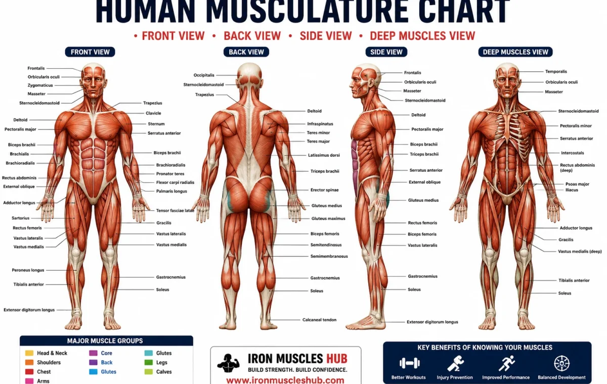
If you’re looking at a human musculature chart and wondering what all those labels mean, start with the major muscle groups rather than trying to memorize every small muscle. A good chart shows where muscles sit, how front and back views differ, and how individual muscles contribute to movement.
- Quick Answer
- What Is a Human Musculature Chart?
- Why Muscle Charts Matter for Lifters
- The Two Views You Need to Understand First
- Anterior View: Front of the Body
- Posterior View: Back of the Body
- Human Musculature Chart: Major Muscle Groups
- Chest Muscles
- Pectoralis Major
- Pectoralis Minor
- Shoulder Muscles
- Deltoid
- Rotator Cuff
- Back Muscles
- Trapezius
- Latissimus Dorsi
- Rhomboids
- Arm Muscles
- Biceps Brachii
- Triceps Brachii
- Core Muscles
- Rectus Abdominis
- External Obliques
- Deeper Core Structures
- Glute Muscles
- Gluteus Maximus
- Gluteus Medius and Minimus
- Thigh Muscles
- Quadriceps
- Hamstrings
- Adductors
- Lower-Leg Muscles
- Gastrocnemius
- Soleus
- Tibialis Anterior
- How to Read a Human Musculature Chart Correctly
- Step 1: Identify the View
- Step 2: Find Large Landmarks
- Step 3: Learn the Major Muscle
- Step 4: Connect Muscle to Movement
- Step 5: Add Smaller Muscles
- Common Mistakes → Why → Fix
- Mistake 1: Memorizing the Whole Chart
- Mistake 2: Using Only a Bodybuilding Chart
- Mistake 3: Thinking Every Muscle Has One Job
- Mistake 4: Confusing “Muscle Group” With “Muscle”
- A 30-Second Muscle Chart Self-Test
- Worked Example: Using a Chart for a 70 kg / 154 lb Beginner
- How Muscle Anatomy Connects to Strength Training
- Three Types of Muscle Tissue
- How Much of the Body Is Skeletal Muscle?
- What Is Skeletal Muscle Made Of?
- Superficial vs. Deep Muscles
- Superficial Muscles
- Deep Muscles
- Who This Is NOT For
- How Beginners Can Memorize Muscle Anatomy Faster
- 1. Location
- 2. Name
- 3. Action
- 4. Exercise
- FAQ: Human Musculature Chart
- 1. What are the major muscle groups in the human body?
- 2. How many muscles are in the human body?
- 3. What are the three types of muscle tissue?
- 4. What is the largest muscle in the human body?
- 5. What is the difference between an anterior and posterior muscle chart?
- Final Takeaway
For beginners, the most useful areas to learn first are the chest, shoulders, back, arms, abdominals, glutes, thighs, and calves. Once you can recognize those regions, smaller muscles and deeper layers become much easier to understand. For more insights read here :- muscular anatomy chart and human muscle chart.
Quick Answer
- A human musculature chart is a visual reference showing the location, names, and sometimes functions of muscles.
- Most beginner charts show anterior (front) and posterior (back) views.
- The major training regions include the chest, shoulders, back, arms, core, glutes, thighs, and calves.
- Skeletal muscle is attached to the skeleton through connective tissues including tendons and produces movement when it contracts.
What Is a Human Musculature Chart?
A human musculature chart is an anatomical reference that maps muscles onto the body. Depending on the chart, it may show only large superficial muscles or include deeper muscles, smaller structures, and anatomical terminology.
For a beginner, the best chart is not necessarily the one with the most labels. A crowded diagram can make learning harder.
A useful chart should help you answer three simple questions:
- Where is the muscle?
- What is the muscle called?
- What movement or function is it associated with?
Anatomy references commonly organize muscles by body region, such as the upper limb, lower limb, head and neck, and trunk. They may also provide information about muscle origins, insertions, innervation, and functions.
Why Muscle Charts Matter for Lifters
You don’t need a medical-school level knowledge of anatomy to train effectively.
However, knowing basic muscle anatomy can make your training more logical.
For example, instead of thinking:
“I need another chest exercise.”
You can start thinking:
“This exercise involves the pectoralis major, while my shoulder and triceps also contribute.”
That difference matters because resistance exercises rarely isolate a muscle completely.
A pressing exercise can involve the chest, front shoulder, and triceps. A rowing movement can involve the back and elbow-flexing muscles. A squat involves several muscles around the hip, knee, and ankle.
Skeletal muscle produces force and movement, while also contributing to posture, joint stability, temperature regulation, and other physiological functions.
The Two Views You Need to Understand First
Most human musculature charts use two primary views:
- Anterior view: looking at the front of the body
- Posterior view: looking at the back of the body
These aren’t different bodies. They’re two perspectives of the same muscular system.
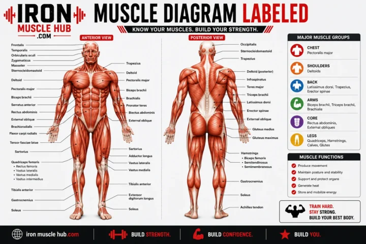
Anterior View: Front of the Body
The anterior view commonly makes it easier to identify:
- Pectoralis major
- Deltoid
- Biceps brachii
- Rectus abdominis
- External oblique
- Quadriceps
- Sartorius
- Adductors
- Tibialis anterior
Anatomical diagrams may show considerably more detail. For example, educational muscular-system diagrams can label muscles such as the sternocleidomastoid, serratus anterior, brachialis, rectus femoris, vastus lateralis, vastus medialis, and gastrocnemius.
Posterior View: Back of the Body
The posterior view commonly shows:
- Trapezius
- Rear deltoid
- Latissimus dorsi
- Triceps brachii
- Rhomboids
- Erector-spinae region
- Gluteus maximus
- Hamstrings
- Gastrocnemius
- Soleus
This view is particularly useful for understanding back training.
For example, the broad latissimus dorsi occupies much of the lateral back, while the trapezius spans a large area across the upper back and neck region.
The posterior leg also contains important muscles such as the hamstrings and calf muscles.
Human Musculature Chart: Major Muscle Groups
Here’s a practical beginner-friendly reference.
| Body region | Major muscles to learn | Main training relevance |
|---|---|---|
| Chest | Pectoralis major, pectoralis minor | Pressing and shoulder movement |
| Shoulders | Deltoid | Raising and positioning the arm |
| Upper back | Trapezius, rhomboids | Scapular movement and upper-back control |
| Back | Latissimus dorsi | Shoulder movement and pulling |
| Front arm | Biceps brachii, brachialis | Elbow flexion |
| Back arm | Triceps brachii | Elbow extension |
| Core | Rectus abdominis, obliques | Trunk movement and stabilization |
| Glutes | Gluteus maximus, medius, minimus | Hip movement and pelvic control |
| Front thigh | Quadriceps | Knee extension |
| Rear thigh | Hamstrings | Knee flexion and hip extension |
| Lower leg | Gastrocnemius, soleus, tibialis anterior | Ankle movement and lower-leg function |
This table is a learning map, not a complete list of every muscle.
A detailed anatomy chart can contain many more structures. For example, professional anatomy references divide muscles into areas such as the rotator cuff, shoulder and arm, forearm and hand, thigh, leg, foot, trunk, and head and neck.
Chest Muscles
Pectoralis Major
The pectoralis major is the large, superficial muscle covering much of the front of the chest.
For lifters, it’s one of the easiest muscles to recognize on an anterior musculature chart.
It contributes to movements of the upper arm and is heavily involved in pressing patterns.
Exercises such as:
- Bench presses
- Push-ups
- Dumbbell presses
- Cable presses
can train the chest while also involving other muscles.
Pectoralis Minor
The pectoralis minor sits deeper than the pectoralis major.
This is a good example of why a basic musculature chart can be misleading if you assume every visible muscle is superficial.
When studying anatomy, think in layers:
surface → deeper muscle → underlying structures
That approach makes a detailed chart much easier to interpret.
Shoulder Muscles
Deltoid
The deltoid forms the rounded contour of the shoulder.
It is commonly divided into three portions:
- Anterior deltoid
- Lateral or middle deltoid
- Posterior deltoid
A chart helps you see that these portions occupy different positions around the shoulder.
The deltoid is involved in movements of the arm, with different portions contributing differently depending on the movement.
Rotator Cuff
The rotator cuff is another important group to recognize.
The four muscles are:
- Supraspinatus
- Infraspinatus
- Teres minor
- Subscapularis
Don’t treat the rotator cuff as simply another “shoulder muscle” on a chart.
These muscles are important for shoulder-joint control and movement.
For beginners, the key lesson is simple: large visible muscles aren’t the whole story around a joint.
Back Muscles
The back is where many beginners benefit most from using both anterior and posterior views.
Trapezius
The trapezius is a large muscle extending across the upper back and neck region.
On a posterior chart, it is usually one of the easiest structures to identify.
Its fibers run in different directions, and the muscle contributes to movements and positioning of the shoulder blades.
Latissimus Dorsi
The latissimus dorsi, often called the “lats,” is a large muscle on the lateral back.
It contributes to movements of the upper limb, particularly pulling actions involving the shoulder.
Common gym exercises associated with the lats include:
- Pull-ups
- Lat pulldowns
- Rows
- Straight-arm pulldowns
The exercise doesn’t make the anatomy disappear, though. Other muscles contribute depending on the movement.
Rhomboids
The rhomboid muscles sit between the spine and shoulder blades.
They’re smaller and less visually obvious than the lats or trapezius, which makes them a good example of why a detailed posterior chart can be useful.
Arm Muscles
Biceps Brachii
The biceps brachii sits on the anterior upper arm.
It’s commonly associated with elbow flexion and forearm movement.
A chart can help you distinguish the biceps from the brachialis, which lies deeper and also contributes to elbow flexion. NCBI’s medical terminology reference identifies the biceps brachii as an anterior upper-arm muscle and the brachialis as an elbow-flexing muscle.
Triceps Brachii
The triceps brachii occupies the posterior upper arm.
Its major training role is elbow extension.
Exercises such as:
- Pushdowns
- Overhead extensions
- Close-grip pressing
- Dips
can involve the triceps.
One common beginner mistake is assuming that “arm training” means only biceps curls.
Your upper arm has muscles on both the front and back, and the triceps make up a major portion of the posterior upper-arm anatomy.
Core Muscles
“Core” is a training term rather than the name of one single muscle.
A human musculature chart can show several muscles around the trunk.
Rectus Abdominis
The rectus abdominis is the long muscle running vertically along the front of the abdomen.
It contributes to trunk flexion and abdominal control.
External Obliques
The external obliques sit on the sides of the abdominal region.
They contribute to trunk movement and control.
Deeper Core Structures
A detailed anatomy chart can also show deeper abdominal and trunk muscles that aren’t obvious from the outside.
This is important for beginners:
Visible muscle doesn’t equal complete muscle anatomy.
Your chart is a map of structures, not simply a picture of what you can see in the mirror.
Glute Muscles
Gluteus Maximus
The gluteus maximus is the large superficial muscle forming much of the buttock region.
It contributes strongly to hip extension.
Common exercises that train the glutes include:
- Squats
- Hip thrusts
- Lunges
- Step-ups
- Deadlift variations
The exact contribution varies with technique, exercise selection, load, range of motion, and individual anatomy.
Gluteus Medius and Minimus
The gluteus medius and gluteus minimus are positioned more laterally and deeper than the gluteus maximus.
They contribute to movement and stabilization around the hip.
If your musculature chart has a superficial and deep layer, these muscles become easier to understand.
Thigh Muscles
The thigh is commonly divided into anterior, medial, and posterior regions.
Quadriceps
The quadriceps group is located on the front of the thigh.
The four major muscles are:
- Rectus femoris
- Vastus lateralis
- Vastus medialis
- Vastus intermedius
The quadriceps are strongly involved in knee extension.
The first three are easier to identify on a superficial chart. The vastus intermedius sits deeper beneath the rectus femoris.
Hamstrings
The hamstrings occupy the posterior thigh.
Major muscles include:
- Biceps femoris
- Semitendinosus
- Semimembranosus
They contribute to knee flexion and hip extension.
A posterior musculature chart makes their location much easier to understand.
Adductors
The medial thigh contains the adductor group.
These muscles contribute to bringing the thigh toward the body’s midline and also assist with other hip movements depending on the muscle and joint position.
Lower-Leg Muscles
Gastrocnemius
The gastrocnemius is the prominent superficial calf muscle.
It crosses both the knee and ankle joints, which helps explain why its function is more complicated than simply “calf movement.”
Soleus
The soleus lies deeper than the gastrocnemius.
Together, these muscles contribute substantially to plantarflexion at the ankle.
Tibialis Anterior
The tibialis anterior is found on the front of the lower leg.
It’s associated with dorsiflexion of the ankle.
This is another useful example of why a front-view chart matters: not every lower-leg muscle belongs to the calf region you can see from behind.
How to Read a Human Musculature Chart Correctly
A chart becomes much more useful when you stop trying to memorize every label at once.
Use this five-step approach.
Step 1: Identify the View
First ask:
Am I looking at the front, back, or side?
Don’t start reading labels until you know the orientation.
Step 2: Find Large Landmarks
Look for major regions:
- Chest
- Shoulder
- Upper arm
- Abdomen
- Thigh
- Lower leg
Large structures give you a reference point.
Step 3: Learn the Major Muscle
Start with the largest recognizable muscle in the region.
For example:
Upper back → trapezius → smaller surrounding muscles
rather than trying to memorize ten names simultaneously.
Step 4: Connect Muscle to Movement
After identifying the muscle, ask:
What does this muscle help me do?
This converts anatomy from memorization into a practical training concept.
Step 5: Add Smaller Muscles
Only after the major structures make sense should you add smaller or deeper muscles.
This approach is consistent with the way many anatomy resources organize muscle charts by body region and muscle group.
Common Mistakes → Why → Fix
| Common mistake | Why it causes problems | Better fix |
| Trying to memorize every label | Too much information at once | Learn major regions first |
| Studying only the front view | You miss major posterior muscles | Alternate front and back views |
| Treating “core” as one muscle | Core contains multiple structures | Learn abdominal and trunk muscles separately |
| Assuming one exercise equals one muscle | Most movements use multiple muscles | Think in movement patterns |
| Ignoring deeper muscles | Some structures are hidden by superficial layers | Use charts with superficial and deep views |
| Learning names without movement | Names become disconnected facts | Pair every muscle with a basic action |
| Confusing a muscle with a muscle group | Some terms describe groups, not single muscles | Check the anatomical label |
Mistake 1: Memorizing the Whole Chart
This is probably the biggest beginner error.
You find a chart containing dozens of labels and decide you’re going to memorize everything.
Don’t.
Start with approximately a dozen major regions or muscle groups.
Once those are familiar, add individual muscles.
Mistake 2: Using Only a Bodybuilding Chart
A bodybuilding chart can be useful, but it may emphasize visible muscles rather than complete anatomical relationships.
For basic anatomy learning, look for charts that include anterior, posterior, and, when available, deeper structures.
Mistake 3: Thinking Every Muscle Has One Job
Muscles can contribute to more than one action, and their contribution can change depending on joint position and the movement being performed.
That’s why anatomy is more useful when you connect the chart to actual movement.
Mistake 4: Confusing “Muscle Group” With “Muscle”
“Quadriceps” refers to a group of four muscles.
“Biceps brachii” refers to a specific muscle.
“Chest” is a common training-region term, not the anatomical name of one muscle.
Keeping these distinctions straight will make advanced anatomy much easier later.
A 30-Second Muscle Chart Self-Test
Here’s a simple test you can do whenever you open an anatomy chart.
Look at the diagram for 30 seconds, then cover the labels.
Try to identify:
- One chest muscle.
- One shoulder muscle.
- One upper-arm muscle.
- One back muscle.
- One abdominal muscle.
- One glute muscle.
- One thigh muscle.
- One lower-leg muscle.
If you can identify most of them, you’re ready to move into smaller muscles.
If you can’t, don’t add more information yet.
Go back to the major regions.
This is a practical decision rule:
If you can’t locate the major muscle groups without labels, keep studying the basic chart before moving to detailed anatomy.
Worked Example: Using a Chart for a 70 kg / 154 lb Beginner
Suppose you’re a beginner weighing 70 kg (154 lb).
You don’t need your body weight to identify muscles on an anatomy chart. Your weight is simply a useful way to show why anatomy and training should be kept separate.
You might organize a basic study session like this:
| Study area | What to identify |
| Chest | Pectoralis major |
| Shoulders | Deltoid |
| Upper back | Trapezius |
| Back | Latissimus dorsi |
| Front arm | Biceps brachii |
| Back arm | Triceps brachii |
| Abdomen | Rectus abdominis |
| Hip | Gluteus maximus |
| Front thigh | Quadriceps |
| Rear thigh | Hamstrings |
| Calf | Gastrocnemius and soleus |
Notice what isn’t required:
- No body-fat calculation
- No maximum lift
- No complicated formula
- No supplement protocol
- No advanced training program
The goal is simply to build an anatomical map.
The same learning method works for someone weighing 50 kg (110 lb) or 100 kg (220 lb) because muscle location doesn’t change simply because body weight changes.
How Muscle Anatomy Connects to Strength Training
As a coach, I want beginners to understand one important point:
You train movements, not isolated pictures.
A musculature chart shows individual structures. A barbell, dumbbell, cable, or bodyweight exercise creates movement involving several structures.
For example, a pressing movement can involve:
- Pectoralis major
- Deltoid
- Triceps
A pulling movement may involve:
- Latissimus dorsi
- Trapezius
- Rhomboids
- Biceps and other elbow-flexing muscles
A squat pattern can involve:
- Quadriceps
- Gluteal muscles
- Hamstrings
- Trunk musculature
- Muscles around the ankle
That doesn’t mean every muscle contributes equally.
The exercise, technique, range of motion, joint positions, external load, and individual anatomy all influence muscular contribution.
Skeletal muscle generates force through contraction, allowing the body to produce movement and maintain position.
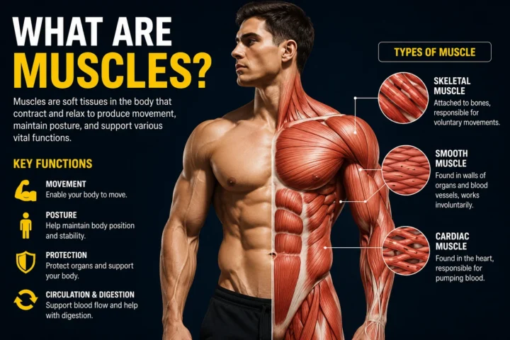
Three Types of Muscle Tissue
A human musculature chart normally focuses on skeletal muscle, but the human body contains three broad types of muscle tissue:
| Muscle type | Main location | Basic characteristic |
| Skeletal | Attached to bones and other structures | Primarily voluntary control |
| Cardiac | Heart | Specialized involuntary muscle |
| Smooth | Walls of many internal organs and vessels | Involuntary control |
Skeletal muscle is the type most relevant to strength training and most full-body musculature charts.
Cardiac muscle makes up the muscular tissue of the heart.
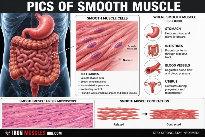
Smooth muscle is found in structures such as the digestive tract and blood vessels.
NCBI references identify skeletal, cardiac, and smooth muscle as the three major muscle-tissue categories.
How Much of the Body Is Skeletal Muscle?
One commonly cited figure is that skeletal muscle represents about 40% of body weight, although the exact proportion varies between individuals and depends on how muscle mass is measured.
A PubMed-indexed review reports approximately 40% of total body weight, while another NCBI/PubMed-indexed reference gives the same approximate figure. Other research reports ranges rather than a single fixed percentage.
That distinction matters.
Don’t look at “40%” as a universal rule for every person.
Muscle mass differs with factors such as age, sex, body composition, physical activity, and health status. Research using measured body composition also demonstrates meaningful differences between individuals.
For a beginner reading a musculature chart, the practical takeaway is simple:
Your skeletal muscles make up a large and functionally important part of your body, but there isn’t one muscle-percentage number that accurately describes everyone.
What Is Skeletal Muscle Made Of?
At a basic level, a skeletal muscle contains bundles of muscle fibers and connective tissue.
A simplified organization looks like this:
Whole muscle → fascicles → muscle fibers → myofibrils → sarcomeres
Connective tissue layers help organize these structures.
The epimysium surrounds the whole muscle, the perimysium surrounds fascicles, and the endomysium surrounds individual muscle fibers.
At an even smaller scale, muscle fibers contain myofibrils with repeating contractile units called sarcomeres.
Actin and myosin are major contractile proteins involved in force production.
You don’t need to memorize this entire chain to use a human musculature chart.
But understanding the hierarchy helps explain why anatomy can be viewed at different levels.
A full-body chart shows the muscle.
A cross-sectional diagram can show fascicles and fibers.
A microscopic diagram can show sarcomeres and proteins.
They’re all describing the same system at different scales.
Superficial vs. Deep Muscles
This distinction is extremely useful when reading anatomy charts.
Superficial Muscles
These are closer to the surface of the body.
They’re generally easier to identify visually and are often emphasized on beginner musculature charts.
Examples include:
- Pectoralis major
- Deltoid
- Biceps brachii
- Rectus abdominis
- Gluteus maximus
- Gastrocnemius
Deep Muscles
These lie underneath superficial structures.
Examples include:
- Pectoralis minor
- Subscapularis
- Vastus intermedius
- Soleus
- Smaller deep back muscles
A chart showing only superficial muscles isn’t necessarily wrong.
It’s simply showing a different layer.
Detailed anatomy references may specifically separate superficial and deep structures to make these relationships clearer.
Who This Is NOT For
A basic human musculature chart is probably not enough if you’re studying for a medical, physiotherapy, anatomy, or other professional examination.
You may need more detailed resources covering:
- Muscle origins
- Muscle insertions
- Innervation
- Blood supply
- Anatomical landmarks
- Joint actions
- Deep anatomical relationships
- Clinical anatomy
For general gym training, however, you usually don’t need to memorize every small muscle before starting.
Learn the major structures first, then expand your knowledge as your training experience grows.
How Beginners Can Memorize Muscle Anatomy Faster
Try the location → name → action → exercise method.
1. Location
Find the muscle on the chart.
2. Name
Say the name out loud.
3. Action
Learn one basic movement or function associated with it.
4. Exercise
Think of one exercise that trains the region.
For example:
Posterior upper arm → triceps brachii → elbow extension → triceps pushdown
Or:
Anterior thigh → quadriceps → knee extension → squat/leg extension
This gives your brain four connected pieces of information instead of one isolated word.
You can also alternate between labeled and blank charts.
A labeled chart teaches recognition.
A blank chart tests recall.
That combination is more useful than repeatedly looking at the same labels.
FAQ: Human Musculature Chart
1. What are the major muscle groups in the human body?
For beginner fitness and anatomy study, the major regions include the chest, shoulders, back, arms, abdomen, glutes, thighs, and lower legs. These regions contain multiple individual muscles. For example, the quadriceps are a group of four muscles, while the biceps brachii is an individual muscle. A detailed chart can break each region into smaller superficial and deep structures.
2. How many muscles are in the human body?
There isn’t one universally accepted number because different sources use different definitions for what counts as an individual muscle or anatomical structure. Some educational references describe more than 400 distinct skeletal muscles, while anatomy resources commonly discuss 600+ muscles depending on classification. For beginners, the exact total isn’t important. Learning the major muscle groups is a much more useful starting point.
3. What are the three types of muscle tissue?
The three major types are skeletal, cardiac, and smooth muscle. Skeletal muscle is primarily responsible for voluntary movement and is the main type shown on fitness-oriented full-body muscle charts. Cardiac muscle forms the muscular tissue of the heart, while smooth muscle is found in many internal organs and blood vessels and operates involuntarily.
4. What is the largest muscle in the human body?
The gluteus maximus is commonly described as the largest muscle by mass, although “largest” can depend on the measurement or definition being used. For beginner training, it’s more useful to recognize the gluteus maximus as the large superficial muscle forming much of the buttock region and contributing strongly to hip extension. A posterior human musculature chart makes its location easy to identify.
5. What is the difference between an anterior and posterior muscle chart?
An anterior chart shows the body from the front, making structures such as the pectorals, biceps, abdominals, quadriceps, and tibialis anterior easier to identify. A posterior chart shows the back, highlighting structures such as the trapezius, latissimus dorsi, triceps, gluteus maximus, hamstrings, and gastrocnemius. Using both views gives you a more complete picture of muscle location.
Final Takeaway
A human musculature chart is one of the simplest tools you can use to build a practical understanding of anatomy. Don’t try to memorize hundreds of labels on your first day. Start with the major regions, learn the difference between anterior and posterior views, then connect each important muscle to its basic function and common training movements.
Once the big picture makes sense, smaller muscles and deeper layers become much easier to learn.
For beginners, the goal isn’t to become an anatomy textbook. It’s to understand your body well enough to recognize the major structures, follow sensible training, and communicate clearly about movement.
If you want to go further, use a labeled chart alongside a blank chart and test yourself regularly. That small habit can make anatomy far less confusing.