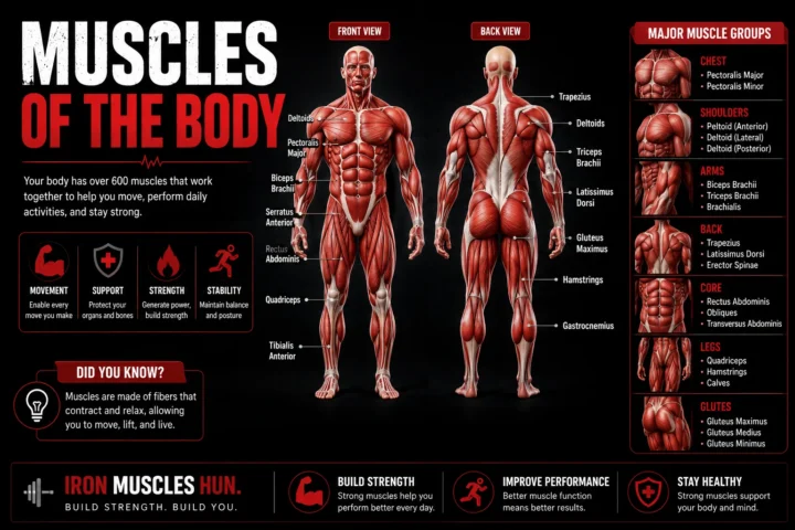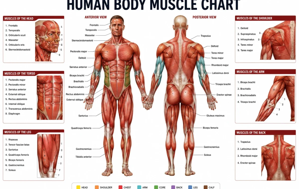
If you’re trying to understand a human body muscle chart, start by learning the major muscles by body region rather than memorising hundreds of individual names. The most useful groups for beginners are the chest, shoulders, back, arms, abdominals, glutes, thighs and calves.
- Quick Answer
- What Is a Human Body Muscle Chart?
- Front View: Major Muscles You Should Know
- 1. Deltoids — Shoulders
- 2. Pectoralis Major — Chest
- 3. Biceps Brachii — Front of Upper Arm
- 4. Triceps Brachii — Back of Upper Arm
- 5. Rectus Abdominis — Front of the Abdomen
- 6. External Obliques — Sides of the Abdomen
- 7. Quadriceps — Front of the Thigh
- 8. Tibialis Anterior — Front of Lower Leg
- Back View: Major Muscles You Should Know
- 1. Trapezius — Upper and Mid Back
- 2. Latissimus Dorsi — Lats
- 3. Triceps — Back of the Upper Arm
- 4. Posterior Deltoid — Rear Shoulder
- 5. Gluteus Maximus — Main Buttock Muscle
- 6. Hamstrings — Back of the Thigh
- 7. Gastrocnemius — Calf
- 8. Soleus — Deep Calf Muscle
- Human Body Muscle Chart: Front vs Back
- How to Read a Human Body Muscle Chart
- Step 1: Learn the body region
- Step 2: Learn one major muscle at a time
- Step 3: Learn the basic action
- Step 4: Connect the muscle to an exercise
- Common Mistakes → Why → Fix
- Mistake 1: Trying to memorise every muscle
- Mistake 2: Treating each muscle as completely separate
- Mistake 3: Assuming visible muscles are the whole story
- Mistake 4: Confusing a muscle with a muscle group
- Mistake 5: Using anatomy as a reason to change technique constantly
- A 30-Second Muscle Chart Self-Test
- Worked Example: Using a Muscle Chart With a Beginner Workout
- How Muscle Anatomy Helps Your Training
- It helps you understand exercise selection
- It helps you spot gaps in a workout
- It improves exercise vocabulary
- Human Body Muscle Chart: Major Muscle Groups at a Glance
- Who This Muscle Chart Is Not For
- How to Memorise the Human Body Muscle Chart Faster
- Day 1: Front
- Day 2: Back
- Day 3: Connect movement
- Day 4: Test yourself
- Day 5: Add detail
- FAQ: Human Body Muscle Chart
- How many muscles are in the human body?
- What are the major muscles of the human body?
- What are the three types of muscle tissue?
- What is the largest muscle in the human body?
- How should a beginner learn human muscle anatomy?
- Final Takeaway
This guide breaks down the major muscles you’ll see on a typical front-and-back muscle chart, what each group does, and how the anatomy relates to basic strength training. For more insights read here :- label skeletal muscle and human muscle chart .
Quick Answer
- A human body muscle chart is a visual map of muscles, usually showing anterior (front) and posterior (back) views.
- The main muscles beginners should recognise include the pectorals, deltoids, latissimus dorsi, trapezius, biceps, triceps, abdominals, glutes, quadriceps, hamstrings and calves.
- Muscles don’t work in isolation. Many exercises involve several muscles working together around one or more joints.
- For training, learn the location + basic action + common exercise for each major muscle group.
What Is a Human Body Muscle Chart?
A human body muscle chart is an anatomical reference that shows where muscles are located on the body. Most beginner charts use an anterior view to show the front of the body and a posterior view to show the back.
The purpose isn’t simply to memorise names. A useful chart helps you connect anatomy to movement.
For example, once you know that the pectoralis major sits across the front of the chest, you can better understand why pressing movements train the chest. Once you recognise the latissimus dorsi along the sides of the back, pulling movements become easier to understand.
The muscular system contains several types of muscle tissue. Skeletal muscle is the type most relevant to gym training because it produces voluntary movement and works with the skeleton. Smooth muscle and cardiac muscle have different roles inside the body.
A full anatomical chart can contain far more detail than a beginner needs. Medical anatomy charts may label smaller muscles of the hands, feet, face and deeper layers. For general fitness, learning the major superficial muscles first is a much more practical starting point.
Front View: Major Muscles You Should Know
When looking at a human body muscle chart from the front, begin at the shoulders and work down toward the legs.
1. Deltoids — Shoulders
The deltoid is the rounded muscle covering the shoulder.
It is commonly divided into anterior, lateral and posterior portions. These portions contribute to different shoulder movements, which is why pressing, raising and pulling exercises can involve the shoulder in different ways.
For a beginner, remember:
Location: Shoulder
Main role: Moving the upper arm at the shoulder
Common exercises: Overhead press, lateral raise, dumbbell press
Don’t make the common mistake of thinking that every shoulder exercise trains exactly the same part of the deltoid.
2. Pectoralis Major — Chest
The pectoralis major is the large, superficial muscle covering much of the front of the chest.
It contributes to movements that bring the upper arm across the body and is heavily involved in pressing exercises.
You will commonly see it highlighted on a muscle chart because it is relatively easy to identify.
Location: Front of chest
Main role: Assists shoulder and upper-arm movement
Common exercises: Push-up, bench press, dumbbell press
The pectoralis major isn’t the only muscle in the chest region. The pectoralis minor lies deeper and has a different anatomical position and function.
3. Biceps Brachii — Front of Upper Arm
The biceps brachii sits on the front of the upper arm.
Its name is useful to remember because “bi” refers to its two heads. It contributes to elbow flexion and also assists with movement at the shoulder and forearm.
Location: Front of upper arm
Main role: Bending the elbow and assisting forearm movement
Common exercises: Curl variations, chin-ups and pulling movements
If you’re studying a chart, don’t confuse the biceps with the entire elbow-flexor group. Other muscles also contribute to elbow flexion.
4. Triceps Brachii — Back of Upper Arm
The triceps brachii occupies much of the back of the upper arm.
It has three heads and is important for elbow extension.
That makes the basic relationship easy to remember:
Biceps → elbow flexion
Triceps → elbow extension
Common exercises: Push-ups, bench pressing, overhead triceps extensions and cable pressdowns.
This biceps-versus-triceps pairing is one of the simplest ways for a beginner to start reading a muscle chart.
5. Rectus Abdominis — Front of the Abdomen
The rectus abdominis is the long muscle running vertically along the front of the abdomen.
It’s often called the “six-pack muscle,” although visible abdominal definition depends on much more than the muscle itself.
Its role includes contributing to trunk flexion and helping stabilise the torso.
Location: Front of abdomen
Main role: Trunk movement and stabilisation
Common exercises: Controlled curl-up variations, loaded carries and compound movements that challenge trunk control
A useful training point is that you don’t need to perform hundreds of repetitions to train your abdominal muscles effectively.
6. External Obliques — Sides of the Abdomen
The external obliques sit on the sides of the trunk.
They contribute to trunk rotation, side-bending and stabilisation.
On a muscle chart, look immediately to either side of the rectus abdominis.
Location: Sides of the torso
Main role: Rotation, side-bending and trunk control
Common exercises: Side plank variations, carries and controlled rotational exercises
Your obliques also work during many compound lifts because your torso has to resist unwanted movement.
7. Quadriceps — Front of the Thigh
The quadriceps femoris is a group of muscles on the front of the thigh.
The group includes four muscles:
- Rectus femoris
- Vastus lateralis
- Vastus medialis
- Vastus intermedius
The quadriceps are heavily involved in extending the knee.
Location: Front of thigh
Main role: Knee extension
Common exercises: Squats, split squats, lunges and leg presses
For beginners, thinking of the quadriceps as one functional group is usually easier than immediately memorising every anatomical detail.
8. Tibialis Anterior — Front of Lower Leg
The tibialis anterior is positioned toward the front of the lower leg.
It contributes to dorsiflexion, which means bringing the foot upward toward the shin.
You may not see as much attention given to it in fitness-focused muscle charts, but it is an important lower-leg muscle.
Location: Front of lower leg
Main role: Dorsiflexion of the ankle
Common exercises: Tibialis raises and movements involving controlled ankle motion
Back View: Major Muscles You Should Know
Turn the chart around and you’ll see several muscles that are especially important for pulling movements, hip extension, posture and lower-body strength.
1. Trapezius — Upper and Mid Back
The trapezius, often called the traps, is a large muscle extending across the upper back and neck region.
It isn’t just one small patch around your shoulders.
Different portions of the trapezius contribute to different movements of the scapula and neck.
Location: Upper back and neck region
Main role: Scapular and shoulder-girdle movement
Common exercises: Shrugs, rows, carries and pulling movements
This is a good example of why a muscle chart should be viewed as an anatomy map rather than an exercise map. One muscle can have multiple functions.
2. Latissimus Dorsi — Lats
The latissimus dorsi, usually shortened to “lats,” is a broad muscle covering much of the side and lower portion of the back.
It’s one of the most recognisable muscles on a posterior muscle chart.
The lats contribute to movements involving the upper arm, particularly pulling the arm downward and toward the body.
Location: Side and lower back
Main role: Shoulder and upper-arm movement
Common exercises: Pull-ups, pulldowns and rows
When performing a back exercise, don’t worry about trying to “feel” one exact muscle on every repetition. Good technique and appropriate loading matter more than chasing a particular sensation.
3. Triceps — Back of the Upper Arm
From the rear view, the triceps become much easier to see.
The muscle occupies much of the posterior upper arm and works primarily to extend the elbow.
A beginner reading a chart can use the arm as a simple study exercise:
Front upper arm = biceps
Back upper arm = triceps
That simple distinction will help you identify many basic anatomy diagrams.
4. Posterior Deltoid — Rear Shoulder
The rear portion of the deltoid sits at the back of the shoulder.
It contributes to shoulder movement and works alongside several muscles of the upper back.
Common exercises: Reverse fly variations, rows and other pulling movements.
One important point: the posterior deltoid isn’t an isolated “back muscle.” Anatomically, it’s part of the deltoid group surrounding the shoulder.
5. Gluteus Maximus — Main Buttock Muscle
The gluteus maximus is the largest and most superficial muscle of the gluteal region.
It is a major hip extensor and plays an important role in movements such as standing up, climbing and jumping.
Location: Buttocks
Main role: Hip extension
Common exercises: Squats, hip thrusts, deadlift variations and step-ups
A human muscle chart often makes the gluteal region look like one large muscle, but the gluteal area contains multiple muscles, including the gluteus medius and gluteus minimus.
6. Hamstrings — Back of the Thigh
The hamstrings are a group of muscles located on the back of the thigh.
Major muscles in this group include the biceps femoris, semitendinosus and semimembranosus.
They contribute to knee flexion and hip extension.
Location: Back of thigh
Main role: Knee flexion and hip extension
Common exercises: Romanian deadlifts, leg curls, hip-hinge exercises
A useful beginner distinction is:
Quadriceps = front of thigh
Hamstrings = back of thigh
That relationship is much easier to remember when you see it on an anterior/posterior chart.
7. Gastrocnemius — Calf
The gastrocnemius is the large, visible muscle forming much of the upper calf.
It crosses both the knee and ankle, which makes its anatomy different from muscles that cross only one joint.
Location: Back of lower leg
Main role: Plantarflexion and assistance with knee movement
Common exercise: Standing calf raise
The deeper soleus also contributes strongly to plantarflexion and is an important part of the calf complex.
8. Soleus — Deep Calf Muscle
The soleus lies deep to the gastrocnemius.
Because it is underneath the gastrocnemius, you won’t see its full shape on a superficial muscle chart.
Its primary action is plantarflexion at the ankle.
This is a useful reminder that what you can see on a muscle chart isn’t the entire muscular system. Some muscles sit underneath larger superficial muscles.
Human Body Muscle Chart: Front vs Back
| Body region | Front/anterior muscles | Back/posterior muscles | Beginner function to remember |
|---|---|---|---|
| Shoulder | Anterior/lateral deltoid | Posterior deltoid | Move and stabilise the upper arm |
| Chest | Pectoralis major | — | Pressing and arm movement |
| Upper arm | Biceps brachii | Triceps brachii | Bend vs extend the elbow |
| Upper back | — | Trapezius, latissimus dorsi | Shoulder-blade and pulling actions |
| Abdomen | Rectus abdominis, obliques | Erector spinae | Trunk movement and control |
| Hip | Hip-flexor region | Gluteus maximus/medius | Move and stabilise the hip |
| Thigh | Quadriceps | Hamstrings | Knee extension vs flexion |
| Lower leg | Tibialis anterior | Gastrocnemius, soleus | Control ankle movement |
This simplified table is not a substitute for a medical anatomy atlas. It’s a beginner’s training map.
A detailed anatomical chart can show many additional muscles, including smaller muscles of the hands, feet, face, neck and deeper layers.
How to Read a Human Body Muscle Chart
Don’t try to memorise every label in one sitting.
Use this four-step method instead.
Step 1: Learn the body region
Start with:
- Head and neck
- Shoulder
- Chest
- Back
- Arm
- Abdomen
- Hip
- Thigh
- Lower leg
This gives you a mental map before you add individual muscle names.
Step 2: Learn one major muscle at a time
For example:
Chest → pectoralis major
Then:
Front upper arm → biceps
Then:
Back upper arm → triceps
Then:
Front thigh → quadriceps
Then:
Back thigh → hamstrings
You’re building associations rather than memorising disconnected Latin terms.
Step 3: Learn the basic action
Ask:
“What does this muscle help me do?”
For example:
- Quadriceps → extend the knee
- Hamstrings → flex the knee and assist hip extension
- Triceps → extend the elbow
- Biceps → flex the elbow and assist forearm movement
- Glutes → extend the hip
- Calves → plantarflex the ankle
This makes anatomy much easier to remember.
Step 4: Connect the muscle to an exercise
Now attach a familiar exercise to the anatomy.
Pectorals → push-up
Lats → pulldown
Quadriceps → squat
Hamstrings → Romanian deadlift
Glutes → hip thrust
Calves → calf raise
Remember that exercises usually train multiple muscles. The table is a learning shortcut, not a claim that one exercise isolates one muscle.
Common Mistakes → Why → Fix
Mistake 1: Trying to memorise every muscle
Why: A detailed chart can contain dozens of names and several layers of anatomy.
Fix: Start with the major muscle groups. Add smaller muscles after you understand the basic regions.
Mistake 2: Treating each muscle as completely separate
Why: Human movement is coordinated. Muscles often work together around joints.
Fix: Learn anatomy alongside movement patterns. Think “push,” “pull,” “squat,” “hinge,” “carry” and “rotate,” then identify the major muscles involved.
Mistake 3: Assuming visible muscles are the whole story
Why: Superficial charts show muscles close to the skin, while deeper muscles may be hidden underneath.
Fix: When you see a detailed anatomy chart, look for labels such as superficial, deep, anterior and posterior.
Mistake 4: Confusing a muscle with a muscle group
Why: Some popular fitness terms describe groups rather than individual muscles.
Fix: Learn the distinction. “Quadriceps” refers to a group of four muscles. “Hamstrings” refers to a group of muscles at the back of the thigh.
Mistake 5: Using anatomy as a reason to change technique constantly
Why: Beginners can become obsessed with finding the “perfect” exercise for each muscle.
Fix: Build your foundation first. Use controlled technique, a sensible range of motion and progressive resistance appropriate to your experience.
NSCA guidance recognises that exercise selection can be based on the muscle groups involved, movement patterns and training goals.
A 30-Second Muscle Chart Self-Test
Look at a blank or unlabeled body diagram.
Give yourself 30 seconds to point to these areas:
- Chest
- Shoulders
- Biceps
- Triceps
- Abs
- Lats
- Glutes
- Quadriceps
- Hamstrings
- Calves
Then name one major muscle associated with each.
If you can identify most of them without looking at the answers, you’re ready to start adding smaller muscles.
If you can’t, don’t add more information yet. Repeat the basic chart until the major regions feel automatic.
Worked Example: Using a Muscle Chart With a Beginner Workout
Suppose a beginner weighs 70 kg (154 lb) and wants to understand which muscles are involved in a simple full-body session.
The exact load isn’t important for this anatomy exercise. The goal is to connect exercises with muscle groups.
| Exercise | Main regions to identify | Other muscles involved |
| Squat | Quadriceps, glutes | Hamstrings, trunk muscles |
| Push-up | Pectoralis major | Triceps, deltoids, trunk |
| Row | Latissimus dorsi, upper back | Biceps, rear deltoids |
| Romanian deadlift | Hamstrings, glutes | Back and trunk muscles |
| Calf raise | Gastrocnemius, soleus | Foot and ankle stabilisers |
The important lesson is that one exercise rarely belongs to one muscle.
A squat isn’t simply a “quad exercise.” A push-up isn’t simply a “chest exercise.” The body coordinates multiple muscles to produce and control movement.
For a beginner, that is a more useful way to read a human body muscle chart than trying to assign every exercise to one anatomical label.
How Muscle Anatomy Helps Your Training
Learning anatomy can improve your training decisions without turning your gym session into a biology exam.
It helps you understand exercise selection
If you know the basic function of a muscle, you can understand why an exercise is being used.
A pulldown involves shoulder movement that strongly involves the latissimus dorsi and other upper-body muscles.
A squat involves coordinated movement at the hip, knee and ankle, requiring several muscle groups to contribute.
This is much more useful than memorising a list of “best exercises.”
It helps you spot gaps in a workout
Imagine a routine containing only pressing exercises.
A muscle chart can quickly show you that the program is heavily focused on the chest, shoulders and triceps while giving less direct attention to pulling muscles.
You don’t need to panic about perfect balance after every workout. Instead, look at the overall training pattern.
It improves exercise vocabulary
When someone says “posterior chain,” “upper back,” “quad-dominant” or “elbow extension,” you’ll have enough anatomy knowledge to understand what they’re discussing.
That makes coaching cues and training information easier to follow.
Human Body Muscle Chart: Major Muscle Groups at a Glance
For quick revision, use this simplified map:
Head and neck
- Facial muscles
- Masseter
- Sternocleidomastoid
Shoulders
- Deltoids
Chest
- Pectoralis major
- Pectoralis minor
Upper back
- Trapezius
- Rhomboids
- Latissimus dorsi
Arms
- Biceps brachii
- Triceps brachii
- Forearm muscle groups
Core
- Rectus abdominis
- External obliques
- Internal obliques
- Transversus abdominis
- Erector spinae
Hips and glutes
- Gluteus maximus
- Gluteus medius
- Gluteus minimus
- Hip-flexor muscles
Thigh
- Quadriceps
- Hamstrings
- Adductor group
Lower leg
- Gastrocnemius
- Soleus
- Tibialis anterior
- Other lower-leg muscles
This regional approach is consistent with how major muscles are commonly organised in anatomy references: head, neck, trunk, upper limb and lower limb.
Who This Muscle Chart Is Not For
A beginner-friendly chart is useful for general fitness and basic anatomy learning, but it isn’t enough for every purpose.
It is not a substitute for formal anatomical education if you’re studying medicine, physiotherapy, sports medicine or another profession requiring detailed anatomical knowledge.
It also isn’t a diagnostic tool.
If you’re trying to identify the cause of persistent pain, numbness, weakness or an injury, don’t use a chart to diagnose yourself. A qualified healthcare professional can assess the problem in context.
For ordinary gym training, though, a simplified muscle chart is an excellent starting point.
How to Memorise the Human Body Muscle Chart Faster
Try this simple routine.
Day 1: Front
Learn:
- Deltoids
- Pectorals
- Biceps
- Abdominals
- Quadriceps
- Tibialis anterior
Day 2: Back
Learn:
- Trapezius
- Latissimus dorsi
- Triceps
- Glutes
- Hamstrings
- Gastrocnemius
Day 3: Connect movement
Review each muscle and ask what movement it contributes to.
Day 4: Test yourself
Use an unlabeled diagram.
Point to each region and name the muscle without looking at the answer.
Day 5: Add detail
Only now start learning smaller muscles such as the rhomboids, rotator-cuff muscles, adductors and deeper abdominal muscles.
This approach reduces the amount you have to remember at one time.
FAQ: Human Body Muscle Chart
How many muscles are in the human body?
There isn’t a single universally useful number because the way individual muscles are counted can vary with anatomical definitions. Medical references commonly describe the body as having more than 600 muscles. For beginners, the exact total matters less than understanding the major muscle groups and how they contribute to movement.
What are the major muscles of the human body?
The major muscles commonly highlighted on beginner charts include the pectoralis major, deltoids, trapezius, latissimus dorsi, biceps, triceps, abdominal muscles, gluteal muscles, quadriceps, hamstrings and calf muscles. A detailed anatomy chart will include many smaller and deeper muscles as well.
What are the three types of muscle tissue?
The three major types are skeletal, smooth and cardiac muscle. Skeletal muscle is associated with voluntary movement and the skeleton. Smooth muscle is found in the walls of various internal organs, while cardiac muscle is the specialised muscle tissue of the heart.
What is the largest muscle in the human body?
The gluteus maximus is generally described as the largest muscle in the human body. It is the largest and most superficial muscle of the gluteal region and is an important hip extensor. For training purposes, it’s also heavily involved in movements such as climbing, jumping and standing from a lower position.
How should a beginner learn human muscle anatomy?
Start with the major muscle groups rather than trying to memorise every anatomical label. Learn each muscle’s location, one basic function and a familiar movement that uses it. Then test yourself using a blank anterior and posterior diagram. Once those large groups are familiar, add smaller and deeper muscles.
Final Takeaway
A human body muscle chart is most useful when you treat it as a map, not a list of names. Start with the major regions: chest, shoulders, back, arms, core, glutes, thighs and calves. Then learn the major muscles within each region and connect them to basic movements.
You don’t need to memorise every muscle before you can train effectively. A solid understanding of the major muscles gives you enough anatomy to follow beginner training programs, understand exercise cues and recognise how different movements work together.
If you’re building your anatomy knowledge for the gym, save this chart as a quick reference and revisit it as you learn new exercises.