If you’re new to anatomy, start with the major muscle groups rather than trying to learn every small muscle.
- Front View: anatomy chart muscles
- 1. Pectoralis Major
- 2. Deltoids
- 3. Biceps Brachii
- 4. Rectus Abdominis
- 5. External Obliques
- 6. Quadriceps
- 7. Tibialis Anterior
- 1. Trapezius
- 2. Latissimus Dorsi
- 3. Triceps Brachii
- 4. Posterior Deltoid
- 5. Erector Spinae
- 6. Gluteus Maximus
- 7. Hamstrings
- 8. Gastrocnemius
- 9. Soleus
- Step 1: Identify the View
- Step 2: Find the Body Region
- Step 3: Learn the Muscle's Main Action
- Step 4: Connect It to an Exercise
- Goblet Squat
- Mistake 1: Trying to Memorize Every Muscle
- Mistake 2: Thinking One Exercise Trains Only One Muscle
- Mistake 3: Confusing a Muscle Group With an Individual Muscle
- Mistake 4: Assuming the Chart Shows Everything
- Mistake 5: Using Anatomy to Chase Perfect Muscle Isolation
- Skeletal Muscle
- Cardiac Muscle
- Smooth Muscle
- Start With These Movement Categories
- Day 1
- Day 2
- Day 3
- Day 4
- 1. What is an anatomy chart muscles diagram?
- 2. What are the major muscles shown on a human muscle chart?
- 3. How many muscles are in the human body?
- 4. What muscles should beginners learn first?
- 5. Is an anatomy chart useful for working out?
There are more than 600 muscles in the human body, although the exact count can vary depending on how individual muscles and muscle subdivisions are classified. NIH’s National Institute of Arthritis and Musculoskeletal and Skin Diseases also describes the body as having more than 650 muscles.
For lifting, a smaller list is much more practical. For more insights read here :- muscle model and muscle man diagram .
Front View: anatomy chart muscles
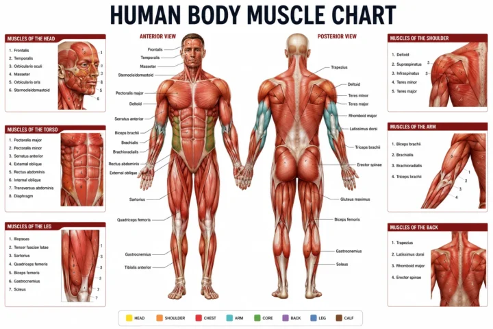
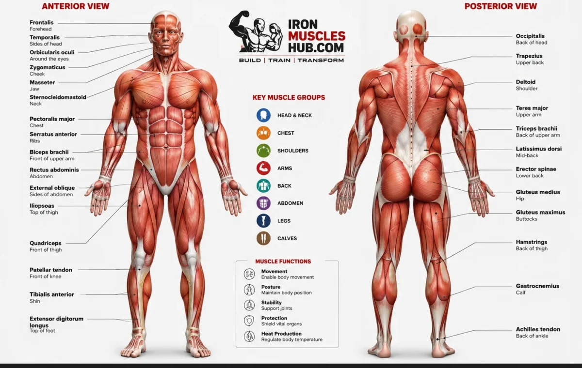
When looking at a muscular system chart from the front, these are the main muscles you should recognize.
1. Pectoralis Major
The pectoralis major, commonly called the chest muscle, covers much of the front of the upper chest.
Its main actions include moving the upper arm toward and across the body.
Common exercises:
- Bench press
- Dumbbell press
- Push-ups
- Cable fly
- Machine chest press
For a beginner, think of the pecs whenever you’re performing a pressing movement for the chest.
2. Deltoids
The deltoid is the large muscle covering the shoulder.
It has three commonly discussed portions:
- Anterior deltoid – front
- Lateral deltoid – side
- Posterior deltoid – rear
The three portions contribute differently to shoulder movement.
Common exercises:
- Overhead press
- Dumbbell lateral raise
- Front raise
- Reverse fly
A common beginner mistake is treating the shoulder as one simple muscle. The deltoid has different sections, so exercise selection can change which portion receives more emphasis.
3. Biceps Brachii
The biceps brachii sits on the front of the upper arm.
Its functions include elbow flexion and assisting with forearm rotation.
Common exercises:
- Dumbbell curl
- Barbell curl
- Hammer curl
- Cable curl
You don’t need dozens of curl variations. A controlled curl performed through a comfortable range of motion is enough to learn the basic movement.
4. Rectus Abdominis
The rectus abdominis is the long abdominal muscle running vertically along the front of the torso.
It’s commonly associated with the “six-pack,” although visible abdominal definition also depends on factors outside the muscle itself.
The rectus abdominis helps with movements involving the trunk and pelvis.
Common exercises:
- Curl-up variations
- Reverse crunches
- Cable crunches
- Controlled hanging knee raises
5. External Obliques
The external obliques are located along the sides of the abdomen.
They contribute to trunk rotation and side-bending movements and help control the torso.
Common exercises:
- Side plank
- Cable rotation
- Controlled carries
- Anti-rotation exercises
For beginners, don’t think only about “training the abs.” Your trunk includes several muscle groups that work together.
6. Quadriceps
The quadriceps femoris is the major muscle group at the front of the thigh.
It contains four muscles:
- Rectus femoris
- Vastus lateralis
- Vastus medialis
- Vastus intermedius
The quadriceps are heavily involved in knee extension.
Common exercises:
- Squats
- Split squats
- Lunges
- Leg press
- Step-ups
A muscle anatomy chart can make this group much easier to understand because the four muscles occupy slightly different positions around the thigh.
7. Tibialis Anterior
The tibialis anterior is located toward the front of the lower leg.
It helps lift the front of the foot upward, a movement called dorsiflexion.
You can see it more clearly on detailed lower-leg anatomy charts than on simplified full-body posters.
Common exercises:
- Heel walks
- Tibialis raises
- Controlled dorsiflexion exercises
Back View: Major Muscles
Turn the anatomy chart around and you’ll see a different set of muscles.
The posterior view is particularly useful for lifters because several large muscles on the back side of the body contribute to pulling, hip extension and posture.
1. Trapezius
The trapezius, or traps, is a large muscle covering part of the upper and middle back and extending toward the neck and shoulders.
It has upper, middle and lower portions with different functions.
Common exercises:
- Shrugs
- Rows
- Loaded carries
- Pulling movements
- Scapular control exercises
Don’t assume that every shrug or row trains the exact same region. Body position and shoulder-blade movement change the muscular emphasis.
2. Latissimus Dorsi
The latissimus dorsi, often shortened to “lats,” is one of the major muscles of the back.
It contributes strongly to movements that pull the upper arm down and toward the body.
Common exercises:
- Pull-ups
- Lat pulldowns
- Rows
- Straight-arm pulldowns
A useful beginner cue is to think about driving the upper arm down rather than simply pulling with the hands.
3. Triceps Brachii
The triceps brachii sits on the back of the upper arm.
It has three heads:
- Long head
- Lateral head
- Medial head
Its primary job is elbow extension.
Common exercises:
- Pushdowns
- Overhead extensions
- Close-grip pressing
- Dips
The triceps are involved in many pressing exercises, so you may already be training them without performing direct isolation work.
4. Posterior Deltoid
The rear portion of the deltoid sits at the back of the shoulder.
It contributes to several shoulder movements, including horizontal abduction.
Common exercises:
- Reverse fly
- Rear-delt cable fly
- Rows
- Face-pull variations
A rear-view muscle chart is especially useful here because the posterior deltoid can be difficult for beginners to locate by feel alone.
5. Erector Spinae
The erector spinae refers to a group of muscles running along the spine.
These muscles contribute to extending and stabilizing the spine.
They work during many compound movements, particularly exercises that require the trunk to resist unwanted movement.
Common exercises:
- Deadlift variations
- Romanian deadlifts
- Back extensions
- Good mornings
This doesn’t mean your lower back should be forced to do all the work. Good technique distributes the load across the hips, legs and trunk.
6. Gluteus Maximus
The gluteus maximus is the largest of the three gluteal muscles and is the major muscle forming the bulk of the buttocks.
It contributes strongly to hip extension.
Common exercises:
- Squats
- Hip thrusts
- Lunges
- Step-ups
- Deadlift variations
Your glutes are involved in many lower-body exercises, not just exercises marketed specifically as “glute workouts.”
7. Hamstrings
The hamstrings are a group of muscles at the back of the thigh.
The major muscles include:
- Biceps femoris
- Semitendinosus
- Semimembranosus
They contribute to knee flexion and hip extension.
Common exercises:
- Romanian deadlifts
- Leg curls
- Good mornings
- Hip hinges
- Sprinting and jumping activities
An anatomy chart makes the difference between the front and back of the thigh much easier to visualize.
8. Gastrocnemius
The gastrocnemius is the large, visible calf muscle.
It crosses both the ankle and knee joints.
Common exercises:
- Standing calf raises
- Single-leg calf raises
- Loaded calf raises
9. Soleus
The soleus sits underneath the gastrocnemius.
It is another important calf muscle and contributes to plantarflexion at the ankle.
Common exercises:
- Seated calf raises
- Bent-knee calf raises
A basic full-body chart may not show the soleus clearly because it lies deeper than the gastrocnemius.
Anatomy Chart Muscles: Front vs Back Comparison
| Body Region | Front View Muscle | Back View Muscle | Basic Function | Common Exercise |
|---|---|---|---|---|
| Chest | Pectoralis major | – | Moves upper arm across body | Bench press |
| Shoulder | Anterior/lateral deltoid | Posterior deltoid | Shoulder movement | Press / reverse fly |
| Upper arm | Biceps brachii | Triceps brachii | Bends/extends elbow | Curl / pushdown |
| Upper back | – | Trapezius | Shoulder-blade movement | Row |
| Back | – | Latissimus dorsi | Pulls upper arm down/back | Lat pulldown |
| Abdomen | Rectus abdominis | – | Trunk control | Curl-up |
| Side torso | External oblique | – | Rotation and trunk control | Side plank |
| Thigh | Quadriceps | Hamstrings | Knee extension/flexion | Squat / RDL |
| Hip | Iliopsoas region | Gluteus maximus | Hip movement | Step-up / hip thrust |
| Lower leg | Tibialis anterior | Gastrocnemius/soleus | Ankle movement | Tibialis raise / calf raise |
This table is one of the most useful ways to use an anatomy chart: muscle + location + function + exercise.
You aren’t just memorizing labels. You’re building a movement map.
How to Read an Anatomy Chart Muscles Diagram
A detailed anatomy chart can look overwhelming because it may contain dozens of labels.
Use this four-step method.
Step 1: Identify the View
First ask:
Am I looking at the front or the back?
Some charts also include a side view.
Front-view charts are usually best for learning:
- Chest
- Front shoulders
- Biceps
- Abdominals
- Quadriceps
- Tibialis anterior
Back-view charts help you identify:
- Trapezius
- Lats
- Rear deltoids
- Triceps
- Glutes
- Hamstrings
- Calves
Step 2: Find the Body Region
Don’t scan the entire chart randomly.
Choose one area.
For example:
Upper arm → biceps and triceps
Then move to:
Thigh → quadriceps and hamstrings
Then:
Lower leg → tibialis, gastrocnemius and soleus
This makes anatomy easier to retain.
Step 3: Learn the Muscle’s Main Action
Once you know the location, ask:
What does this muscle help me do?
For example:
- Pec → pressing and bringing the arm across the body
- Biceps → elbow flexion
- Triceps → elbow extension
- Lats → pulling the upper arm down/back
- Quadriceps → knee extension
- Hamstrings → knee flexion and hip extension
- Glutes → hip extension
You don’t need to memorize every anatomical detail on day one.
Step 4: Connect It to an Exercise
This is where anatomy becomes useful for training.
For example:
Quadriceps → knee extension → squat
Hamstrings → hip extension/knee flexion → Romanian deadlift or leg curl
Triceps → elbow extension → pushdown
Lats → pulling the upper arm down → pulldown
This simple chain is enough to make a muscle chart useful in the gym.
A Worked Example: From Anatomy Chart to Workout
Suppose a beginner weighs 70 kg (154 lb) and is 175 cm (69 inches) tall.
They perform a goblet squat using a 20 kg (44 lb) dumbbell.
The important point isn’t the person’s body size. The useful information is what the movement asks the muscles to do.
Goblet Squat
Primary areas:
- Quadriceps
- Gluteus maximus
Supporting areas:
- Hamstrings
- Calves
- Trunk musculature
The exercise involves coordinated movement across the hips, knees and ankles.
So instead of looking at an anatomy chart and saying, “I need one exercise for every muscle,” the beginner can think:
One compound movement can involve several muscle groups at once.
That is a much better way to organize beginner training.
The 20 kg (44 lb) example is only an illustration, not a recommended starting weight. Your appropriate load depends on experience, technique, available equipment and individual circumstances.
Common Anatomy Chart Mistakes → Why → Fix
Mistake 1: Trying to Memorize Every Muscle
Why: Detailed charts can contain a huge amount of information. Trying to memorize every label at once creates unnecessary mental overload.
Fix: Start with about 15-20 major muscles or muscle groups that you actually encounter during training.
Learn their location, basic action and common exercises first.
Mistake 2: Thinking One Exercise Trains Only One Muscle
Why: Exercise names often encourage people to think in isolation.
A bench press isn’t simply a “chest exercise.” Several muscles contribute to the movement.
Fix: Think in terms of primary muscles and supporting muscles.
A squat, for example, involves several muscles around the hips, knees and trunk.
Mistake 3: Confusing a Muscle Group With an Individual Muscle
Why: Fitness language often uses broad terms such as “quads,” “hamstrings” or “abs.”
Fix: Learn the difference.
The quadriceps are a group containing four muscles. The hamstrings also refer to a group. “Abs” can refer broadly to several abdominal muscles.
This distinction becomes more useful as your training experience increases.
Mistake 4: Assuming the Chart Shows Everything
Why: Simplified charts are designed for quick identification.
A full anatomy reference may show deeper muscles that aren’t visible on a basic poster.
Fix: Use a beginner chart for the big picture, then move to a more detailed anatomy reference when you need to study individual muscles, attachments or actions.
Mistake 5: Using Anatomy to Chase Perfect Muscle Isolation
Why: Beginners sometimes believe every muscle requires its own exercise.
That’s rarely necessary.
Resistance training can improve strength, muscle size and physical function, and the 2026 ACSM position stand found that many different resistance-training prescriptions can work depending on the goal.
Fix: Build your training around basic movement patterns first, then add isolation work where it makes sense.
A 30-Second Muscle Chart Self-Test
Look at your anatomy chart and point to these areas without reading the labels:
- Chest
- Front shoulder
- Back shoulder
- Biceps
- Triceps
- Abdominals
- Lats
- Trapezius
- Glutes
- Quadriceps
- Hamstrings
- Calves
Now test yourself again.
For each muscle, answer:
Where is it?
What movement does it help with?
Name one exercise that trains it.
If you can answer those three questions for most of the list, you have enough basic anatomy knowledge to start applying the chart to your workouts.
You don’t need a perfect score.
Who This Anatomy Chart Is Not For
A beginner muscle chart is not a replacement for a detailed medical anatomy textbook.
It also isn’t enough for someone who needs to study:
- Surgical anatomy
- Clinical anatomy
- Detailed nerve pathways
- Individual muscle origins and insertions
- Complex injury assessment
- Rehabilitation programming
Those subjects require much more detailed resources and, where appropriate, professional education.
For a beginner lifter, however, a major-muscle chart is an excellent starting point.
How Many Muscles Are in the Human Body?
You will commonly see the answer given as more than 600 muscles.
NIH’s NIAMS states that there are more than 650 muscles in the human body, while Cleveland Clinic describes the body as having more than 600 muscles. The difference illustrates why you shouldn’t treat the number as a perfectly fixed count. Anatomical classification can vary depending on how individual muscles are defined and grouped.
For gym training, the exact total isn’t particularly important.
You can become a competent beginner lifter without memorizing hundreds of muscle names.
Start with the major groups and build from there.
What Are the Three Types of Muscle Tissue?
There are three major types of muscle tissue:
Skeletal Muscle
Skeletal muscle attaches to bones through tendons and produces voluntary body movement.
This is the main type shown on most fitness anatomy charts.
Cardiac Muscle
Cardiac muscle makes up the muscular tissue of the heart and contracts involuntarily.
Smooth Muscle
Smooth muscle is found in various internal organs and structures and also operates involuntarily.
The three types have different structures and jobs, so a “muscle chart” designed for lifters usually concentrates on skeletal muscle.
Why Muscle Anatomy Matters for Strength Training
You don’t need an anatomy degree to lift weights.
But basic anatomy can improve the way you think about exercise.
Instead of asking:
“What’s the best exercise for this muscle?”
you can ask:
“What movement does this muscle perform, and which exercises train that movement?”
That change matters.
For example, the triceps extend the elbow. A pushdown trains elbow extension. A pressing exercise also requires elbow extension, so the triceps can contribute even when you’re not doing a direct triceps exercise.
This is one reason compound movements can train multiple muscles simultaneously.
Current evidence supports resistance training as an effective way to improve muscle strength, muscle size and physical function. A 2026 ACSM overview synthesized 137 systematic reviews involving more than 30,000 participants and found significant benefits from resistance training across several outcomes.
How Often Should You Train the Muscles?
There isn’t one perfect schedule for every beginner.
The current ACSM position stand reports that strength can be improved with progressive resistance training and notes that training at least 2 sessions per week was among the variables associated with greater strength improvements in the evidence it reviewed.
That doesn’t mean you need to train every muscle hard every day.
A beginner could organize training around full-body sessions, for example, while allowing adequate recovery between harder sessions.
The goal is consistency, progressive training and good technique rather than trying to hit every muscle with as much work as possible.
How to Use a Muscle Anatomy Chart for Your Own Program
Here’s the simple coaching approach I’d use with a beginner.
Start With These Movement Categories
Push
- Chest
- Shoulders
- Triceps
Pull
- Upper back
- Lats
- Rear shoulders
- Biceps
Knee-dominant lower body
- Quadriceps
- Glutes
Hip-dominant lower body
- Glutes
- Hamstrings
- Trunk
Core
- Abdominals
- Obliques
- Other trunk stabilizers
Lower leg
- Calves
- Tibialis anterior
You can then choose exercises that cover these areas rather than building a separate workout for every anatomical label.
Anatomy Chart Muscles: The Best Way to Study It
If you’re using a printed or digital chart, try this simple routine.
Day 1
Learn:
- Pectoralis major
- Deltoids
- Biceps
- Triceps
Day 2
Learn:
- Trapezius
- Latissimus dorsi
- Posterior deltoid
- Erector spinae
Day 3
Learn:
- Rectus abdominis
- Obliques
- Gluteus maximus
- Hip muscles
Day 4
Learn:
- Quadriceps
- Hamstrings
- Gastrocnemius
- Soleus
- Tibialis anterior
Then repeat the chart without looking at the labels.
This approach turns anatomy study into a short daily practice rather than a giant memorization project.
FAQ: Anatomy Chart Muscles
1. What is an anatomy chart muscles diagram?
An anatomy chart muscles diagram is a visual reference showing the location and names of muscles in the human body. Beginner charts usually focus on larger, easily identified skeletal muscles from the front and back. More detailed charts can include smaller or deeper muscles and additional information such as origins, insertions, actions and nerve supply.
2. What are the major muscles shown on a human muscle chart?
A beginner human muscle chart commonly shows the pectorals, deltoids, biceps, triceps, trapezius, latissimus dorsi, abdominal muscles, glutes, quadriceps, hamstrings and calf muscles. Detailed charts may include many more structures. You don’t need to learn every muscle immediately to understand basic strength-training anatomy.
3. How many muscles are in the human body?
Most health and anatomy references describe the human body as having more than 600 muscles, with some sources giving a figure above 650. The exact number isn’t completely fixed because anatomical classification can vary. NIH’s NIAMS uses the figure of more than 650 muscles, while Cleveland Clinic uses more than 600.
4. What muscles should beginners learn first?
Start with the large muscle groups you encounter during common exercises: chest, shoulders, upper back, lats, arms, abdominals, glutes, quadriceps, hamstrings and calves. Learn each muscle’s location, basic function and one or two exercises that involve it. This gives you practical knowledge without requiring you to memorize an entire anatomy textbook.
5. Is an anatomy chart useful for working out?
Yes. An anatomy chart can help you understand which muscles contribute to a movement and how different exercises overlap. It can also stop you from thinking that every exercise isolates one muscle. Resistance training works through coordinated movement, with several muscles often contributing to the same exercise. Current evidence supports resistance training for improving strength, muscle size and physical function.
Final Takeaway
An anatomy chart muscles reference is most useful when you treat it as a training map rather than a memorization test. Learn the major muscles from the front and back, understand their basic actions, and connect those actions to exercises you already perform.
Start with the chest, shoulders, back, arms, core, glutes, thighs and lower legs. Once those are familiar, you can study smaller and deeper muscles as needed.
For most beginners, knowing where a muscle is, what it does and how an exercise trains it is enough to make anatomy practical. Keep your training progressive, use good technique and give your body time to adapt.
If you want to study anatomy further, use a detailed anatomical reference alongside your beginner chart rather than trying to put every label onto one crowded poster.