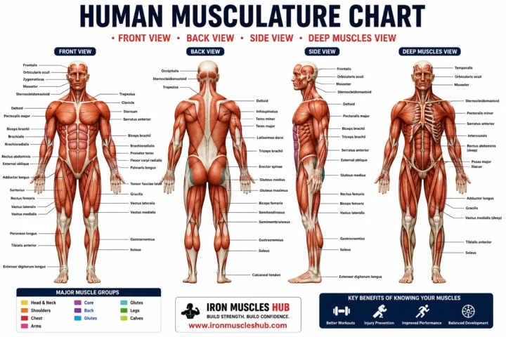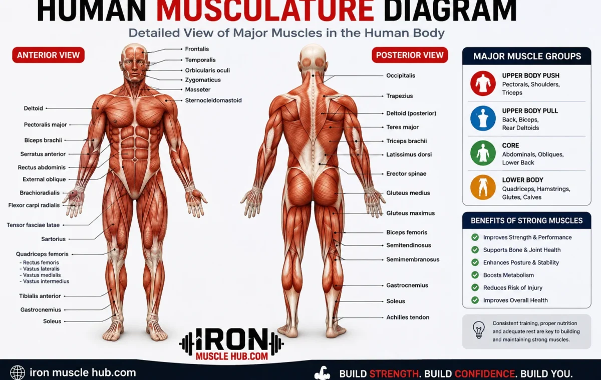
A human musculature diagram is a visual map of the body’s muscles, usually showing where major muscles are located and how they are arranged. A good diagram can help beginners identify muscles such as the biceps, triceps, deltoids, pectorals, abdominals, quadriceps, hamstrings and calf muscles.
- Human Musculature Diagram at a Glance
- What Is a Human Musculature Diagram?
- The Three Types of Muscle Tissue
- 1. Skeletal Muscle
- 2. Cardiac Muscle
- 3. Smooth Muscle
- Major Muscles on a Front-View Diagram
- Deltoids
- Pectoralis Major
- Biceps Brachii
- Rectus Abdominis
- External Obliques
- Quadriceps
- Tibialis Anterior
- Major Muscles on a Back-View Diagram
- Trapezius
- Latissimus Dorsi
- Triceps Brachii
- Gluteus Maximus
- Hamstrings
- Gastrocnemius
- Soleus
- How to Read a Human Musculature Diagram
- Step 1: Check the View
- Step 2: Start With Large Muscle Groups
- Step 3: Connect the Name With the Location
- Step 4: Test Yourself Without the Labels
- Step 5: Study One Region at a Time
- Human Musculature Diagram: Front vs. Back
- Why Muscle Diagrams Are Useful
- A Simple Muscle-Study Example
- Common Mistakes When Using a Muscle Diagram
- Mistake 1: Assuming Every Diagram Shows Every Muscle
- Mistake 2: Confusing Muscle Groups With Individual Muscles
- Mistake 3: Mixing Up Front and Back Views
- Mistake 4: Treating a Fitness Chart as a Medical Reference
- Where to Find a Reliable Human Musculature Diagram
- Internal Link Ideas for Iron Muscle Hub
- Frequently Asked Questions
- What does a human musculature diagram show?
- What are the major muscles shown on a human muscle diagram?
- How many types of muscle are in the human body?
- What is the difference between anterior and posterior muscle diagrams?
- What is the easiest way to learn human muscles?
- Is a human musculature diagram useful for fitness?
- Build Your Muscle-Anatomy Knowledge One Region at a Time
- Quick Guide to Major Muscle Groups
- How Skeletal Muscles Create Movement
- What Is an Agonist?
- What Is an Antagonist?
- What Is a Synergist?
- Understanding Muscle Attachments
- Origin
- Insertion
- Tendons
- Superficial vs. Deep Muscles
- Superficial Muscles
- Deep Muscles
- Major Upper-Body Muscles to Learn
- Deltoid
- Pectoralis Major
- Trapezius
- Latissimus Dorsi
- Biceps Brachii
- Triceps Brachii
- Major Core and Trunk Muscles
- Rectus Abdominis
- External Obliques
- Erector Spinae
- Diaphragm
- Major Lower-Body Muscles
- Gluteus Maximus
- Gluteus Medius
- Quadriceps
- Hamstrings
- Gastrocnemius
- Soleus
- Tibialis Anterior
- Why Muscle Groups Work Together
- A Better Way to Memorize a Muscle Diagram
- 1. Learn the Body Regions
- 2. Learn the Major Landmarks
- 3. Add the Function
- 4. Use an Unlabeled Diagram
- 5. Repeat at Increasing Difficulty
- Human Musculature Diagram vs. Muscle Anatomy Chart
- How Muscle Diagrams Relate to Exercise
- Common Diagram Labels Beginners Should Know
- Where to Get a Copyright-Safe Muscle Diagram
- A Simple Checklist for Choosing a Good Muscle Diagram
- Final Takeaway: Learn the Map Before the Details
- Quick Reference: The Human Muscular System
- How to Identify Muscles on a Diagram
- 1. Identify the View
- 2. Find the Major Landmarks
- 3. Follow the Label
- Why Some Muscle Diagrams Look Different
- Understanding the Front of the Body
- Head and Face
- Neck
- Chest
- Upper Arm
- Abdomen
- Thigh
- Understanding the Back of the Body
- Trapezius
- Latissimus Dorsi
- Triceps
- Gluteal Muscles
- Hamstrings
- Calf
- How Muscles Attach to Bones
- What Are Origin and Insertion?
- Why Muscle Names Can Help You Learn Anatomy
- Muscle Fiber Organization
- The Three Types of Muscle Tissue
- Skeletal Muscle
- Cardiac Muscle
- Smooth Muscle
- How Muscles Work Together
- A Practical Way to Study a Human Muscle Diagram
- Day 1: Shoulders and Arms
- Day 2: Chest and Abdomen
- Day 3: Back
- Day 4: Hips and Glutes
- Day 5: Thighs
- Day 6: Lower Legs
- Day 7: Test Yourself
- A Simple Self-Test
- How to Choose a Good Diagram for Your Website
- Copyright and Image Licensing
- Human Musculature Diagram: What Beginners Should Remember
- Frequently Asked Questions
- How many muscles are in the human body?
- What is the largest muscle shown on a muscle diagram?
- What is the difference between a muscle group and a muscle?
- Why do anatomy diagrams show muscles in different colors?
- Can a muscle diagram show deep muscles?
- Is a muscle diagram enough to diagnose an injury?
- The Best Way to Use a Muscle Anatomy Chart
- Final Thoughts
You don’t need to memorize every anatomical name at once. Start by learning the major muscle groups, understand whether you’re looking at the front or back of the body, and then connect each muscle with its basic role. This guide makes that process much easier. for more insights read here :-how to get bigger and how to gain muscle fast .
Quick answer: A human musculature diagram shows the location and names of muscles throughout the body. Most beginner diagrams focus on skeletal muscles, the muscles that work with bones to produce movement and help maintain posture.
Human Musculature Diagram at a Glance
| Body area | Major muscles to learn first | What they mainly help with |
|---|---|---|
| Shoulders | Deltoids | Moving the arm |
| Chest | Pectoralis major | Moving the upper arm |
| Upper back | Trapezius, latissimus dorsi | Shoulder and arm movement |
| Upper arms | Biceps, triceps | Bending and straightening the elbow |
| Abdomen | Rectus abdominis, obliques | Trunk movement and stability |
| Hips and buttocks | Gluteus maximus, gluteus medius | Hip movement and stability |
| Front of thighs | Quadriceps | Straightening the knee |
| Back of thighs | Hamstrings | Bending the knee and moving the hip |
| Lower legs | Gastrocnemius, soleus | Ankle and foot movement |
These are only some of the major muscles. A complete anatomical diagram contains many more structures.
What Is a Human Musculature Diagram?
A human musculature diagram is an anatomical illustration that identifies muscles by their location, shape or name. Diagrams can show the anterior view, meaning the front of the body, the posterior view, meaning the back, or both.
Many educational diagrams also distinguish between superficial muscles that are closer to the surface and deeper muscles underneath them. OpenStax, for example, presents anterior and posterior views with both superficial and deeper structures represented.
For beginners, this is more useful than trying to study a long list of muscle names without seeing where they actually sit.
The Three Types of Muscle Tissue
Before studying a labeled muscle diagram, it’s important to understand that not every muscle in your body is a large skeletal muscle that you can see moving.
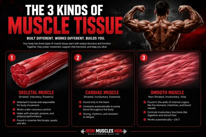
There are three main types of muscle tissue: skeletal, cardiac and smooth muscle.
1. Skeletal Muscle
Skeletal muscles attach to bones, generally through tendons, and are responsible for voluntary movement. They help you walk, lift objects, change position and maintain posture.
Most human musculature diagrams used for fitness or basic anatomy focus primarily on these muscles.
2. Cardiac Muscle
Cardiac muscle is the specialized muscle tissue found in the heart. Its contractions help pump blood around the body. Unlike skeletal muscle, you don’t consciously tell your heart to contract.
3. Smooth Muscle
Smooth muscle is found in the walls of many internal organs and passageways. It helps control involuntary processes such as movement through parts of the digestive system and changes in the diameter of blood vessels.
So, when you search for a human body muscle diagram, check what the illustration actually covers. A chart of visible skeletal muscles isn’t a complete picture of every muscle tissue in the human body.
Major Muscles on a Front-View Diagram
The front view, also called the anterior view, makes several familiar muscles easier to identify.
Deltoids
The deltoids form the rounded muscles around the shoulders. They contribute to several movements of the upper arm.
Pectoralis Major
The pectoralis major is the large muscle across the front of the chest. It helps produce movements of the upper arm.
Biceps Brachii
The biceps brachii is located on the front of the upper arm. It contributes to elbow flexion and forearm movement.
Rectus Abdominis
This muscle runs vertically along the front of the abdomen. It’s commonly associated with the appearance of the abdominal “six-pack.”
External Obliques
The external obliques sit along the sides of the abdomen. They contribute to movements and rotation of the trunk.
Quadriceps
The quadriceps group occupies the front of the thigh. It includes four major muscles and plays an important role in extending the knee.
Tibialis Anterior
The tibialis anterior is located toward the front of the lower leg. It helps move the foot upward at the ankle.
These structures appear among the major muscles identified in standard anterior muscular-system diagrams.
Major Muscles on a Back-View Diagram
A posterior view shows muscles that are easier to see from behind.
Trapezius
The trapezius spans the upper back and neck region. It participates in movements of the shoulder blades and upper limbs.
Latissimus Dorsi
Often called the “lats,” these large muscles occupy much of the middle and lower back and contribute to upper-arm movement.
Triceps Brachii
The triceps sit on the back of the upper arm. Their primary role is helping extend the elbow.
Gluteus Maximus
The gluteus maximus is the large muscle forming much of the buttock region. It contributes strongly to movements of the hip.
Hamstrings
The hamstrings are a group of muscles on the back of the thigh. They help bend the knee and contribute to hip movement.
Gastrocnemius
The gastrocnemius is one of the prominent muscles of the calf. It contributes to movement at the ankle.
Soleus
The soleus lies deep to the gastrocnemius in the calf region and also contributes to plantar flexion of the foot.
Standard anatomical diagrams commonly identify these structures in posterior views.
How to Read a Human Musculature Diagram
A complicated muscle chart can look intimidating at first. Use this simple method.
Step 1: Check the View
First determine whether the illustration shows the front, back or side of the body.
Don’t try to identify a muscle until you know which direction you’re viewing.
Step 2: Start With Large Muscle Groups
Learn the biggest and easiest landmarks first:
- Shoulders
- Chest
- Upper back
- Arms
- Abdominals
- Glutes
- Thighs
- Calves
Once those locations become familiar, smaller muscles are much easier to place.
Step 3: Connect the Name With the Location
Don’t memorize “deltoid” as just a word. Think: deltoid = shoulder.
Likewise:
- Biceps = front of upper arm
- Triceps = back of upper arm
- Pectorals = chest
- Quadriceps = front of thigh
- Hamstrings = back of thigh
- Gastrocnemius = prominent calf muscle
This location-based approach is also useful because many anatomical muscle names provide clues about location, size, shape or action.
Step 4: Test Yourself Without the Labels
Once you’ve studied a labeled diagram, cover the names and try to identify each muscle yourself.
An unlabeled chart is often more useful for learning than repeatedly looking at the answers.
Step 5: Study One Region at a Time
You don’t have to learn the entire muscular system in one sitting.
Try this order:
Shoulder → arm → chest → abdomen → back → hip → thigh → lower leg
Regional learning is also recommended by anatomy education resources because it gives you a manageable starting point rather than presenting the whole body as one enormous list.
Human Musculature Diagram: Front vs. Back
| Feature | Front / Anterior View | Back / Posterior View |
| Shoulders | Deltoids | Deltoids and upper-back muscles |
| Chest | Pectoralis major | Not directly visible |
| Arms | Biceps prominent | Triceps prominent |
| Abdomen | Rectus abdominis and obliques | Back muscles |
| Thighs | Quadriceps | Hamstrings |
| Buttocks | Less prominent | Gluteal muscles |
| Lower leg | Tibialis anterior | Gastrocnemius and soleus |
The exact appearance of a diagram depends on whether it shows superficial muscles, deeper muscles, or both. OpenStax’s standard figure deliberately shows superficial and deeper structures in different portions of the illustration.
Why Muscle Diagrams Are Useful
A good muscle anatomy chart isn’t only for medical students.
It can help:
- Students learn basic anatomy.
- Beginners understand fitness terminology.
- Trainers and coaches communicate about muscle groups.
- Exercise learners connect movements with body regions.
- Readers understand anatomy articles and educational material.
- Teachers create labeling exercises and revision activities.
The biggest benefit is visual context. Instead of memorizing isolated names, you can see how muscles are positioned in relation to neighboring structures.
A Simple Muscle-Study Example
Suppose you’re learning the muscles of the arm.
Don’t start by memorizing every small forearm muscle.
Begin with three landmarks:
Biceps → front of upper arm
Triceps → back of upper arm
Deltoid → shoulder
Next, learn what each broadly does. The biceps contributes to bending the elbow, while the triceps contributes to straightening it. The deltoid contributes to movement of the upper arm.
Once those three locations are familiar, you can gradually add the brachialis, brachioradialis and forearm muscles.
That’s a much more manageable learning path than trying to memorize an entire anatomy chart in one go.
Common Mistakes When Using a Muscle Diagram
Mistake 1: Assuming Every Diagram Shows Every Muscle
A simplified chart is designed for quick learning. It won’t necessarily show every muscle or deep anatomical structure.
Fix: Check the diagram’s title, labels and source before treating it as a complete reference.
Mistake 2: Confusing Muscle Groups With Individual Muscles
“Quadriceps” refers to a group of four muscles, while “rectus abdominis” identifies a particular muscle.
Fix: Pay attention to whether a label describes one muscle, a muscle group or a broader region.
Mistake 3: Mixing Up Front and Back Views
A muscle can look completely different depending on the viewing angle.
Fix: Always identify the anatomical view first.
Mistake 4: Treating a Fitness Chart as a Medical Reference
Fitness diagrams often simplify anatomy for easier understanding.
Fix: For medical, injury or clinical questions, use authoritative medical resources and speak with an appropriately qualified healthcare professional.
Where to Find a Reliable Human Musculature Diagram
For educational use, look for diagrams from established anatomy textbooks, universities, medical organizations or clearly licensed educational repositories.
OpenStax provides a detailed muscular-system figure with anterior and posterior views, including superficial and deeper muscles. Its material is published under an open license, but you should follow the attribution requirements when reusing the actual image.
OpenStax Anatomy and Physiology
The National Institute of Arthritis and Musculoskeletal and Skin Diseases (NIAMS) also provides beginner-friendly educational material explaining the three main muscle types and the role of skeletal muscle.
Internal Link Ideas for Iron Muscle Hub
Consider linking this article to related articles such as:
- What Are Muscles? A Beginner’s Guide to Muscle Anatomy
- What Are the Major Muscles in the Human Body?
- Skeletal vs. Cardiac vs. Smooth Muscle: What’s the Difference?
These links can create a useful learning path for readers who want to move from a basic diagram to more detailed muscle anatomy.
Frequently Asked Questions
What does a human musculature diagram show?
A human musculature diagram shows the location and names of muscles, usually skeletal muscles. Depending on the illustration, it may show the front, back or side of the body and may include superficial and deeper muscles. A beginner diagram normally focuses on recognizable major muscle groups rather than every anatomical structure.
What are the major muscles shown on a human muscle diagram?
Commonly labeled muscles include the deltoids, pectoralis major, biceps, triceps, trapezius, latissimus dorsi, rectus abdominis, obliques, gluteus maximus, quadriceps, hamstrings and gastrocnemius. The exact labels vary depending on how detailed the diagram is.
How many types of muscle are in the human body?
There are three main types of muscle tissue: skeletal, cardiac and smooth muscle. Skeletal muscle generally produces voluntary movement, cardiac muscle forms the muscular tissue of the heart, and smooth muscle is found in many internal organs and passageways.
What is the difference between anterior and posterior muscle diagrams?
An anterior diagram shows the front of the body, while a posterior diagram shows the back. This matters because different muscles are easier to see from each direction. For example, the biceps are prominent on the front of the upper arm, while the triceps are on the back.
What is the easiest way to learn human muscles?
Start with large muscle groups and study one body region at a time. Use a labeled diagram first, then switch to an unlabeled version and test yourself. Learning a muscle’s location alongside its basic action is generally more useful than memorizing names alone.
Is a human musculature diagram useful for fitness?
Yes. A diagram can help you understand basic muscle-group terminology used in exercise and fitness. However, an anatomy chart shouldn’t be used to diagnose an injury or determine the cause of persistent pain. For those situations, seek appropriate medical advice.
Build Your Muscle-Anatomy Knowledge One Region at a Time
A human musculature diagram becomes much easier to understand once you stop treating it as one giant collection of unfamiliar names. Begin with the major regions, learn the front and back views, and connect each muscle to its location and basic function.
For most beginners, the best starting point is the shoulders, chest, arms, abdomen, back, hips, thighs and calves. After those landmarks become familiar, you can move into smaller and deeper muscles.
A clear diagram is a learning tool, not a substitute for a medical assessment. Use reputable anatomy sources, pay attention to licensing when reusing images, and keep building your knowledge step by step.
Human Musculature Diagram: Deeper Anatomy, Muscle Functions, and Study Guide
A human musculature diagram becomes much more useful when you understand what the labeled muscles actually do. Instead of memorizing a long list of names, you can organize the body into regions, identify the major muscles, and then learn how those muscles work with bones, joints, tendons, and other muscles.
This second part goes beyond basic labels. It explains deeper muscle anatomy, common muscle actions, muscle groups that work together, and a simple way to study a muscular-system diagram without getting overwhelmed.
Quick Guide to Major Muscle Groups
| Region | Important muscles or groups | Basic role |
|---|---|---|
| Neck | Sternocleidomastoid | Helps move and position the head |
| Shoulder | Deltoid | Moves the upper arm |
| Chest | Pectoralis major | Helps move the upper arm |
| Upper back | Trapezius | Helps control the shoulder blades and neck region |
| Back | Latissimus dorsi | Helps move the upper arm |
| Upper arm | Biceps brachii, triceps brachii | Elbow movement |
| Abdomen | Rectus abdominis, obliques | Trunk movement and stability |
| Lower back | Erector spinae group | Helps extend and stabilize the spine |
| Hip | Gluteal muscles | Hip movement and stability |
| Front thigh | Quadriceps | Extends the knee |
| Back thigh | Hamstrings | Flexes the knee and assists hip movement |
| Lower leg | Gastrocnemius, soleus, tibialis anterior | Foot and ankle movement |
A diagram won’t necessarily show every muscle in these regions. The amount of detail depends on its purpose.
How Skeletal Muscles Create Movement
A muscle doesn’t simply “push” a bone. Skeletal muscles generally create movement by contracting and producing tension that is transmitted through tendons to bones. Muscles and bones then work together as a mechanical system around joints.
This is why a labeled muscle diagram should ideally be studied alongside the skeleton.
For example, when you bend your elbow, several muscles can contribute to the movement. One muscle may act as the primary mover while other muscles assist or stabilize the movement.
OpenStax describes these relationships using terms such as agonist, antagonist, synergist, origin, and insertion.
What Is an Agonist?
An agonist, also called a prime mover, is the muscle that provides the main force for a particular movement.
The exact prime mover depends on the action being performed.
What Is an Antagonist?
An antagonist is a muscle that produces an opposing action to another muscle.
A simple example is the relationship between the biceps and triceps during movements around the elbow. Their actions are different, allowing the joint to move in opposite directions.
What Is a Synergist?
A synergist helps another muscle perform a movement.
This matters because real human movement rarely depends on one isolated muscle. Several muscles can cooperate to produce controlled movement and stability.
Understanding Muscle Attachments
A detailed musculature diagram may label more than just muscle names.
You may also see origin, insertion, and tendon.
Origin
The origin is generally described as the attachment at the more stabilized end of a muscle during a particular movement.
Insertion
The insertion is generally the attachment at the end associated with the bone being moved.
These definitions are useful for studying anatomy, but remember that the way a muscle functions can change depending on the movement and which part of the body is stabilized.
Tendons
Tendons connect muscles to bones. When a skeletal muscle contracts, the resulting tension can be transferred through the tendon to influence movement at a joint.
This is one reason a complete anatomy chart may show tendons as well as the larger muscle bodies.
Superficial vs. Deep Muscles
One of the most confusing things about a human body muscle diagram is that not every muscle sits directly on the surface.
Superficial Muscles
Superficial muscles are closer to the surface of the body.
Many beginner charts focus heavily on these because they’re easier to identify visually.
Examples include:
- Deltoids
- Pectoralis major
- Biceps brachii
- Rectus abdominis
- Gluteus maximus
- Gastrocnemius
Deep Muscles
Deep muscles sit underneath other structures.
A diagram showing deeper anatomy may look quite different from a basic fitness poster because some superficial muscles are removed or partially hidden to reveal structures underneath.
That’s why two diagrams can both be accurate while showing different labels.
Tip: Before comparing two muscle charts, check whether they show superficial anatomy, deep anatomy, or a combination of both.
Major Upper-Body Muscles to Learn
Deltoid
The deltoid covers the shoulder and contributes to movement of the upper arm. Its triangular shape is also reflected in the origin of its name, which relates to the Greek letter delta.
Pectoralis Major
The pectoralis major is the large muscle on the front of the chest. It contributes to movement of the upper arm and is a common landmark on anterior muscle diagrams.
Trapezius
The trapezius occupies the upper and middle back region. It contributes to movements involving the shoulder blades and upper limbs.
Latissimus Dorsi
The latissimus dorsi is a broad muscle of the back that contributes to movements of the upper arm.
Biceps Brachii
The biceps brachii is positioned on the front of the upper arm. It’s an important muscle to recognize when studying elbow movement.
Triceps Brachii
The triceps brachii occupies the back of the upper arm and contributes to elbow extension.
Major Core and Trunk Muscles
The word “core” is often used broadly in fitness, while anatomy diagrams use specific muscle names.
Rectus Abdominis
The rectus abdominis is positioned vertically along the front of the abdomen. It’s one of the most recognizable muscles on an anterior muscular-system chart.
External Obliques
The external obliques are located along the sides of the abdomen. They contribute to movement and control of the trunk.
Erector Spinae
The erector spinae refers to a group of muscles associated with the back of the spine. These muscles play important roles in extending and stabilizing the trunk.
Diaphragm
The diaphragm deserves special attention because it is a muscle involved in breathing. NIAMS explains that the diaphragm helps the lungs fill with air during breathing.
This is a good reminder that muscles aren’t only about lifting weights or moving the limbs.
Major Lower-Body Muscles
Gluteus Maximus
The gluteus maximus is a major muscle of the buttock region and contributes to hip movement.
Gluteus Medius
The gluteus medius is positioned on the outer portion of the hip and contributes to movement and stabilization of the hip.
Quadriceps
The quadriceps are a group of four muscles located at the front of the thigh. Their major shared role is extension of the knee.
Hamstrings
The hamstrings are a group of muscles located toward the back of the thigh. They contribute to knee flexion and hip movement.
Gastrocnemius
The gastrocnemius is a prominent calf muscle and contributes to movement at the ankle.
Soleus
The soleus lies deeper than the gastrocnemius in the calf and also contributes to ankle movement.
Tibialis Anterior
The tibialis anterior sits toward the front of the lower leg and helps move the foot upward at the ankle.
Why Muscle Groups Work Together
It’s tempting to look at a diagram and imagine each muscle working independently.
Human movement is more coordinated than that.
During a simple activity such as standing up from a chair, multiple joints and muscle groups contribute to the movement. The nervous system coordinates muscle contractions, while bones and joints provide the framework through which movement occurs.
The musculoskeletal system includes more than muscles alone. It involves structures such as bones, tendons, ligaments, joints and cartilage.
That bigger picture can help you understand why an isolated muscle label doesn’t tell the whole story.
A Better Way to Memorize a Muscle Diagram
If you’re studying for school, training, or personal interest, try this five-stage method.
1. Learn the Body Regions
Start with:
Head → neck → shoulder → chest → back → arm → abdomen → hip → thigh → lower leg
Don’t worry about small muscles yet.
2. Learn the Major Landmarks
Memorize a few recognizable muscles in each region.
For example:
- Shoulder: deltoid
- Chest: pectoralis major
- Upper arm: biceps and triceps
- Abdomen: rectus abdominis
- Back: trapezius and latissimus dorsi
- Buttocks: gluteus maximus
- Front thigh: quadriceps
- Back thigh: hamstrings
- Calf: gastrocnemius
3. Add the Function
After learning a name, ask:
“What does this muscle help me do?”
This gives the name a practical meaning instead of leaving it as a vocabulary word.
4. Use an Unlabeled Diagram
After studying a labeled chart, switch to an unlabeled version.
Point to a structure and name it without looking at the answer.
5. Repeat at Increasing Difficulty
Once major muscles are easy, start learning smaller and deeper structures.
This gradual approach is much more manageable than trying to memorize an entire anatomical chart in one session.
Human Musculature Diagram vs. Muscle Anatomy Chart
These terms are often used interchangeably, but they can describe slightly different resources.
| Resource | Usually emphasizes | Best for |
| Human musculature diagram | Overall muscle locations | Beginners |
| Labeled muscle diagram | Names and positions | Studying |
| Muscle anatomy chart | Detailed anatomical information | Students and fitness learners |
| Muscular system diagram | The overall muscular system | General education |
| Deep muscle diagram | Muscles beneath superficial layers | More advanced study |
| Unlabeled muscle diagram | Identification practice | Testing yourself |
The important thing isn’t the title of the resource. Check what structures it actually contains.
How Muscle Diagrams Relate to Exercise
A muscle chart can help you understand the names used in exercise descriptions.
For example, an exercise might be described as involving the chest, shoulders and triceps. A diagram lets a beginner see where those structures are located.
But avoid assuming that an exercise isolates only one muscle.
Muscles often work together, and movement depends on joint position, technique, resistance, range of motion and other factors.
NIAMS also emphasizes that healthy muscles aren’t defined simply by size. Physical activity can help muscles become stronger, and people can have healthy, strong muscles without having very large muscles.
Common Diagram Labels Beginners Should Know
Here’s a useful starter vocabulary list:
- Deltoid — shoulder
- Pectoralis major — chest
- Biceps brachii — front upper arm
- Triceps brachii — back upper arm
- Trapezius — upper and middle back
- Latissimus dorsi — broad back muscle
- Rectus abdominis — front abdominal region
- External oblique — side of abdomen
- Erector spinae — back muscle group along the spine
- Gluteus maximus — major buttock muscle
- Quadriceps — front thigh muscle group
- Hamstrings — back thigh muscle group
- Gastrocnemius — major calf muscle
- Soleus — deeper calf muscle
- Tibialis anterior — front of lower leg
This list isn’t a complete anatomical inventory. It’s a practical starting point for someone learning to read a basic muscle diagram.
Where to Get a Copyright-Safe Muscle Diagram
If you’re publishing an anatomy image on a website, don’t simply copy an image from Google Images.
Instead, use an image that you’ve created yourself or one whose license clearly permits the intended reuse.
One example is the OpenStax muscle diagram available through Wikimedia Commons. The listed file is identified as an OpenStax illustration and is licensed under Creative Commons Attribution 4.0 International, subject to the license terms and appropriate attribution.
For your own website, an original illustration is often the cleanest option because you control its design, labeling, branding and usage rights.
A Simple Checklist for Choosing a Good Muscle Diagram
Before using a diagram, check:
- Does it clearly identify the body view?
- Are the labels readable?
- Does it distinguish superficial and deep structures if necessary?
- Are the anatomical names spelled correctly?
- Is the source trustworthy?
- Is the image license clear?
- Does the image match your reader’s knowledge level?
- Is the image sharp enough for mobile screens?
- Does the alt text accurately describe the image?
- Have you avoided presenting an educational diagram as a diagnostic tool?
Final Takeaway: Learn the Map Before the Details
The easiest way to understand a human musculature diagram is to treat it like a map. First learn the major regions, then identify the biggest muscles, then connect each muscle with its basic movement or function.
Once those landmarks are familiar, deeper anatomy becomes much easier.
Remember that the muscular system includes far more than the muscles visible on a typical fitness chart. Skeletal muscles work with bones and tendons to produce movement, while cardiac and smooth muscle perform essential involuntary functions elsewhere in the body.
For beginners, consistent practice with labeled and unlabeled diagrams is more useful than trying to memorize everything at once.
Human Musculature Diagram: Advanced Beginner Guide to Muscle Anatomy
A human musculature diagram is much easier to understand once you know how muscles are organized, how they attach to the skeleton, and why different diagrams show different layers. The visible muscles on a chart are only part of the muscular system. Your body contains more than 600 muscles, including skeletal, cardiac and smooth muscle tissue.
For beginners, the smartest approach is to learn the body region by region. Start with the large muscles you can easily locate, then move toward smaller and deeper structures.
Quick Reference: The Human Muscular System
| Region | Muscle examples | Main purpose |
|---|---|---|
| Head | Temporalis, masseter | Chewing and jaw movement |
| Neck | Sternocleidomastoid | Head and neck movement |
| Shoulder | Deltoid | Upper-arm movement |
| Chest | Pectoralis major | Upper-arm movement |
| Upper back | Trapezius | Shoulder and upper-back movement |
| Middle/lower back | Latissimus dorsi | Upper-arm movement |
| Upper arm | Biceps, triceps | Elbow movement |
| Abdomen | Rectus abdominis, obliques | Trunk movement and stability |
| Lower back | Erector spinae | Spinal extension and posture |
| Hip | Gluteal muscles | Hip movement and stability |
| Front thigh | Quadriceps | Knee extension |
| Back thigh | Hamstrings | Knee and hip movement |
| Calf | Gastrocnemius, soleus | Ankle movement |
| Front lower leg | Tibialis anterior | Foot movement |
This is a learning guide rather than a complete list of every muscle.
How to Identify Muscles on a Diagram
When you first look at a labeled muscle chart, don’t start reading every label.
Use a simple three-step system.
1. Identify the View
Look for a heading such as:
- Anterior = front
- Posterior = back
- Lateral = side
- Superficial = closer to the surface
- Deep = underneath more superficial structures
This matters because the same body region can look very different from another angle.
2. Find the Major Landmarks
Start with the obvious areas:
Shoulder → chest → arm → abdomen → back → hip → thigh → calf
Once you’ve identified the region, look for the largest muscle nearby.
3. Follow the Label
Anatomy diagrams usually use lines or arrows to connect a name with a specific structure.
Follow the line all the way to its endpoint. Don’t assume that a label belongs to the muscle closest to the printed word.
Why Some Muscle Diagrams Look Different
You may find two diagrams online that appear to disagree about where a muscle is located.
Often, they aren’t actually contradictory.
One diagram may show the body’s superficial muscles, while another exposes deeper muscles by removing or separating structures. Educational anatomy resources commonly use this approach to show different layers.
A detailed chart might therefore show:
- Surface muscles
- Deeper muscles
- Tendons
- Muscle attachments
- Muscle groups
- Fiber direction
- Anatomical landmarks
A beginner poster might show only the major surface muscles.
Understanding the Front of the Body
The anterior view is one of the most common formats for a human musculature diagram.
Head and Face
The muscles of the head include structures involved in facial expression, eye movement, chewing and other functions.
Two useful beginner landmarks are the temporalis and masseter. Both are associated with chewing.
Neck
The sternocleidomastoid is a prominent muscle on each side of the neck. It contributes to movement of the head and neck.
Chest
The pectoralis major is the large muscle across the front of the chest.
It contributes to movement of the upper arm and is one of the easiest chest muscles to recognize on a basic diagram.
Upper Arm
The biceps brachii is positioned toward the front of the upper arm.
The triceps brachii is positioned toward the back.
Learning this front-versus-back relationship is a useful first step for understanding the arm.
Abdomen
The rectus abdominis runs vertically along the front of the abdominal region.
The external obliques are positioned toward the sides of the trunk.
Together with other muscles, these structures help control and move the trunk.
Thigh
The quadriceps occupy the front of the thigh.
The name refers to the group’s four muscles, making “quadriceps” a group name rather than the name of one individual muscle.
Understanding the Back of the Body
A posterior diagram reveals muscles that aren’t as obvious from the front.
Trapezius
The trapezius covers a large portion of the upper back and contributes to movements involving the shoulders and shoulder blades.
Latissimus Dorsi
The latissimus dorsi is a broad muscle of the back that contributes to upper-arm movement.
It’s commonly labeled “latissimus dorsi” or simply “lat” on fitness-oriented materials.
Triceps
From behind, the triceps become a major landmark of the upper arm.
The triceps contributes to extension at the elbow.
Gluteal Muscles
The gluteal region contains several muscles, including the gluteus maximus and gluteus medius.
The gluteus maximus is the large muscle that forms much of the buttock region.
Hamstrings
The hamstrings are located on the back of the thigh.
They’re a group rather than one single muscle and contribute to movements involving the hip and knee.
Calf
The gastrocnemius is one of the most prominent calf muscles.
The soleus lies underneath it and also contributes to movement at the ankle.
How Muscles Attach to Bones
A muscle diagram makes more sense when you understand the connection between muscles and bones.
Skeletal muscles generally attach to bones through tendons. When the muscle contracts, tension is transmitted through these attachments, allowing movement at joints.
This creates a basic chain:
Muscle contracts → tendon transmits force → bone moves → joint changes position
The actual mechanics are more complicated, but this simplified model is useful for beginners.
What Are Origin and Insertion?
You may see these terms on an advanced anatomy chart.
Origin generally refers to the attachment on the more stabilized side of a muscle during a particular action.
Insertion generally refers to the attachment associated with the structure being moved.
OpenStax explains that these terms describe how skeletal muscles attach to the skeleton and participate in movement.
Don’t worry if these terms feel confusing at first. Learn muscle location and basic action before trying to memorize every attachment.
Why Muscle Names Can Help You Learn Anatomy
Muscle names aren’t always random.
Anatomical names can describe characteristics such as:
- Location
- Shape
- Size
- Direction of fibers
- Number of divisions
- Action
For example, OpenStax explains that the deltoid gets its name from its triangular shape, while “rectus” relates to a straight orientation.
This gives you a useful memory trick: instead of memorizing a name as an isolated word, look for clues in the name.
Muscle Fiber Organization
A detailed muscle anatomy chart may also show the internal structure of skeletal muscle.
A skeletal muscle contains bundles called fascicles, which contain muscle fibers. Connective-tissue layers organize and support these structures.
At a much smaller scale, skeletal muscle fibers contain myofibrils, which contain the contractile structures responsible for muscle contraction.
This is why a full anatomy textbook can have several different diagrams for “muscle anatomy.” One might show the whole body, while another zooms from a complete muscle down to microscopic structures.
The Three Types of Muscle Tissue
A whole-body skeletal muscle chart doesn’t represent every type of muscle in the body.
There are three major types:
Skeletal Muscle
Skeletal muscle is connected with the skeleton and produces many voluntary movements. It also helps maintain posture and stabilize joints.
Cardiac Muscle
Cardiac muscle is the specialized muscle tissue found in the heart. It contracts involuntarily to help pump blood.
Smooth Muscle
Smooth muscle is found in the walls of various organs and other internal structures. It performs involuntary functions.
Cleveland Clinic identifies skeletal, cardiac and smooth muscle as the three major muscle types.
How Muscles Work Together
One of the biggest beginner mistakes is assuming that one movement equals one muscle.
That’s rarely how the body works.
Muscles can act as:
- Agonists — primary movers
- Antagonists — muscles that oppose a movement
- Synergists — muscles that assist another muscle
- Fixators — muscles that stabilize a body part
OpenStax uses these relationships to explain how skeletal muscles cooperate during movement.
For example, bending your elbow isn’t simply a matter of “turning on the biceps.” Other muscles can contribute to the movement or stabilize the joint and surrounding structures.
A Practical Way to Study a Human Muscle Diagram
If you’re learning anatomy for the first time, try this seven-day approach.
Day 1: Shoulders and Arms
Learn:
- Deltoid
- Biceps brachii
- Triceps brachii
- Major forearm landmarks
Day 2: Chest and Abdomen
Focus on:
- Pectoralis major
- Rectus abdominis
- External obliques
Day 3: Back
Learn:
- Trapezius
- Latissimus dorsi
- Erector spinae
Day 4: Hips and Glutes
Focus on:
- Gluteus maximus
- Gluteus medius
- Other major hip landmarks
Day 5: Thighs
Learn:
- Quadriceps
- Hamstrings
- Major inner-thigh landmarks
Day 6: Lower Legs
Focus on:
- Gastrocnemius
- Soleus
- Tibialis anterior
Day 7: Test Yourself
Use an unlabeled diagram.
Point to each muscle and try to name it without looking at the answer.
Then repeat the exercise from the opposite view.
A Simple Self-Test
Cover the labels on your diagram and answer these questions:
- What muscle covers the shoulder?
- Which major muscle is on the front of the upper arm?
- Which major muscle is on the back of the upper arm?
- What large muscle covers much of the chest?
- What muscle group occupies the front of the thigh?
- What muscle group occupies the back of the thigh?
- What prominent muscle is found in the calf?
- Which large muscle group is located across the upper back?
If you can answer these consistently, you’re ready to add smaller structures.
How to Choose a Good Diagram for Your Website
If you’re using a human musculature diagram on Iron Muscle Hub, clarity matters more than the number of labels.
A useful beginner image should have:
- A clean front view
- A clean back view
- Large, readable labels
- Correct anatomical terminology
- A simple layout
- Good contrast between structures
- Mobile-friendly resolution
- Clear licensing or original ownership
Don’t crowd the image with dozens of tiny labels simply to make it appear more comprehensive.
A clean diagram that teaches 20 important muscles can be more useful to a beginner than an overcrowded chart containing hundreds of labels.
Copyright and Image Licensing
Don’t assume that an image is free to use because it appears in Google Images.
For example, Wikimedia Commons hosts an OpenStax illustration showing anterior and posterior views of major skeletal muscles. The listed image is licensed CC BY 4.0, which permits reuse subject to its attribution requirements.
If you reuse a Creative Commons image, follow the applicable license requirements, including appropriate credit and a link to the license where required.
For maximum control over your website’s branding, an original diagram created specifically for Iron Muscle Hub is another option.
Human Musculature Diagram: What Beginners Should Remember
You don’t need to memorize hundreds of names to understand basic muscle anatomy.
Start with the major landmarks:
Deltoids → shoulders
Pectorals → chest
Biceps → front upper arm
Triceps → back upper arm
Trapezius → upper back
Latissimus dorsi → broad back region
Rectus abdominis → front abdomen
Gluteus maximus → buttock region
Quadriceps → front thigh
Hamstrings → back thigh
Gastrocnemius → calf
Once these become familiar, more detailed anatomy becomes much easier to learn.
Frequently Asked Questions
How many muscles are in the human body?
The human body has more than 600 muscles, according to Cleveland Clinic. The exact number can vary depending on how structures are classified, but “more than 600” is a useful general figure for beginners.
What is the largest muscle shown on a muscle diagram?
The gluteus maximus is commonly described as the largest muscle of the human body. However, “largest” can depend on the measurement being discussed, so avoid assuming that a diagram’s visually largest structure is automatically the largest muscle.
What is the difference between a muscle group and a muscle?
An individual muscle is one anatomical structure, while a muscle group is a collection of muscles that are commonly discussed together because of their location or function. The quadriceps and hamstrings are familiar examples of muscle groups.
Why do anatomy diagrams show muscles in different colors?
Colors are usually used to make individual structures easier to distinguish. They don’t necessarily represent a biological color difference. The meaning depends on the particular diagram and its legend.
Can a muscle diagram show deep muscles?
Yes. A detailed diagram can show deeper muscles by separating, removing or visually exposing superficial structures. Some anatomy resources deliberately present both superficial and deep views.
Is a muscle diagram enough to diagnose an injury?
No. A diagram can help you understand anatomy, but it can’t determine what’s causing pain, weakness, swelling or loss of movement. Persistent or concerning symptoms should be assessed by an appropriate healthcare professional.
The Best Way to Use a Muscle Anatomy Chart
A good human musculature diagram should help you build a mental map of the body.
Start broad. Learn the front and back views, recognize major muscle groups, and understand the basic action of each region. Then add deeper structures and more detailed terminology as your knowledge improves.
Remember that muscles don’t work in isolation. Skeletal muscles interact with tendons, bones, joints and the nervous system to create controlled movement, while cardiac and smooth muscle perform essential involuntary functions.
If you’re creating an anatomy resource for beginners, focus on accuracy, readable labeling and a logical learning sequence. A diagram should make anatomy easier to understand, not turn it into a wall of unfamiliar words.
Final Thoughts
A human musculature diagram is one of the simplest ways to build a basic understanding of muscle anatomy. Instead of trying to memorize every anatomical term at once, start with the major regions of the body and learn the muscles you can easily identify from the front and back.
Focus first on landmarks such as the deltoids, pectorals, biceps, triceps, trapezius, latissimus dorsi, abdominals, glutes, quadriceps, hamstrings and calf muscles. Once these become familiar, you can gradually study smaller and deeper muscles.
Remember that muscles work as part of a larger system. Skeletal muscles interact with bones, tendons and joints to create movement, while cardiac and smooth muscles perform important involuntary functions elsewhere in the body.
For the best learning results, use a labeled diagram first, then test yourself with an unlabeled version. Choose reliable anatomy sources and, if you’re publishing an image online, make sure you have permission to use it or that its license clearly allows reuse.
A clear diagram doesn’t need to show everything. It simply needs to help you understand where muscles are, what they’re called, and how they contribute to movement. That’s the foundation you can build on as your anatomy knowledge grows.