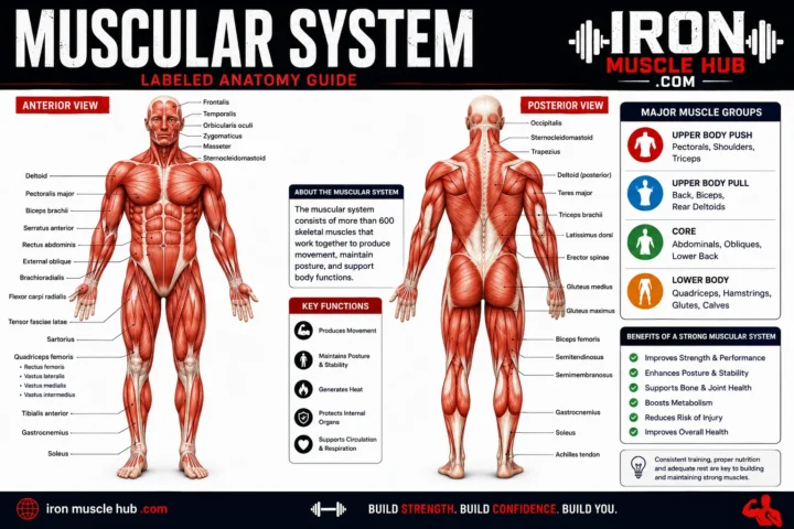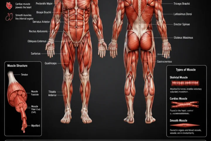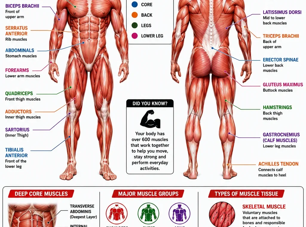
A muscular system diagram is a visual guide that helps you identify where muscles are located and understand what they do. For beginners, a labeled diagram can make human anatomy much easier to learn because you can connect each muscle’s name with its position and function.
- Muscular System Diagram at a Glance
- How to Read a Muscular System Diagram
- 1. Start with the front view
- 2. Study the back view
- 3. Look for superficial versus deep muscles
- Major Muscles of the Human Body
- Head and Neck
- Shoulders: Deltoids
- Chest: Pectoralis Major
- Back: Trapezius and Latissimus Dorsi
- Arms: Biceps and Triceps
- Abdomen: Rectus Abdominis and Obliques
- Hips and Glutes
- Thighs: Quadriceps and Hamstrings
- Lower Leg: Calf Muscles
- The Three Types of Muscle Tissue
- Skeletal Muscle
- Cardiac Muscle
- Smooth Muscle
- Why Muscles Work With Bones
- A Simple Way to Memorize Muscle Anatomy
- The "Location + Action" Trick
- Common Mistakes When Using a Muscle Diagram
- Mistake 1: Assuming every diagram shows every muscle
- Mistake 2: Treating muscle groups as single muscles
- Mistake 3: Confusing muscles with tendons
- Mistake 4: Memorizing names without learning function
- Mistake 5: Assuming diagrams are perfectly to scale
- Where to Find Reliable Muscle Anatomy Diagrams
- Frequently Asked Questions
- What does a muscular system diagram show?
- How many muscles are in the human body?
- What are the three types of muscle?
- What is the largest muscle in the body?
- What is the difference between skeletal and smooth muscle?
- Why do anatomy diagrams show front and back views?
- Related Resources to Explore
- A Clearer Way to Learn the Muscular System
- Major Muscle Groups to Learn First
- Front View: How to Identify the Main Muscles
- Deltoid
- Pectoralis Major
- Biceps Brachii
- Rectus Abdominis
- Quadriceps
- Back View: The Muscles You Should Recognize
- Trapezius
- Latissimus Dorsi
- Triceps Brachii
- Gluteus Maximus
- Hamstrings
- Gastrocnemius
- Why Muscle Names Can Help You Read a Diagram
- Superficial vs. Deep Muscles
- Muscular System vs. Musculoskeletal System
- How Muscles Produce Movement
- An Easy Muscle Diagram Study Routine
- Step 1: Study one region
- Step 2: Look at the labeled version
- Step 3: Hide the labels
- Step 4: Say the answer aloud
- Step 5: Add function
- Step 6: Review later
- A Useful Front-and-Back Memory Map
- Where an Anatomy Diagram Can Be Useful
- School and anatomy study
- Fitness education
- General body knowledge
- Training or rehabilitation education
- What Makes a Good Muscular System Diagram?
- Common Diagram Labels Worth Memorizing
- Frequently Asked Questions
- What is the easiest way to learn a muscular system diagram?
- What muscles should I learn first?
- Why do different muscle diagrams have different labels?
- Is the biceps a single muscle?
- Is the quadriceps one muscle?
- Are all muscles under conscious control?
- Related Iron Muscle Hub Topics
- Final Takeaway
- Muscle Attachments: Origin and Insertion
- Agonists, Antagonists and Synergists
- Agonist
- Antagonist
- Synergist
- Muscle Fibers and Their Arrangement
- Why Some Muscles Look Bigger on a Diagram
- Muscles That Are Easy to Confuse
- Biceps vs. Triceps
- Quadriceps vs. Hamstrings
- Gastrocnemius vs. Soleus
- Pectoralis Major vs. Deltoid
- A 10-Minute Muscle Diagram Practice Method
- A Beginner's Muscle Identification Checklist
- How to Use a Muscular System Diagram for Exercise Education
- How to Choose Between a Simple and Detailed Diagram
- Printable Study Idea
- Important Anatomy Terms to Know
- How to Build Your Own Muscle Anatomy Reference
- Common Questions About Muscle Anatomy
- Can I learn all the muscles from one diagram?
- Should I memorize muscle names or functions first?
- Why are some muscles missing from a basic chart?
- Are muscle diagrams useful for gym beginners?
- What is the best way to test muscle anatomy knowledge?
The muscular system includes three main types of muscle tissue: skeletal, cardiac and smooth muscle. Skeletal muscles are the ones most commonly shown on full-body muscle diagrams because they attach to bones and produce voluntary movement. For more insights read here :- lean and muscle body and how to grow muscle .
You don’t need to memorize hundreds of muscle names at once. Start with the large, easy-to-recognize muscle groups, then add smaller muscles as your understanding improves.
Quick answer: A muscular system diagram shows the location and arrangement of muscles in the body. A useful beginner diagram should identify major muscles such as the pectorals, deltoids, biceps, triceps, abdominal muscles, quadriceps, hamstrings and calf muscles, while explaining their basic roles.
Muscular System Diagram at a Glance
| Muscle area | Examples of major muscles | Basic role |
|---|---|---|
| Head and neck | Sternocleidomastoid, facial muscles | Head movement and facial expression |
| Shoulders | Deltoids | Moving and stabilizing the shoulder |
| Chest | Pectoralis major | Moving the upper arm |
| Upper back | Trapezius, latissimus dorsi | Shoulder and arm movement |
| Arms | Biceps, triceps | Bending and straightening the elbow |
| Abdomen | Rectus abdominis, obliques | Trunk movement and stability |
| Hips | Gluteal muscles | Hip movement and stability |
| Front thighs | Quadriceps | Straightening the knee |
| Back thighs | Hamstrings | Bending the knee and moving the hip |
| Lower legs | Gastrocnemius, soleus | Ankle and foot movement |
This is a simplified study guide rather than a complete anatomical inventory. The body contains more than 600 muscles, according to Cleveland Clinic, and many are too small or deep to appear on a basic full-body diagram.
How to Read a Muscular System Diagram
A good way to study a muscle anatomy chart is to work from large regions to smaller structures.
1. Start with the front view
A front, or anterior, view lets you recognize muscles across the chest, abdomen, arms and front of the legs.
Look first for:
- Deltoids — rounded muscles covering the shoulders.
- Pectoralis major — large chest muscle.
- Biceps brachii — prominent muscle at the front of the upper arm.
- Rectus abdominis — vertical muscle running along the front of the abdomen.
- Quadriceps — large muscle group at the front of the thigh.
- Tibialis anterior — muscle along the front of the lower leg.
Don’t worry about learning every label immediately. First learn where each region begins and ends.
2. Study the back view
A posterior, or back, view reveals several muscles that aren’t easy to see from the front.
Common labels include:
- Trapezius
- Latissimus dorsi
- Triceps brachii
- Gluteus maximus
- Hamstrings
- Gastrocnemius
OpenStax uses regional study of skeletal muscles, working from the head downward, as one approach to learning muscular anatomy.
3. Look for superficial versus deep muscles
This is one detail that many beginner diagrams don’t explain clearly.
A superficial muscle is closer to the body’s surface. A deep muscle lies underneath other structures. That’s why an anatomy chart may show different muscles depending on whether the illustration is displaying superficial or deeper layers.
Some detailed anatomical illustrations deliberately show superficial muscles on one side and deeper structures on the other to make the layers easier to study.
Major Muscles of the Human Body
Head and Neck
Muscles around the head and neck help control facial expression, chewing, swallowing and head movement.
One easily recognizable neck muscle is the sternocleidomastoid, which runs along the side of the neck. It contributes to movements of the head and neck.
A basic muscular system diagram may simplify the head considerably because there are many smaller facial and neck muscles.
Shoulders: Deltoids
The deltoid is the rounded muscle covering much of the shoulder.
It works with other muscles to move the upper arm. On a full-body muscle diagram, it is usually easy to spot because it forms the shoulder’s outer contour.
Chest: Pectoralis Major
The pectoralis major is the large superficial muscle covering much of the front of the chest.
It contributes to movements of the upper arm. Beneath it are deeper structures, including the pectoralis minor.
Back: Trapezius and Latissimus Dorsi
The trapezius occupies a broad region across the upper back and neck.
The latissimus dorsi is a large back muscle extending across much of the lower and middle back. Together with other muscles, these structures contribute to movements of the shoulder and upper limb.
Detailed anatomy references show that the back contains several layers and muscle groups, so a simple diagram won’t represent every structure.
Arms: Biceps and Triceps
The biceps brachii is located toward the front of the upper arm. One of its best-known actions is helping bend the elbow.
The triceps brachii is on the back of the upper arm and plays a major role in straightening the elbow.
A useful way to remember them is:
Biceps → front of upper arm → elbow flexion
Triceps → back of upper arm → elbow extension
These are simplified descriptions; real movements usually involve multiple muscles working together.
Abdomen: Rectus Abdominis and Obliques
The rectus abdominis runs vertically along the front of the abdomen.
The oblique muscles occupy the sides of the abdominal region and help with trunk movement and stability.
A common mistake is to think of the abdominal muscles as one single muscle. In reality, the abdominal wall contains several muscles arranged in different directions.
Hips and Glutes
The gluteal muscles form a major muscle group around the buttocks and hip.
The gluteus maximus is the largest and most superficial of the three main gluteal muscles. Other gluteal muscles contribute to hip stability and movement.
Thighs: Quadriceps and Hamstrings
The quadriceps occupy the front of the thigh. As the name suggests, this group contains four major muscles.
Their primary combined action is extending the knee.
The hamstrings are located toward the back of the thigh. They contribute to knee flexion and hip movement.
This front-versus-back distinction makes the thighs a useful place to begin studying muscle groups.
Lower Leg: Calf Muscles
The gastrocnemius is the large, visible muscle that forms much of the calf.
The soleus lies deeper beneath the gastrocnemius and also contributes to movements at the ankle.
The lower leg contains several additional muscles, so a simplified muscular system diagram may show only the most prominent ones.
The Three Types of Muscle Tissue
Not every muscle appears on a standard full-body muscle chart. That’s because “muscle” refers to different types of tissue.
| Type | Where it’s found | Under conscious control? | Main job |
| Skeletal | Attached to bones | Generally yes | Body movement and posture |
| Cardiac | Heart | No | Pumps blood |
| Smooth | Walls of many organs and blood vessels | No | Moves substances and regulates passageways |
Skeletal muscle is associated with voluntary movement, while cardiac muscle makes up the heart and smooth muscle is found in structures such as the digestive tract and blood vessels.
Skeletal Muscle
Skeletal muscles attach to bones through tendons and work with the skeleton to create movement. They also help maintain posture and contribute to heat production.
When you see a typical human muscular system diagram, you’re usually looking primarily at skeletal muscles.
Cardiac Muscle
Cardiac muscle is found in the heart.
Unlike skeletal muscle, you don’t consciously tell your heart to contract every time it beats. Specialized cardiac tissue helps coordinate the heart’s contractions.
Smooth Muscle
Smooth muscle is found in the walls of many internal organs and blood vessels.
It operates involuntarily and helps move substances through structures such as the digestive tract and regulate the diameter of blood vessels.
Why Muscles Work With Bones
Muscles don’t move your skeleton by pushing bones around.
Instead, skeletal muscles generate force through contraction and transmit that force to bones through tendons. The resulting pull can move a joint.
Think of the system as a team:
Nervous system → muscle activation → muscle contraction → tendon force → bone movement
This is why learning a muscular system diagram alongside a skeleton diagram can be more useful than studying either one alone. The muscular and skeletal systems work together to support and move the body.
A Simple Way to Memorize Muscle Anatomy
If you’re studying for school, training, or simply trying to understand your body, don’t attempt to memorize every label in one sitting.
Try this five-step method:
- Learn the body regions first. Head, neck, shoulder, chest, back, arm, abdomen, hip, thigh and lower leg.
- Pick two or three major muscles per region.
- Connect the name to its location. For example, biceps = front of upper arm.
- Add the main action. Ask what movement the muscle helps produce.
- Test yourself without looking. Cover the labels and try to identify each structure.
The “Location + Action” Trick
For every muscle you learn, write down:
Name → Location → Main action
For example:
Triceps → back of upper arm → helps straighten the elbow
This approach is often more useful than memorizing a long alphabetical list because it gives the name a physical context.
Common Mistakes When Using a Muscle Diagram
Mistake 1: Assuming every diagram shows every muscle
It doesn’t. A beginner chart may show only major superficial muscles, while an anatomy textbook may show layers, attachments and smaller structures.
Mistake 2: Treating muscle groups as single muscles
“Quadriceps” and “hamstrings,” for example, refer to groups rather than one individual muscle.
Mistake 3: Confusing muscles with tendons
A muscle is contractile tissue. A tendon is connective tissue that connects muscle to bone.
A diagram that labels both can make this distinction much easier to understand.
Mistake 4: Memorizing names without learning function
Knowing that a structure is called the deltoid is useful, but understanding its location and role makes the information easier to remember.
Mistake 5: Assuming diagrams are perfectly to scale
Educational illustrations simplify anatomy. They may enlarge a small structure or remove surrounding tissues so a particular muscle is easier to see.
Where to Find Reliable Muscle Anatomy Diagrams
For studying, prioritize educational and medical sources rather than random images copied across websites.
MedlinePlus provides anatomy resources and links to muscle images covering areas such as the head and neck, trunk, upper extremity and lower extremity.
The NCBI Bookshelf also contains anatomy and medical education resources, including illustrations of muscle types and skeletal muscles. Some Open RN figures are published under Creative Commons licenses, but you should still check the specific license and attribution requirements before reusing an image.
For a website such as Iron Muscle Hub, an even safer featured image is an original diagram commissioned or created specifically for the site, rather than downloading an anatomy image from a random search result.
Frequently Asked Questions
What does a muscular system diagram show?
A muscular system diagram is a visual representation of muscles and their locations in the body. Basic diagrams usually focus on major skeletal muscles, often showing front and back views. More advanced charts can include deeper muscles, attachment points, tendons and individual muscle actions.
How many muscles are in the human body?
The human body has more than 600 muscles, according to Cleveland Clinic. The exact number isn’t usually useful for beginner study because anatomy references can organize and count muscles differently. Start by learning major muscle groups before worrying about the complete list.
What are the three types of muscle?
The three major types are skeletal, cardiac and smooth muscle. Skeletal muscle is associated mainly with voluntary movement, cardiac muscle makes up the heart, and smooth muscle is found in many internal organs and blood vessels.
What is the largest muscle in the body?
The gluteus maximus is generally recognized as the largest individual skeletal muscle by mass. It is located in the buttock region and contributes strongly to hip movement.
What is the difference between skeletal and smooth muscle?
Skeletal muscle is attached to bones and is generally under voluntary control. Smooth muscle is found in the walls of internal organs and blood vessels and works involuntarily. Their microscopic structures and methods of contraction also differ.
Why do anatomy diagrams show front and back views?
A single view can’t clearly display all major muscles. Front and back views allow you to see structures on the anterior and posterior sides of the body, making it easier to identify muscles such as the pectorals and quadriceps from the front and the trapezius, latissimus dorsi and hamstrings from the back.
Related Resources to Explore
For readers building their anatomy knowledge, useful related articles on Iron Muscle Hub could include:
- What Are Muscles? A Beginner’s Guide to Muscle Anatomy
- Major Muscles of the Human Body: Names, Locations and Functions
- Skeletal vs. Cardiac vs. Smooth Muscle: What’s the Difference?
These topics naturally build on a muscular system diagram and help readers move from visual identification to understanding how muscles work.
A Clearer Way to Learn the Muscular System
A muscular system diagram is most useful when you treat it as a map rather than a list of words. Learn the major body regions first, identify the most prominent muscles, and then connect each muscle to its location and basic function.
The most important beginner lesson is that muscles work as coordinated groups. Skeletal muscles attach to bones and generate the force needed for movement, while cardiac and smooth muscle perform essential involuntary jobs inside the body.
Once you can recognize the major muscles from both front and back views, more detailed anatomy charts become much easier to understand.
Muscular System Diagram: Major Muscles, Functions and Study Guide — Part 2
If you’re using a muscular system diagram to study anatomy, the next useful step is learning how individual muscles are organized into regions and how their names often give you clues about their location, shape or function. Medical anatomy resources commonly organize skeletal muscles by body region and by characteristics such as location, size, shape, action and fiber direction.
This makes anatomy less overwhelming. Instead of memorizing a huge list, you can use the diagram as a visual map and build your knowledge one region at a time.
Major Muscle Groups to Learn First
A beginner doesn’t need to start with every small muscle. Focus on the large and commonly labeled structures first.
| Body region | Muscles worth learning first | What to remember |
|---|---|---|
| Shoulder | Deltoid | Covers the shoulder |
| Chest | Pectoralis major | Large front-chest muscle |
| Upper back | Trapezius | Broad upper-back/neck region |
| Back | Latissimus dorsi | Large muscle of the back |
| Upper arm | Biceps brachii, triceps brachii | Front and back of upper arm |
| Abdomen | Rectus abdominis, obliques | Front and sides of trunk |
| Buttocks | Gluteus maximus | Major hip muscle |
| Front thigh | Quadriceps | Four-muscle group |
| Back thigh | Hamstrings | Posterior thigh group |
| Calf | Gastrocnemius, soleus | Major muscles of lower leg |
These are simplified study categories. Individual muscles can have multiple functions and work with other muscles during movement.
Front View: How to Identify the Main Muscles
When looking at an anterior, or front, view of a muscle chart, start at the top and work downward.
Deltoid
The deltoid forms the rounded outer portion of the shoulder.
Its position makes it one of the easiest muscles to recognize on a labeled muscular system diagram. Anatomy references describe it as a large triangular muscle covering the shoulder joint.
Pectoralis Major
The pectoralis major is the large superficial muscle across the front of the chest.
Rather than thinking of it simply as the “chest muscle,” remember that it helps produce movements of the upper limb and works with other muscles around the shoulder.
Biceps Brachii
The biceps brachii sits on the anterior side of the upper arm.
The word “biceps” can help you remember that the muscle has two heads. Its location makes it easy to identify on most basic front-view anatomy diagrams.
Rectus Abdominis
The rectus abdominis runs vertically along the front of the abdomen.
It’s commonly shown as a series of segmented sections in fitness illustrations, but those visible sections don’t represent separate muscles. They are parts of the same muscle separated by connective-tissue structures.
Quadriceps
The quadriceps occupy the front of the thigh.
The name is useful because it indicates a group containing four muscles. On a front-view diagram, this is one of the most important lower-body regions to identify.
Back View: The Muscles You Should Recognize
Turn the diagram around and several major structures become much easier to understand.
Trapezius
The trapezius extends across the upper back and neck area.
Its name comes from its shape, one example of how anatomical muscle names can provide clues about structure. Medical terminology references explain that muscles may be named for characteristics such as shape, location or action.
Latissimus Dorsi
The latissimus dorsi is a broad muscle covering a large portion of the back.
It contributes to movements involving the upper limb. In a muscular system diagram, it can be recognized by its broad position below the upper-back region.
Triceps Brachii
The triceps brachii is positioned on the posterior side of the upper arm.
A simple memory trick is:
Biceps = front of upper arm
Triceps = back of upper arm
This isn’t a complete description of their anatomy, but it’s useful for identifying them on a beginner diagram.
Gluteus Maximus
The gluteus maximus forms much of the visible buttock region.
It is an important muscle for hip movement and is one of the most prominent structures on a posterior body diagram.
Hamstrings
The hamstrings occupy the back of the thigh.
They aren’t one individual muscle. Instead, the term refers to a group of muscles located in the posterior thigh.
Gastrocnemius
The gastrocnemius is the prominent superficial muscle of the calf.
NCBI’s anatomy resources identify it as the chief muscle of the calf region.
Why Muscle Names Can Help You Read a Diagram
Anatomical names can look complicated at first, but many names contain useful information.
For example:
- Biceps and triceps relate to the number of heads.
- Maximus indicates a larger muscle within a related naming group.
- Minimus indicates a smaller one.
- Deltoid refers to a triangular shape.
- Flexor indicates a muscle associated with flexion.
- Extensor indicates a muscle associated with extension.
- Adductor refers to movement toward the body’s midline.
This naming system gives you clues before you’ve even memorized the muscle.
Superficial vs. Deep Muscles
One of the biggest reasons muscular anatomy diagrams can look different from one another is depth.
A superficial diagram emphasizes muscles that are closer to the body’s surface. A deeper anatomy illustration may remove or make transparent some superficial muscles so structures underneath can be studied.
This matters because you might look at two diagrams and think one of them is incorrect simply because they show different anatomical layers.
For example, the soleus is positioned deep to the gastrocnemius in the calf. A basic chart may emphasize the gastrocnemius while a deeper-layer illustration reveals the soleus.
So before comparing two charts, check whether they’re showing:
- Superficial muscles
- Deep muscles
- Both layers
- A particular body region
Muscular System vs. Musculoskeletal System
These terms are related but aren’t identical.
The muscular system refers to the body’s muscles. The musculoskeletal system considers muscles together with structures such as bones and connective tissues that support movement. Skeletal muscles attach to bones through tendons and work with the skeleton to produce movement.
That’s why a combined muscle-and-skeleton diagram can sometimes be more useful than a muscle-only illustration.
You can see not only where the muscle is, but also what structure it acts on.
How Muscles Produce Movement
A muscle doesn’t simply “push” a bone.
Skeletal muscle produces force by contracting. Tendons connect skeletal muscles to bones, allowing that force to contribute to movement at joints.
A simplified example is the elbow:
Muscle contraction → tendon transmits force → bone moves at the joint
Movement usually involves several muscles working together rather than one muscle acting completely by itself.
Some muscles can act as prime movers in a movement while others stabilize the joint or assist the action.
An Easy Muscle Diagram Study Routine
If you’re preparing for an anatomy class, don’t stare at a labeled chart and hope the names stick.
Try this routine instead.
Step 1: Study one region
Start with the upper body or lower body rather than the entire figure.
Step 2: Look at the labeled version
Spend a few minutes connecting each name with its physical location.
Step 3: Hide the labels
Use a blank version of the diagram and test yourself.
Step 4: Say the answer aloud
For example:
“Deltoid — shoulder.”
“Quadriceps — front of thigh.”
“Trapezius — upper back.”
Step 5: Add function
Once you know the location, add a basic action.
Step 6: Review later
Short repeated sessions are usually easier than trying to memorize the entire muscular system in one sitting.
A Useful Front-and-Back Memory Map
Here’s a quick way to organize a full-body diagram:
Front
Head → neck → shoulders → chest → upper arm → abdomen → front thigh → shin
Back
Neck → upper back → middle/lower back → back of upper arm → glutes → hamstrings → calf
This isn’t a substitute for an anatomical reference, but it gives you a mental framework for locating major muscles.
Where an Anatomy Diagram Can Be Useful
A labeled muscle chart can help with several different learning goals.
School and anatomy study
Students can use diagrams to associate anatomical terms with physical locations.
Fitness education
A muscle chart can help beginners understand which broad muscle regions are involved in common exercises.
However, exercise descriptions should not be reduced to “one exercise equals one muscle.” Most movements involve multiple muscles and joints.
General body knowledge
Even if you’re not studying medicine, knowing basic muscle locations can make anatomy terminology much easier to understand.
Training or rehabilitation education
Diagrams can provide useful visual context, but they shouldn’t replace instructions from a qualified healthcare professional when you’re dealing with an injury, pain or a rehabilitation program.
What Makes a Good Muscular System Diagram?
Not every anatomy image is equally useful.
A high-quality beginner diagram should ideally have:
- Clear front and back views
- Readable labels
- Consistent terminology
- Major muscles grouped logically
- Enough white space to follow leader lines
- A clear distinction between superficial and deeper structures
- Accurate anatomical proportions
- A source or licensing statement when the image is reused
OpenStax has published a labeled anterior-and-posterior muscle illustration under a Creative Commons Attribution 4.0 license through Wikimedia Commons. If you reuse a Creative Commons image, follow the license’s attribution requirements rather than assuming that “free to view” means “free to copy.”
For a commercial website, creating an original illustration is another practical option because it gives you greater control over branding, labeling and presentation.
Common Diagram Labels Worth Memorizing
If you’re starting from zero, put these on your first study list:
- Deltoid
- Pectoralis major
- Biceps brachii
- Triceps brachii
- Rectus abdominis
- External oblique
- Trapezius
- Latissimus dorsi
- Gluteus maximus
- Quadriceps
- Hamstrings
- Gastrocnemius
- Soleus
Once these become familiar, you can move into smaller muscles and deeper anatomical layers.
Frequently Asked Questions
What is the easiest way to learn a muscular system diagram?
Start with major regions rather than individual muscles. Learn the shoulder, chest, back, arm, abdomen, hip and legs first. Then memorize two or three important muscles in each region. Finally, add basic actions and use blank diagrams to test your recall.
What muscles should I learn first?
Start with easily recognized major muscles such as the deltoid, pectoralis major, biceps brachii, triceps brachii, trapezius, latissimus dorsi, rectus abdominis, gluteus maximus, quadriceps, hamstrings and gastrocnemius. These provide a useful foundation for more detailed anatomy study.
Why do different muscle diagrams have different labels?
Diagrams are created for different purposes. One may show major superficial muscles, while another may focus on deeper anatomy or a particular region. Some also use different naming conventions or levels of detail. Always check the diagram’s source and what anatomical layer it is intended to show.
Is the biceps a single muscle?
The term “biceps brachii” identifies one muscle with two heads, rather than two completely separate muscles. The word “biceps” is part of the anatomical naming system that describes the muscle’s two heads.
Is the quadriceps one muscle?
No. The quadriceps is a group of four muscles in the anterior thigh. The group works together to produce important movements around the knee.
Are all muscles under conscious control?
No. Skeletal muscle is generally under voluntary control, but cardiac and smooth muscle operate involuntarily. Your heart, for example, contracts without you consciously directing every heartbeat.
Related Iron Muscle Hub Topics
If you’re building a beginner-friendly anatomy section, useful internal links could include:
- What Are Muscles? A Beginner’s Guide to Muscle Anatomy
- Major Muscles of the Human Body and Their Functions
- Skeletal vs. Cardiac vs. Smooth Muscle
For external study, reputable resources from Cleveland Clinic, NCBI Bookshelf and OpenStax provide useful anatomy information.
Final Takeaway
A muscular system diagram becomes much easier to understand when you stop treating it as a giant vocabulary test. Think in regions first, then learn the major muscles within each region, and finally connect each muscle with its basic action.
The three broad muscle tissue types are skeletal, cardiac and smooth. For most full-body anatomy charts, your attention will be on skeletal muscles because they attach to bones and help produce movement.
Use front and back views together, check whether a diagram shows superficial or deep structures, and practice with unlabeled charts. With that approach, even a detailed muscle anatomy chart becomes much less intimidating.
Muscular System Diagram: Parts 3
A muscular system diagram becomes much more useful once you understand that muscles aren’t simply separate shapes drawn on a body. Each muscle has a location, structure, attachment points and one or more actions. Learning these relationships can help you read anatomy charts more confidently.
Muscle Attachments: Origin and Insertion
Two terms often appear in anatomy textbooks:
- Origin: commonly describes the attachment that is relatively more fixed during a particular movement.
- Insertion: commonly describes the attachment that moves more during that movement.
These terms are useful for studying muscle actions, but they shouldn’t be treated as absolute rules for every movement. Depending on the exercise or movement, the roles of the attachments can effectively reverse.
For beginners, the simplest approach is to learn the muscle’s location and primary action first. Add origin and insertion details later.
Agonists, Antagonists and Synergists
Muscles often work together rather than independently.
Agonist
An agonist, sometimes called a prime mover, is a muscle that makes an important contribution to a particular movement.
Antagonist
An antagonist produces an opposing action and can help control movement.
A familiar example is the relationship between the biceps brachii and triceps brachii during movements of the elbow.
Synergist
A synergist helps another muscle perform a movement or contributes to controlling unwanted movement.
This is why simply labeling one muscle as “the muscle used for an exercise” can be misleading. Human movement is coordinated and usually involves several muscles.
Muscle Fibers and Their Arrangement
Look closely at a detailed anatomy illustration and you’ll notice that muscle fibers aren’t always arranged in the same direction.
Muscles can have different architectural arrangements, including:
- Parallel
- Convergent
- Pennate
- Circular
The arrangement affects how a muscle can produce force and move a body part.
For a beginner, you don’t need to memorize every architecture type immediately. The important point is that the internal organization of a muscle helps explain why different muscles have different shapes and functions.
Why Some Muscles Look Bigger on a Diagram
Anatomical illustrations aren’t photographs.
An illustrator may enlarge a structure, separate muscles slightly, or remove surrounding tissues so a particular feature is easier to identify.
That means you shouldn’t assume that the visual size of a muscle on a study chart represents its exact real-world proportions.
The purpose of an educational diagram is clarity, not necessarily photographic realism.
Muscles That Are Easy to Confuse
Some structures can be confusing when you’re learning anatomy.
Biceps vs. Triceps
The biceps brachii is primarily associated with the front of the upper arm, while the triceps brachii is positioned on the back.
Memory clue:
Front = biceps
Back = triceps
Quadriceps vs. Hamstrings
The quadriceps are located at the front of the thigh.
The hamstrings are located toward the back of the thigh.
Gastrocnemius vs. Soleus
Both are important calf muscles, but they occupy different layers.
The gastrocnemius is more superficial, while the soleus lies deeper.
Pectoralis Major vs. Deltoid
The pectoralis major occupies the front of the chest, while the deltoid covers much of the shoulder.
On a front-view muscular system diagram, their boundaries can be easier to understand once you identify the shoulder joint.
A 10-Minute Muscle Diagram Practice Method
If you want a simple study routine, try this:
Minute 1-2: Look at a labeled front-view diagram.
Minute 3-4: Cover the labels and identify the major muscles yourself.
Minute 5-6: Repeat with the back view.
Minute 7: Pick five muscles and say their locations aloud.
Minute 8: Add one basic action for each.
Minute 9: Review the muscles you missed.
Minute 10: Test yourself again without looking.
You can repeat this routine several times across different days instead of trying to memorize the entire chart in one session.
A Beginner’s Muscle Identification Checklist
When looking at an unlabeled diagram, ask yourself these questions in order:
- Is this the front or back of the body?
- Which body region am I looking at?
- Is the structure superficial or deep?
- What major muscle group is nearby?
- Does the muscle’s name provide a clue about its shape, location or action?
- What movement is it commonly associated with?
- Could another nearby muscle be confused with it?
This turns a confusing picture into a series of manageable questions.
How to Use a Muscular System Diagram for Exercise Education
Muscle diagrams are common in fitness because they help people understand which body regions are involved in different movements.
However, an exercise rarely works only one muscle.
For example, a pressing movement can involve muscles around the chest, shoulder and arm, while the exact contribution can change according to technique, joint position, resistance and individual anatomy.
So use a diagram to understand muscle involvement, not to assume that an exercise isolates one muscle perfectly.
If you’re dealing with pain or an injury, an anatomy diagram shouldn’t be used to diagnose the cause. A qualified healthcare professional can provide appropriate assessment.
How to Choose Between a Simple and Detailed Diagram
The best diagram depends on what you’re trying to learn.
| Your goal | Best diagram type |
|---|---|
| First introduction to muscles | Simple labeled full-body chart |
| Learning major muscle groups | Front and back superficial views |
| Anatomy coursework | Detailed regional diagrams |
| Studying muscle layers | Superficial and deep views |
| Learning movement | Muscle-action illustrations |
| Learning attachments | Origin-and-insertion diagrams |
A beginner usually benefits more from a clear diagram with 20 useful labels than from a crowded chart containing hundreds of names.
Printable Study Idea
If you’re creating your own study material, make two versions:
Version 1: Labeled
Use it as your reference.
Version 2: Unlabeled
Use it as your test.
Keep the labels in exactly the same positions between the two versions. This makes self-testing quick and reduces the temptation to simply reread the answers.
You can also divide the body into four study sections:
- Head, neck and shoulders
- Chest, back and arms
- Abdomen and hips
- Thighs and lower legs
Once you can identify the major structures in each section, combine them into one full-body review.
Important Anatomy Terms to Know
A few directional terms make muscle diagrams much easier to understand.
| Term | Simple meaning |
| Anterior | Toward the front |
| Posterior | Toward the back |
| Medial | Toward the body’s midline |
| Lateral | Away from the body’s midline |
| Proximal | Closer to the point of attachment or trunk |
| Distal | Farther from the point of attachment or trunk |
| Superficial | Closer to the surface |
| Deep | Farther beneath the surface |
For example, the biceps brachii is on the anterior side of the upper arm, while the triceps brachii is on its posterior side.
Learning these terms can make anatomy textbooks and diagrams much easier to follow.
How to Build Your Own Muscle Anatomy Reference
If you’re making a study sheet, don’t try to include everything.
A useful beginner reference can contain:
Column 1: Muscle name
Column 2: Body region
Column 3: Location
Column 4: Main action
Column 5: Front or back view
For example:
| Muscle | Region | Basic location | Main role |
| Deltoid | Shoulder | Outer shoulder | Helps move the upper arm |
| Biceps brachii | Arm | Front upper arm | Helps flex the elbow |
| Triceps brachii | Arm | Back upper arm | Helps extend the elbow |
| Rectus abdominis | Abdomen | Front trunk | Helps flex the trunk |
| Gluteus maximus | Hip | Buttock region | Powerful hip movement |
| Quadriceps | Thigh | Front thigh | Extends the knee |
| Hamstrings | Thigh | Back thigh | Helps flex the knee |
| Gastrocnemius | Lower leg | Calf | Helps move the foot at the ankle |
These descriptions are intentionally simplified for beginners. Detailed anatomy references provide more complete information about individual muscles, attachments and actions.
Common Questions About Muscle Anatomy
Can I learn all the muscles from one diagram?
A single diagram can provide a useful overview, but it won’t normally show every muscle clearly. Larger charts focus on major superficial muscles, while detailed anatomy resources separate the body into regions and layers.
Should I memorize muscle names or functions first?
Start with location and name, then add the basic function. Knowing where a muscle is makes its name easier to remember, and learning its action gives the information practical meaning.
Why are some muscles missing from a basic chart?
The illustrator may have intentionally left out smaller or deeper structures to keep the diagram readable. A full anatomy reference can contain considerably more detail than a beginner chart.
Are muscle diagrams useful for gym beginners?
Yes. They can help beginners understand broad muscle groups and basic anatomy. But they shouldn’t be used to assume that an exercise trains only one muscle or to diagnose an injury.
What is the best way to test muscle anatomy knowledge?
Use an unlabeled diagram. Point to a structure, name it, identify its location, and state one basic function. This is more effective for self-testing than repeatedly looking at a labeled image.
Final Thoughts: Making Sense of the Muscular System Diagram
Learning a muscular system diagram doesn’t have to mean memorizing a massive list of unfamiliar Latin terms. Start with the major regions of the body, learn the most prominent muscles, and connect each name with its location and basic movement.
Once those foundations are comfortable, add deeper muscles, anatomical directions, attachments and muscle actions. Front and back diagrams are especially useful because they reveal different groups of muscles, while layered illustrations help explain why one chart can look different from another.
The best anatomy study method is the one you can repeat. Spend a few minutes identifying muscles, cover the labels, test yourself, and return to the structures you missed. Over time, a complicated full-body chart starts looking much more like a familiar map.