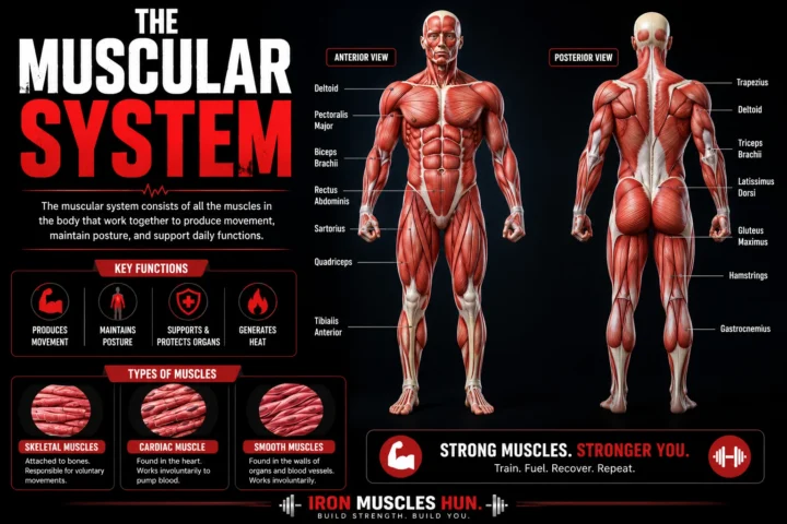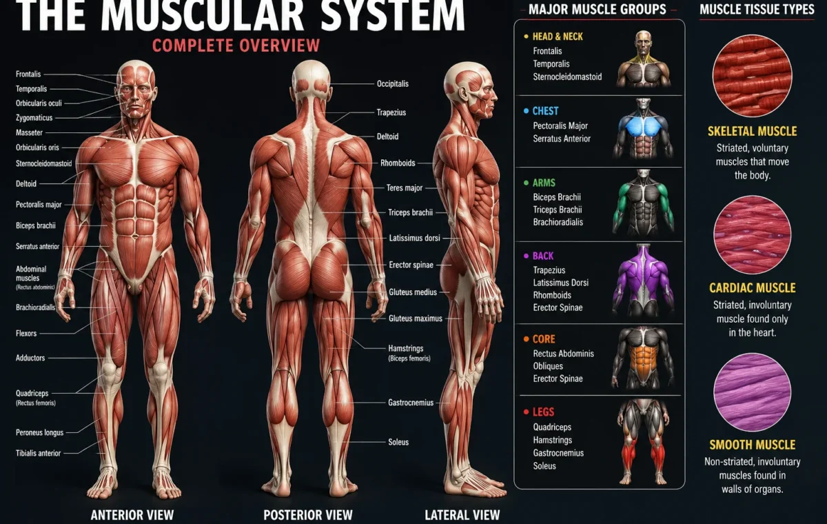
Photos of the muscular system help you understand how muscles are arranged throughout the human body and how they create movement. A good muscular system image usually shows major muscle groups such as the chest, shoulders, arms, abdomen, back, and legs with clear labels.
- Quick Answer: Photos of the Muscular System Explained
- 1. Front View Muscular System Photos
- 2. Back View Muscular System Photos
- 3. Labeled Muscular System Diagrams
- Skeletal Muscle
- Cardiac Muscle
- Smooth Muscle
- Step 1: Start With Large Muscle Groups
- Step 2: Match Muscles With Movement
- Step 3: Learn Common Muscle Names
- Mistake 1: Thinking Bigger Muscles Always Mean More Strength
- Mistake 2: Ignoring Smaller Muscles
- Mistake 3: Confusing Muscle Location
- For Exercise Learning
- For Studying Anatomy
- For Fitness Planning
- What are photos of the muscular system?
- How many muscles are in the human body?
- What is the largest muscle shown in muscular system pictures?
- What is the difference between a muscle photo and a muscle diagram?
- Why are labeled muscle pictures useful for fitness?
- Can I learn all muscles from one muscular system image?
- Understanding Muscle Layers in Photos of the Muscular System
- Superficial Muscles
- Deep Muscles
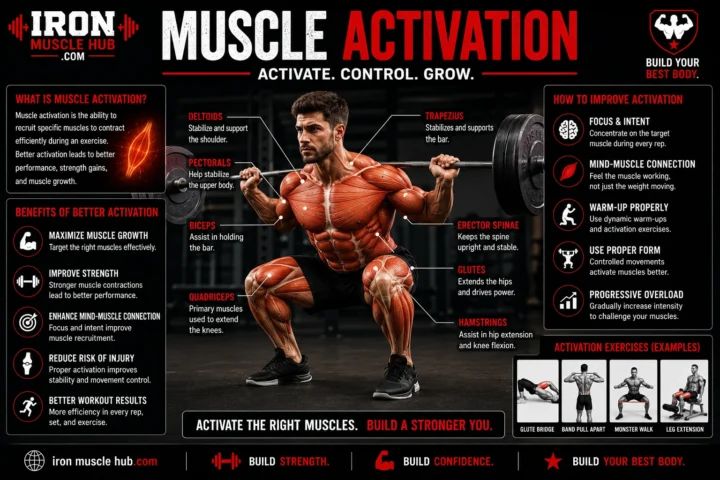
- Example: Push-Up Muscle Activation
- Example: Squat Muscle Activation
- 1. Only Training Visible Muscles
- 2. Thinking Every Muscle Has a Separate Exercise
- 3. Ignoring Muscle Balance
- Week 1: Upper Body Front
- Week 2: Upper Body Back
- Week 3: Lower Body
- Week 4: Review and Connect
Whether you are a student, fitness beginner, or simply curious about your body, looking at labeled muscle anatomy pictures makes it easier to learn where muscles are located and what they do. The muscular system includes different types of muscle tissue, including skeletal muscles that help you move, along with cardiac and smooth muscles that work automatically inside your body. For more insights read here :- how to build muscle and how to gain muscle fast .
Quick Answer: Photos of the Muscular System Explained
| View | What You Can Learn |
|---|---|
| Front muscular system photo | Chest, abs, biceps, quadriceps, and front shoulder muscles |
| Back muscular system photo | Trapezius, latissimus dorsi, glutes, and hamstrings |
| Labeled muscle diagram | Names and locations of major muscles |
| 3D muscle model | Muscle depth, layers, and movement patterns |
Key Takeaways:
- The human muscular system contains hundreds of muscles that support movement and body functions.
- Labeled photos make it easier to identify muscles by location and function.
- The largest visible muscle groups include the chest, back, shoulders, arms, and legs.
- Muscle anatomy images are useful for fitness training, education, and understanding injuries.
Why Photos of the Muscular System Are Helpful
A picture can make anatomy much easier to understand than a list of muscle names. When you see a labeled muscular system image, you can connect a muscle’s name with its actual location on the body.
For beginners, this is especially useful because many muscle names sound complicated at first. Seeing the muscle on a diagram helps you remember both its position and purpose.
For example:
- Biceps brachii is located on the front of the upper arm.
- Triceps brachii is located on the back of the upper arm.
- Deltoids cover the shoulder area.
- Quadriceps are the large muscles on the front of the thigh.
Types of Muscular System Images You Will Find
Different photos and diagrams show different levels of detail. Choosing the right type depends on what you want to learn.
1. Front View Muscular System Photos
Front-view images show muscles visible from the front of the body.
These pictures commonly highlight:
- Chest muscles (pectorals)
- Front shoulders (anterior deltoids)
- Biceps
- Abdominal muscles
- Quadriceps
- Shin muscles
Fitness beginners often use front-view images because many popular exercises target these visible muscle groups.
2. Back View Muscular System Photos
Back-view diagrams show muscles that are harder to see without anatomy knowledge.
Important muscles shown include:
| Muscle | Location | Main Function |
|---|---|---|
| Trapezius | Upper back and neck | Helps move and stabilize shoulders |
| Latissimus dorsi | Middle and lower back | Helps pull movements |
| Gluteus maximus | Buttocks | Supports hip movement |
| Hamstrings | Back of thighs | Helps bend the knee |
Back anatomy images are especially useful for people learning exercises such as rows, pull-ups, and deadlifts.
3. Labeled Muscular System Diagrams
A labeled muscular system diagram adds names to each muscle.
These diagrams are useful for:
- Biology students
- Fitness professionals
- Personal trainers
- Beginners learning exercise technique
A quality labeled diagram should show:
- Muscle names
- Body position
- Front and back views
- Major muscle groups
Educational anatomy collections, such as those from medical and academic resources, provide examples of labeled human muscle illustrations.
Major Muscle Groups Shown in Human Anatomy Photos
The human body contains many muscles, but beginners usually start by learning the major groups.
| Muscle Group | Main Muscles | Where They Are |
|---|---|---|
| Chest | Pectoralis major and minor | Front upper body |
| Back | Latissimus dorsi, trapezius | Rear upper body |
| Shoulders | Deltoids | Shoulder joints |
| Arms | Biceps and triceps | Upper arms |
| Core | Rectus abdominis and obliques | Abdomen |
| Legs | Quadriceps, hamstrings, calves | Lower body |
Understanding these groups helps you connect exercises with the muscles they train.
Three Main Types of Muscle Tissue
Many muscular system photos focus on skeletal muscles, but the body has three major muscle tissue types.
Skeletal Muscle
Skeletal muscles attach to bones and allow voluntary movement, such as walking, lifting, and running. They are the muscles people usually see in fitness anatomy pictures.
Examples:
- Biceps
- Quadriceps
- Chest muscles
- Calf muscles
Cardiac Muscle
Cardiac muscle is found only in the heart. It contracts automatically to help pump blood throughout the body.
Smooth Muscle
Smooth muscle works inside organs and blood vessels. Unlike skeletal muscle, it is controlled without conscious effort.
How to Read a Muscular System Picture
A beginner can understand most muscle diagrams by following a few simple steps.
Step 1: Start With Large Muscle Groups
Learn the biggest areas first:
- Chest
- Back
- Shoulders
- Arms
- Legs
- Core
Large groups provide a foundation before learning smaller muscles.
Step 2: Match Muscles With Movement
Try connecting each muscle with common actions.
Examples:
- Chest muscles help with pushing movements.
- Back muscles help with pulling movements.
- Leg muscles help with walking, jumping, and squatting.
Step 3: Learn Common Muscle Names
Start with frequently used names:
- Biceps
- Triceps
- Deltoids
- Pectorals
- Abdominals
- Glutes
- Hamstrings
- Calves
Common Mistakes When Looking at Muscle Anatomy Images
Many beginners misunderstand anatomy pictures because they focus only on appearance.
Mistake 1: Thinking Bigger Muscles Always Mean More Strength
Muscle size and strength are related, but they are not exactly the same. Training experience, nervous system adaptation, technique, and other factors also affect strength.
Mistake 2: Ignoring Smaller Muscles
Smaller muscles help stabilize joints and support movement.
Examples include:
- Rotator cuff muscles
- Forearm muscles
- Deep core muscles
Mistake 3: Confusing Muscle Location
Some muscles overlap or sit underneath others.
A labeled image shows the approximate location, but real human anatomy has multiple layers.
Best Ways to Use Photos of the Muscular System for Learning
You can use muscle anatomy pictures in several practical ways:
For Exercise Learning
Look at the muscles used during an exercise before performing it.
Example:
A push-up mainly involves:
- Chest muscles
- Triceps
- Front shoulders
- Core stabilizers
For Studying Anatomy
Students can:
- Memorize muscle names
- Compare front and back views
- Practice labeling diagrams
For Fitness Planning
Understanding muscle locations can help you organize workouts by body region.
Example weekly split:
| Day | Focus |
|---|---|
| Monday | Chest and triceps |
| Tuesday | Back and biceps |
| Wednesday | Legs |
| Thursday | Shoulders and core |
Where to Find Copyright-Safe Muscular System Images
When using photos or diagrams online, always check usage rights.
Good options include:
- Public-domain anatomy illustrations
- Educational resources with permission
- Original diagrams created for your website
- Licensed medical illustrations
Avoid copying random images from search results because many anatomy images are protected by copyright.
For educational reference, resources such as Wikimedia Commons and academic anatomy collections provide examples of openly available or educational images.
Internal Link Ideas for Iron Muscle Hub
Suggested related articles:
- What Are Muscles? A Beginner’s Guide to Human Muscle Function
- Major Muscles of the Human Body and Their Functions
- How the Muscular System Works With Other Body Systems
Frequently Asked Questions
What are photos of the muscular system?
Photos of the muscular system are images or diagrams that show the muscles of the human body. They may include front views, back views, labeled illustrations, or 3D models to help people understand muscle locations and functions.
How many muscles are in the human body?
The human body has hundreds of muscles. Some anatomy references describe approximately 700 named skeletal muscles, although the exact count can vary depending on how muscles are classified.
What is the largest muscle shown in muscular system pictures?
The gluteus maximus is commonly recognized as the largest skeletal muscle by mass. It is located in the buttocks and plays an important role in hip movement.
What is the difference between a muscle photo and a muscle diagram?
A muscle photo shows a realistic view of anatomy, while a diagram simplifies structures using labels and colors. Diagrams are often easier for beginners because they clearly identify each muscle.
Why are labeled muscle pictures useful for fitness?
Labeled muscle pictures help people understand which muscles are involved during exercises. This knowledge can improve exercise awareness and make workout planning easier.
Can I learn all muscles from one muscular system image?
A single image can teach the major muscles, but learning every muscle requires studying multiple diagrams, layers, and anatomy resources.
Understanding Your Body Starts With Anatomy
Photos of the muscular system are one of the simplest ways to understand how the human body moves. A clear labeled image helps you identify major muscles, connect them with everyday movements, and build a stronger foundation for fitness or anatomy learning.
Start by learning the major muscle groups first, then gradually explore smaller muscles and deeper structures. With consistent study, even complex anatomy becomes easier to understand.
Understanding Muscle Layers in Photos of the Muscular System
When you look at photos of the muscular system, it is important to remember that the human body is not made of a single flat layer of muscles. Muscles exist at different depths, with some muscles located close to the skin and others positioned underneath larger muscle groups.
A basic anatomy image often shows the most visible muscles, but advanced diagrams may reveal deeper muscles that support posture, stability, and movement.
Superficial Muscles
Superficial muscles are the muscles closest to the skin. These are usually the muscles people recognize in fitness pictures.
Examples include:
- Pectoralis major (chest)
- Biceps brachii (front upper arm)
- Triceps brachii (back upper arm)
- Rectus abdominis (abs)
- Quadriceps (front thigh)
These muscles are often highlighted in beginner-friendly muscular system images.
Deep Muscles
Deep muscles sit underneath other muscles and play important roles in stability and movement.
Examples include:
- Rotator cuff muscles around the shoulder
- Deep spinal muscles
- Transverse abdominis in the core
A detailed muscular system diagram can help show how these hidden muscles support the larger visible muscles.
Front vs Back Photos of the Muscular System
Many beginners search for front and back muscular system photos because each view teaches different information.
| View | Main Muscles Visible | Why It Helps |
|---|---|---|
| Front view | Chest, shoulders, abs, biceps, quadriceps | Helps identify muscles used in pushing and front-body movements |
| Back view | Traps, lats, triceps, glutes, hamstrings | Shows pulling muscles and posture-supporting areas |
| Side view | Shoulder shape, arms, legs, posture muscles | Shows muscle depth and body alignment |
Using multiple views creates a more complete understanding of human anatomy.
Muscular System Photos for Fitness Beginners
Many people use muscle anatomy pictures when starting exercise because they want to understand what they are training.
A workout does not isolate only one muscle. Most movements involve several muscles working together.
Example: Push-Up Muscle Activation
A push-up mainly involves:
| Muscle | Role |
|---|---|
| Chest muscles | Main pushing force |
| Triceps | Straighten the elbow |
| Front shoulders | Assist the movement |
| Core muscles | Keep the body stable |
A muscular system image helps you visualize these connections.
Example: Squat Muscle Activation
Squats are often associated with legs, but several muscles contribute.
Main muscles involved:
- Quadriceps
- Gluteus maximus
- Hamstrings
- Calves
- Core muscles
Looking at a lower-body muscular system picture makes these relationships easier to understand.
Common Muscle Names You See in Anatomy Pictures
Beginners often see scientific names on labeled diagrams. Here are some common ones explained simply.
| Scientific Name | Common Name | Location |
|---|---|---|
| Pectoralis major | Chest muscle | Upper front chest |
| Deltoid | Shoulder muscle | Around shoulder joint |
| Biceps brachii | Biceps | Front upper arm |
| Triceps brachii | Triceps | Back upper arm |
| Latissimus dorsi | Lats | Upper and middle back |
| Rectus abdominis | Abs | Front stomach area |
| Gluteus maximus | Glutes | Buttocks |
| Gastrocnemius | Calf muscle | Back lower leg |
Learning these names makes it easier to understand exercise programs and fitness articles.
How Muscular System Images Help With Exercise Form
A major benefit of studying muscle pictures is improving your understanding of movement.
For example, if you know where the shoulder muscles are located, you can better understand exercises like:
- Shoulder presses
- Lateral raises
- Push-ups
If you understand back muscles, exercises like:
- Pull-ups
- Rows
- Lat pulldowns
become easier to visualize.
However, anatomy knowledge should support proper training technique, not replace professional guidance when dealing with injuries or pain.
Muscle Anatomy Mistakes Beginners Should Avoid
1. Only Training Visible Muscles
Some people focus only on muscles they can see in a mirror.
Common examples:
- Chest
- Arms
- Abs
A balanced approach also includes:
- Back muscles
- Leg muscles
- Supporting muscles
A complete muscular system photo reminds you that the body works as one connected system.
2. Thinking Every Muscle Has a Separate Exercise
Many exercises train multiple muscles together.
For example:
A bench press does not only work the chest. It also involves the shoulders and triceps.
A deadlift does not only work the back. It also uses muscles in the legs, hips, and core.
3. Ignoring Muscle Balance
Balanced muscle development supports movement quality.
Examples:
- Strong chest muscles with weak back muscles may affect posture.
- Strong front thigh muscles with weak supporting muscles may create imbalance.
Muscle diagrams can help beginners remember to train different body regions.
How to Memorize the Muscular System Faster
Learning hundreds of muscles at once can feel overwhelming. A better approach is to study in groups.
Week 1: Upper Body Front
Learn:
- Chest
- Shoulders
- Biceps
- Abs
Week 2: Upper Body Back
Learn:
- Traps
- Lats
- Triceps
- Lower back muscles
Week 3: Lower Body
Learn:
- Glutes
- Quadriceps
- Hamstrings
- Calves
Week 4: Review and Connect
Practice:
- Naming muscles from pictures
- Matching muscles with exercises
- Understanding basic functions
Small steps make anatomy easier to remember.
How Artists and Trainers Create Muscle System Images
Professional anatomy illustrations are usually created using medical references, digital modeling, and careful study of human structures.
A quality muscular system illustration should:
- Show accurate muscle placement
- Use clear labels
- Avoid misleading proportions
- Separate major muscle groups clearly
For fitness websites, original or properly licensed images are important because copying protected anatomy artwork may violate copyright rules.
Simple Guide: Choosing the Right Muscular System Picture
| Your Goal | Best Image Type |
|---|---|
| Learning basic muscles | Simple labeled diagram |
| Studying anatomy | Detailed medical illustration |
| Planning workouts | Muscle activation images |
| Teaching beginners | Clear front/back comparison |
| Understanding movement | 3D anatomy model |
Final Thoughts on Photos of the Muscular System
Photos of the muscular system are powerful learning tools because they turn complicated anatomy into something visual and understandable. A labeled image can help beginners recognize major muscles, understand movement, and create a stronger foundation for fitness education.
Start with the large muscle groups, learn their functions, and slowly add more detail. You do not need to memorize every muscle immediately. Understanding the basics first will make advanced anatomy much easier.