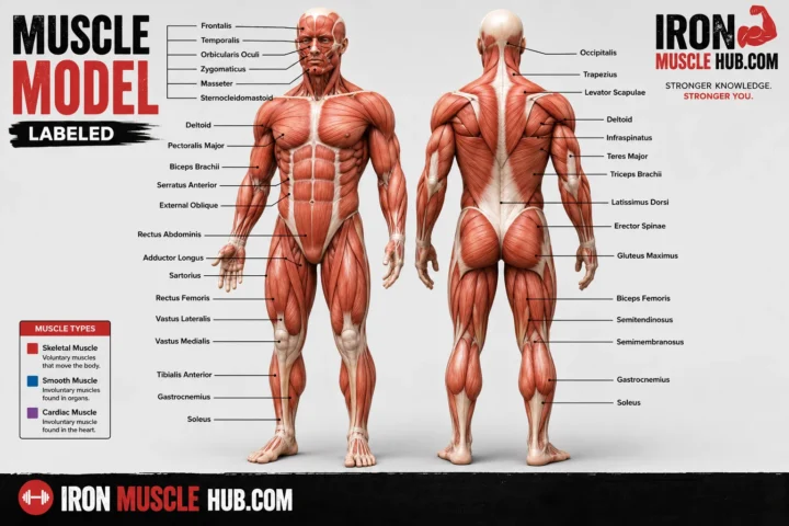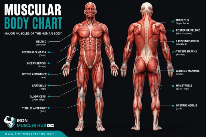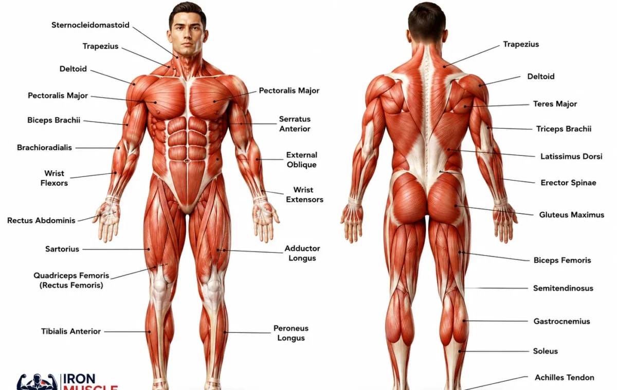
A muscle man labeled diagram is a visual chart that identifies the major muscles of the human body, usually from the front, back, or both views. It can help beginners quickly see where muscles such as the biceps, triceps, pectorals, quadriceps, hamstrings, and gastrocnemius are located.
- Muscle Man Labeled: Quick Reference
- What Does a Muscle Man Labeled Diagram Show?
- Front View: Major Muscles to Learn
- 1. Deltoids
- 2. Pectoralis Major
- 3. Biceps Brachii
- 4. Rectus Abdominis
- 5. External Obliques
- 6. Quadriceps
- Back View: Major Muscles to Learn
- 1. Trapezius
- 2. Latissimus Dorsi
- 3. Triceps Brachii
- 4. Gluteus Maximus
- 5. Hamstrings
- 6. Gastrocnemius
- How to Read a Labeled Muscle Diagram
- Muscle Man Labeled: Muscle Groups vs. Individual Muscles
- How Muscles Actually Move the Body
- Three Types of Muscle Tissue
- A Better Way to Memorize Muscle Anatomy
- Upper body
- Core
- Lower body
- Common Mistakes With Labeled Muscle Charts
- Where to Find a Reliable Labeled Muscle Diagram
- Internal Link Ideas
- Frequently Asked Questions
- What is a muscle man labeled diagram?
- What are the main muscles to learn first?
- What is the difference between biceps and triceps?
- Are quadriceps one muscle or four muscles?
- What is the largest muscle shown on a human muscle chart?
- Why are front and back muscle diagrams different?
- A Simple Starting Point for Learning Muscle Anatomy
- Muscle Man Labeled: A Closer Look at the Major Muscle Groups
- Upper Body Muscles
- Chest and Abdominal Muscles
- Rectus Abdominis
- External Oblique
- Serratus Anterior
- Back Muscles
- Trapezius
- Latissimus Dorsi
- Erector Spinae
- Hip and Thigh Muscles
- Gluteus Maximus
- Quadriceps
- Hamstrings
- Lower-Leg Muscles
- Gastrocnemius
- Soleus
- Tibialis Anterior
- How to Study a Muscle Man Labeled Diagram Efficiently
- Use the five-region method
- Add movement to every label
- Compare similar muscles
- A Beginner's Muscle Identification Test
- Superficial vs. Deep Muscles
- Why Muscle Diagrams Use Different Names
- Practical Study Routine
- What a Good Labeled Muscle Chart Should Include
- Key Takeaway
- Muscle Man Labeled: Understanding Muscle Names, Locations, and Functions
- How Anatomical Muscle Names Help You
- Common Terms on Muscle Charts
- Major Muscles of the Head and Neck
- Sternocleidomastoid
- Masseter
- Shoulder Muscles
- Forearm Muscles
- Hand Muscles
- Pelvic and Hip Muscles
- The Four Muscles of the Quadriceps
- Rectus Femoris
- Vastus Lateralis
- Vastus Medialis
- Vastus Intermedius
- The Main Hamstring Muscles
- Understanding Muscle Attachments
- Why One Muscle Can Have Several Actions
- Agonists and Antagonists
- Why Muscle Size on a Diagram Can Be Misleading
- Anterior vs. Posterior: A Quick Memory Trick
- Muscle Man Labeled Study Table
- A Five-Minute Muscle Diagram Exercise
- What to Learn After the Major Muscles
- Final Study Checklist
- The Best Way to Use a Muscle Man Labeled Chart
- Frequently Asked Questions
- Keep Building Your Anatomy Knowledge
- Final Thoughts
If you’re studying anatomy, learning exercise terminology, or simply trying to understand a human muscle chart, labels make the subject much easier. You don’t need to memorize hundreds of anatomical names at once. Start with the large, easy-to-identify muscles and learn what each one generally does. For more insights read here :- how to build muscle quickly for skinny guys and lean build .
Quick answer: A labeled muscle man diagram typically shows major superficial skeletal muscles on the front and back of the body. The most useful beginner labels include the deltoids, pectorals, biceps, triceps, abdominals, quadriceps, hamstrings, gluteus maximus, trapezius, latissimus dorsi, and calf muscles.
Muscle Man Labeled: Quick Reference
| Body area | Major muscle | Basic role |
|---|---|---|
| Neck | Sternocleidomastoid | Helps move and turn the head |
| Shoulders | Deltoid | Helps raise and move the arm |
| Chest | Pectoralis major | Helps move the upper arm |
| Upper back | Trapezius | Helps move and stabilize the shoulder area |
| Upper arm, front | Biceps brachii | Helps bend the elbow |
| Upper arm, back | Triceps brachii | Helps straighten the elbow |
| Abdomen | Rectus abdominis | Helps flex and stabilize the trunk |
| Side of abdomen | External oblique | Helps rotate and bend the trunk |
| Back | Latissimus dorsi | Helps move the upper arm |
| Buttocks | Gluteus maximus | Helps extend the hip |
| Front thigh | Quadriceps | Helps straighten the knee |
| Back thigh | Hamstrings | Help bend the knee and extend the hip |
| Calf | Gastrocnemius | Helps point the foot downward |
These are simplified descriptions for beginners. Individual muscles can have several actions and often work together rather than operating alone.
What Does a Muscle Man Labeled Diagram Show?
A labeled muscle diagram identifies muscles by name and places those names next to the corresponding structures. A detailed chart may show dozens of individual muscles, while a beginner chart usually concentrates on the larger muscles that are easiest to recognize.
For example, the front view commonly identifies the pectoralis major, deltoid, biceps brachii, rectus abdominis, external oblique, sartorius, and quadriceps. The back view may identify the trapezius, triceps brachii, latissimus dorsi, gluteus maximus, hamstrings, and gastrocnemius.
This front-and-back approach is useful because some muscles are much easier to see from one direction than another.
Front View: Major Muscles to Learn
Start at the top of the body and work downward. This makes a complicated diagram easier to remember.
1. Deltoids
The deltoids form the rounded muscle mass around the shoulders. They help move the upper arm in several directions.
On a labeled muscle man diagram, look for them on either side of the upper torso, directly below the shoulder area.
2. Pectoralis Major
The pectoralis major is the large superficial muscle covering much of the front of the chest.
It contributes to movements of the upper arm and is one of the most recognizable muscles on a front-view anatomy chart.
3. Biceps Brachii
The biceps brachii sits on the front of the upper arm.
One of its best-known actions is helping flex the elbow. If you bend your elbow and bring your forearm toward your upper arm, the biceps is involved in that movement.
4. Rectus Abdominis
The rectus abdominis is located along the front of the abdomen.
It’s commonly called the “abs.” Anatomy diagrams often divide the visible abdominal area into several labeled regions, but the rectus abdominis is one muscle running along the front of the trunk.
5. External Obliques
The external obliques are positioned on the sides of the abdomen.
They contribute to trunk rotation and side-bending movements. On a front-facing diagram, they sit to either side of the rectus abdominis.
6. Quadriceps
The quadriceps are the major muscle group on the front of the thigh.
Rather than being a single muscle, the quadriceps group contains four muscles. They work together to extend the knee, making them important for movements such as standing, walking, jumping, and climbing stairs.
Back View: Major Muscles to Learn
The back view can look more complicated because several overlapping muscles are visible.
1. Trapezius
The trapezius is a large muscle across the upper back and neck region.
It contributes to movement and stabilization of the shoulder blades and helps position the neck and shoulders.
2. Latissimus Dorsi
The latissimus dorsi, often called the “lats,” covers a substantial portion of the middle and lower back.
It helps move the upper arm, including movements that bring the arm toward the body.
3. Triceps Brachii
The triceps brachii is located on the back of the upper arm.
Its primary action is opposite the elbow-bending action associated with the biceps: it helps extend the elbow.
That’s why a labeled anatomy chart often places the biceps and triceps on opposite sides of the upper arm.
4. Gluteus Maximus
The gluteus maximus is the large superficial muscle of the buttock region.
It plays an important role in hip movement and is involved in activities such as climbing, rising from a seated position, and running.
5. Hamstrings
The hamstrings are a group of muscles at the back of the thigh.
They help with knee flexion and hip extension. A detailed muscle diagram may label individual hamstring muscles rather than simply using the group name.
6. Gastrocnemius
The gastrocnemius is the prominent superficial muscle of the calf.
It contributes to plantar flexion, the movement involved in pressing the foot downward. A labeled back-view diagram makes its location particularly easy to recognize.
How to Read a Labeled Muscle Diagram
If you’re looking at a detailed anatomy chart and the names seem overwhelming, use this simple method.
Step 1: Identify the view.
Determine whether you’re looking at the anterior, or front, view or the posterior, or back, view.
Step 2: Find the large landmarks.
Locate the head, neck, shoulders, chest, abdomen, hips, thighs, and lower legs.
Step 3: Learn the biggest muscles first.
Start with deltoids, pectorals, biceps, triceps, abdominals, quadriceps, hamstrings, gluteus maximus, and calf muscles.
Step 4: Connect each name with a movement.
Don’t memorize names alone. Associate each muscle with a simple action.
Step 5: Test yourself without looking.
Cover the labels and try to identify each muscle. Then check your answers.
This approach turns a diagram from a picture you recognize into a study tool you can actually use.
Muscle Man Labeled: Muscle Groups vs. Individual Muscles
One common beginner mistake is treating every label as though it represents one muscle.
Some terms describe groups of muscles.
For example:
- Quadriceps refers to a group of four muscles at the front of the thigh.
- Hamstrings refers to a group of muscles at the back of the thigh.
- Abdominals can refer broadly to muscles of the abdominal region.
- Calf muscles can refer to more than one muscle in the lower leg.
A detailed anatomy textbook may therefore use specific names such as rectus femoris, vastus lateralis, semitendinosus, or biceps femoris instead of a broad group name.
Understanding this distinction makes more advanced muscle charts much easier to follow.
How Muscles Actually Move the Body
Skeletal muscles don’t push bones into position. They produce tension by contracting and pulling through their attachments.
In many cases, the force created by a muscle contraction is transferred through a tendon to a bone. The bones and joints then work as a mechanical system that allows movement.
Muscles also commonly work in coordinated pairs or groups. For example, the biceps and triceps have opposing actions at the elbow: the biceps helps flex the elbow, while the triceps helps extend it.
This is why a labeled diagram is more useful when you study both location and function rather than memorizing names alone.
Three Types of Muscle Tissue
A human muscle chart normally focuses on skeletal muscle, but the body actually contains three major types of muscle tissue.
| Muscle type | Where it’s found | Main characteristic |
| Skeletal muscle | Attached to bones and other skeletal structures | Produces voluntary movement |
| Cardiac muscle | Heart | Contracts to pump blood |
| Smooth muscle | Walls of organs and blood vessels | Produces involuntary movement |
Skeletal muscle is the type most commonly shown in a full-body “muscle man labeled” illustration. Cardiac and smooth muscle aren’t normally displayed as large surface muscles on these charts.
A Better Way to Memorize Muscle Anatomy
Instead of trying to memorize an entire chart in one sitting, break it into regions.
Upper body
Learn:
- Deltoid
- Pectoralis major
- Biceps brachii
- Triceps brachii
- Trapezius
- Latissimus dorsi
Core
Learn:
- Rectus abdominis
- External obliques
- Serratus anterior
Lower body
Learn:
- Gluteus maximus
- Quadriceps
- Hamstrings
- Gastrocnemius
- Tibialis anterior
Once those names become familiar, you can add smaller muscles from a more detailed chart.
A useful study trick is to look at the blank version of a muscle diagram first. Try labeling it from memory, then compare your answers with the completed chart. This gives you active practice instead of simply rereading the same labels.
Common Mistakes With Labeled Muscle Charts
Mistake 1: Confusing biceps and triceps.
The biceps is on the front of the upper arm; the triceps is on the back.
Mistake 2: Treating quadriceps as one muscle.
The quadriceps is a four-muscle group.
Mistake 3: Assuming every visible muscle is superficial.
Anatomy diagrams can include deeper structures that aren’t visible on the body’s surface.
Mistake 4: Memorizing names without learning location.
Knowing that the latissimus dorsi is a muscle isn’t enough. You should also know that it’s located across much of the back.
Mistake 5: Assuming a diagram is a complete map of every muscle.
Most educational charts simplify anatomy. A detailed anatomy reference can contain many more structures than a beginner chart. OpenStax, for example, presents both superficial and deeper muscles in its full-body illustrations.
Where to Find a Reliable Labeled Muscle Diagram
For study purposes, look for anatomy diagrams from educational or academic sources rather than relying on an unattributed image.
The OpenStax Anatomy and Physiology textbook provides an anterior-and-posterior muscle figure with English labels and extensive supporting anatomy material. Its educational content is available under a Creative Commons license, subject to the applicable attribution requirements.
OpenStax Anatomy and Physiology 2e
Another useful reference is AnatomyTOOL’s presentation of the OpenStax labeled muscle figure, which identifies numerous superficial and deeper structures and records the applicable Creative Commons Attribution licensing information.
For a printable classroom-style reference, Kuraplan also provides a labeled muscular-system illustration under a CC BY-NC 4.0 license, meaning its permitted use is different from OpenStax’s CC BY material. Check the license before using any image commercially.
Internal Link Ideas
For a broader anatomy or education section, consider linking this article to:
- Human Muscle Chart: Major Muscles Explained
- Muscle Diagram Labeled: Front and Back Views
- Skeletal Muscle vs. Smooth Muscle vs. Cardiac Muscle
These related pages can help readers move from basic muscle identification to more detailed anatomy concepts.
Frequently Asked Questions
What is a muscle man labeled diagram?
A muscle man labeled diagram is an illustration of the human body with names pointing to specific muscles. Most beginner diagrams focus on major skeletal muscles such as the deltoids, pectorals, biceps, triceps, abdominals, quadriceps, hamstrings, gluteus maximus, and calf muscles.
What are the main muscles to learn first?
Beginners can start with the deltoids, pectoralis major, biceps, triceps, trapezius, latissimus dorsi, rectus abdominis, gluteus maximus, quadriceps, hamstrings, and gastrocnemius. Learning their locations and basic actions provides a useful foundation before moving to smaller or deeper muscles.
What is the difference between biceps and triceps?
The biceps brachii is on the front of the upper arm and helps flex the elbow. The triceps brachii is on the back of the upper arm and helps extend the elbow. They therefore have opposing primary actions at the elbow and are commonly shown together on muscle anatomy charts.
Are quadriceps one muscle or four muscles?
The quadriceps is a group of four muscles located at the front of the thigh. A detailed anatomy chart can identify each individual muscle, while a simpler educational diagram may label the entire group as “quadriceps.”
What is the largest muscle shown on a human muscle chart?
The gluteus maximus is generally identified as the largest skeletal muscle by mass. However, comparisons can depend on how muscles are defined and measured, so a basic anatomy chart should be treated as an educational overview rather than a precise ranking of every muscle.
Why are front and back muscle diagrams different?
Many muscles are positioned or most clearly visible from one side of the body. The front view highlights structures such as the pectorals, biceps, abdominals, and quadriceps, while the back view makes muscles such as the trapezius, latissimus dorsi, triceps, gluteus maximus, hamstrings, and gastrocnemius easier to identify.
A Simple Starting Point for Learning Muscle Anatomy
A muscle man labeled chart becomes much less intimidating once you stop trying to memorize everything at once. Learn the major muscles by body region, connect each name to its location, and then add a simple movement or function to your memory.
For beginners, a front-and-back diagram is usually the best starting point because it gives you a clear visual map of the major skeletal muscles. Once those landmarks are familiar, you can move on to individual muscles, deeper anatomy, origins and insertions, and the mechanics of movement.
Use a reputable educational reference, practice with unlabeled diagrams, and check unfamiliar terms rather than guessing. That approach will give you a much stronger foundation than memorizing a long list of names.
Muscle Man Labeled: A Closer Look at the Major Muscle Groups
If you’re using a muscle man labeled chart for study, the next step is to move beyond simply recognizing names. A useful anatomy reference should help you understand where each muscle sits, what movement it supports, and how it relates to nearby structures.
You don’t need to memorize every muscle in the body in one session. Focus first on the major regions, then gradually add smaller muscles as your familiarity improves.
Upper Body Muscles
The upper body contains several large muscle groups that appear frequently on anatomy charts.
Deltoid
The deltoid covers the shoulder and gives the shoulder much of its rounded appearance. It helps move the arm at the shoulder joint.
On a front-view diagram, look for the deltoid immediately below and around the shoulder.
Pectoralis Major
The pectoralis major is the large superficial muscle across the front of the chest. It contributes to movement of the upper arm.
A labeled front-view chart usually makes this muscle easy to identify because of its broad position across the chest.
Biceps Brachii
The biceps brachii is positioned on the front of the upper arm. It helps flex the elbow and also contributes to movement at the shoulder.
A simple memory aid is:
Biceps = front of upper arm.
Triceps Brachii
The triceps brachii occupies the back of the upper arm. Its main action at the elbow is extension.
A good way to study these two muscles is to compare them directly rather than learning each one separately.
| Muscle | Location | Main action |
|---|---|---|
| Biceps brachii | Front of upper arm | Elbow flexion |
| Triceps brachii | Back of upper arm | Elbow extension |
This simple comparison is one of the easiest ways to make a labeled muscle diagram easier to remember.
Chest and Abdominal Muscles
A front-facing muscle chart normally shows several important muscles around the torso.
Rectus Abdominis
The rectus abdominis runs vertically along the front of the abdomen. It contributes to flexion of the trunk.
The visible horizontal divisions often associated with the “six-pack” appearance don’t mean that each section is a separate muscle.
External Oblique
The external oblique sits along the sides of the abdominal region.
It contributes to trunk movement, including rotation and lateral flexion. On a labeled diagram, the fibers and location help distinguish the external obliques from the central rectus abdominis.
Serratus Anterior
The serratus anterior is located along the side of the chest near the ribs. It has an important relationship with the shoulder blade and contributes to its movement and stabilization.
Because it sits partly underneath other structures, a simplified muscle man chart may not show it clearly.
Back Muscles
The back contains some of the largest and most recognizable muscles on a posterior anatomy diagram.
Trapezius
The trapezius extends across the upper back and neck region.
It helps with movements and stabilization of the scapula. Depending on the diagram, the muscle may appear as a large diamond-like structure across the upper back.
Latissimus Dorsi
The latissimus dorsi is a broad muscle covering much of the lower and middle back.
It’s commonly called the “lat.” It helps move the upper arm and is particularly easy to identify on a posterior muscle diagram because of its broad shape.
Erector Spinae
The erector spinae refers to a group of muscles running along the back of the spine.
These muscles contribute to maintaining posture and extending or controlling movement of the vertebral column.
A beginner chart may simplify this area, while a more detailed anatomy diagram can separate the individual muscles within the group.
Hip and Thigh Muscles
The lower body contains several major muscle groups that are important for walking, running, standing and other movements.
Gluteus Maximus
The gluteus maximus is the large superficial muscle of the buttock.
It contributes strongly to hip extension. This makes it important for movements such as standing from a seated position, climbing stairs and running.
Quadriceps
The quadriceps femoris is a group of four muscles on the front of the thigh.
The four muscles are:
- Rectus femoris
- Vastus lateralis
- Vastus medialis
- Vastus intermedius
Together, they play a major role in extending the knee.
This is an important distinction for anyone studying a detailed muscle man labeled diagram: “quadriceps” is a group name rather than the name of one individual muscle.
Hamstrings
The hamstrings are a group of muscles positioned on the back of the thigh.
They help bend the knee and contribute to hip extension.
A basic chart may simply label the area “hamstrings,” while a more advanced anatomy chart may identify individual muscles such as the biceps femoris, semitendinosus and semimembranosus.
Lower-Leg Muscles
The lower leg contains muscles involved in movements of the ankle and foot.
Gastrocnemius
The gastrocnemius is one of the most prominent superficial calf muscles.
It crosses the knee and ankle joints and contributes to plantar flexion of the foot.
Soleus
The soleus is another important calf muscle. It lies deep to the gastrocnemius.
Because it is underneath the gastrocnemius, a simple surface muscle chart may not display it as clearly as a detailed anatomy illustration.
Tibialis Anterior
The tibialis anterior is located toward the front of the lower leg.
It contributes to dorsiflexion of the foot, helping bring the foot upward at the ankle.
How to Study a Muscle Man Labeled Diagram Efficiently
A common problem with anatomy charts is trying to memorize every label immediately. A better strategy is to work from large regions toward smaller structures.
Use the five-region method
Divide the body into:
- Head and neck
- Upper limbs
- Chest and back
- Abdomen and pelvis
- Legs and feet
Learn a small group of muscles from each region before moving deeper.
Add movement to every label
Instead of remembering:
“Triceps = back of arm.”
Try remembering:
“Triceps = back of arm + helps straighten the elbow.”
The second version gives your brain both a location and a function.
Compare similar muscles
Some muscles are easier to remember as pairs or groups.
For example:
- Biceps vs. triceps
- Quadriceps vs. hamstrings
- Pectoralis major vs. latissimus dorsi
- Tibialis anterior vs. gastrocnemius
Comparing muscles helps prevent common labeling mistakes.
A Beginner’s Muscle Identification Test
Once you’ve studied a labeled diagram, test yourself without looking at the answers.
1. Which muscle is on the front of the upper arm?
Answer: Biceps brachii.
2. Which muscle is on the back of the upper arm?
Answer: Triceps brachii.
3. What group occupies the front of the thigh?
Answer: Quadriceps.
4. What group is located at the back of the thigh?
Answer: Hamstrings.
5. Which large muscle covers much of the upper and middle back?
Answer: Latissimus dorsi, although the trapezius also occupies a substantial portion of the upper back.
6. Which major muscle is located across the front of the chest?
Answer: Pectoralis major.
If you can answer these without looking at a chart, you’ve already built a useful foundation.
Superficial vs. Deep Muscles
One detail that can make anatomy diagrams confusing is the difference between superficial and deep muscles.
A superficial muscle is closer to the body’s surface. A deep muscle lies underneath another structure.
For example, the gastrocnemius is superficial to the soleus in the calf. A detailed diagram may remove or partially expose superficial muscles to show structures underneath.
This explains why two legitimate muscle charts can look different even though both are anatomically accurate.
Why Muscle Diagrams Use Different Names
You may encounter slightly different labeling styles depending on the textbook or educational source.
For example, one chart may use:
“quadriceps”
while another uses:
“quadriceps femoris.”
A more detailed chart may skip the group label entirely and identify individual muscles.
The important thing is to understand the anatomical hierarchy:
Muscle group → individual muscle → specific location and action.
Learning that hierarchy makes it much easier to progress from a beginner muscle chart to more advanced anatomy resources.
Practical Study Routine
If you’re preparing for an anatomy class, quiz or personal study session, try this short routine:
Day 1: Learn the major front-view muscles.
Day 2: Learn the major back-view muscles.
Day 3: Review the names without looking at the labels.
Day 4: Add basic functions.
Day 5: Practice with a blank diagram.
Day 6: Focus on muscles you repeatedly confuse.
Day 7: Take another blank-diagram test.
The goal isn’t to stare at the same picture repeatedly. Retrieval practice, where you try to recall the information before checking the answer, can make study sessions more active and useful.
What a Good Labeled Muscle Chart Should Include
Before using an anatomy diagram as a study reference, check whether it gives you enough information.
A useful beginner chart should ideally have:
- Clear front and back views
- Readable muscle names
- Consistent labeling
- Major superficial muscles
- Basic anatomical terminology
- A reputable educational source
- Licensing information if you plan to reuse the image
If you’re publishing the image on a website, don’t assume that finding it through Google makes it free to use. Check the original source and its stated license.
For educational reference, OpenStax provides anatomy material under Creative Commons licensing, with specific attribution requirements. Always check the current license information before republishing an illustration.
Key Takeaway
A muscle man labeled diagram is most useful when you treat it as a map rather than a list of words.
Start with the major muscles:
deltoids → pectorals → biceps → triceps → abdominals → trapezius → latissimus dorsi → glutes → quadriceps → hamstrings → calves.
Then add individual muscles and deeper structures as your knowledge improves.
You don’t need to know every anatomical term on your first attempt. Learn the location, name and basic function of the major muscles first. Once those landmarks become familiar, detailed anatomy becomes much easier to understand.
Muscle Man Labeled: Understanding Muscle Names, Locations, and Functions
A muscle man labeled diagram becomes much easier to study once you understand the naming system behind the labels. Instead of memorizing unfamiliar Latin terms as isolated words, connect each muscle with three things: where it is, what it does, and what nearby muscles look like.
This approach is especially helpful if you’re moving from a basic muscle chart to a more detailed anatomy reference.
How Anatomical Muscle Names Help You
Muscle names often contain clues about a muscle’s location, shape, size, direction, or attachment.
For example, rectus generally relates to a straight direction, while lateralis indicates a position toward the side. Terms such as major, minor, maximus, and minimus can indicate relative size.
You don’t have to become fluent in anatomical Latin. Learning a handful of recurring terms can make unfamiliar labels much easier to decode.
Common Terms on Muscle Charts
| Term | General meaning | Example |
|---|---|---|
| Major | Larger | Pectoralis major |
| Minor | Smaller | Pectoralis minor |
| Maximus | Largest | Gluteus maximus |
| Minimus | Smallest | Gluteus minimus |
| Medialis | Toward the middle | Vastus medialis |
| Lateralis | Toward the side | Vastus lateralis |
| Anterior | Toward the front | Tibialis anterior |
| Posterior | Toward the back | Anatomical direction |
| Rectus | Straight | Rectus abdominis |
| Oblique | Slanted | External oblique |
These terms don’t tell you everything about a muscle, but they can provide useful clues when you’re reading a detailed anatomy diagram.
Major Muscles of the Head and Neck
Full-body muscle charts often give less attention to the face and neck than to the trunk and limbs, but these regions contain many important muscles.
Sternocleidomastoid
The sternocleidomastoid is a prominent muscle on each side of the neck.
Its name can look intimidating, but it actually contains clues about its attachments: the sternum, clavicle and mastoid region.
It contributes to movements of the head and neck.
Masseter
The masseter is one of the major muscles involved in chewing.
It sits along the side of the jaw and can become noticeable when you clench your teeth.
A detailed facial-muscle diagram may show several additional muscles involved in facial expression and chewing.
Shoulder Muscles
The shoulder region can be confusing because several muscles contribute to movement and stability.
The deltoid is the large superficial muscle covering the shoulder.
Underneath and around the shoulder are smaller muscles that are important in shoulder-joint movement. These include muscles associated with the rotator cuff.
The four rotator cuff muscles are:
- Supraspinatus
- Infraspinatus
- Teres minor
- Subscapularis
A beginner muscle man diagram may label only the deltoid, while an advanced anatomy chart may show the individual rotator cuff muscles.
Forearm Muscles
A basic human muscle chart may label the forearm as a broad region, but the forearm actually contains many muscles.
These muscles are often grouped according to their actions and anatomical compartments.
Some contribute to:
- Wrist flexion
- Wrist extension
- Finger movement
- Forearm rotation
This is one reason detailed anatomy charts can suddenly become much more complicated than beginner diagrams.
You don’t need to memorize every forearm muscle to understand a basic full-body chart. Learn the major landmarks first and add individual muscles later if your course or study goal requires them.
Hand Muscles
The hand contains numerous small muscles that allow precise movement.
A simplified muscle man labeled illustration usually doesn’t attempt to identify all of them.
Instead, more detailed diagrams may focus on muscles of the thumb, little finger and spaces between the hand bones.
If your goal is general anatomy knowledge, the hand can be left until you’ve mastered the larger muscles of the body.
Pelvic and Hip Muscles
The hip is another area where superficial and deeper muscles overlap.
The gluteus maximus is the major superficial muscle of the buttock.
Other gluteal muscles include:
- Gluteus medius
- Gluteus minimus
The gluteus medius and gluteus minimus lie deeper than the gluteus maximus and are important in hip movement and stabilization.
A detailed diagram may therefore show structures that aren’t obvious on a simple front-and-back muscle chart.
The Four Muscles of the Quadriceps
The quadriceps deserves extra attention because it’s frequently misunderstood.
The quadriceps femoris consists of four muscles:
Rectus Femoris
The rectus femoris runs down the central front of the thigh and crosses both the hip and knee joints.
Vastus Lateralis
The vastus lateralis is positioned toward the outer side of the thigh.
Vastus Medialis
The vastus medialis lies toward the inner side of the thigh.
Vastus Intermedius
The vastus intermedius lies deep to the rectus femoris.
Together, these muscles contribute to extension of the knee.
This is a good example of why a basic label such as “quadriceps” can eventually lead to several more detailed labels.
The Main Hamstring Muscles
The hamstrings are also a muscle group rather than one individual muscle.
The commonly identified hamstring muscles include:
- Biceps femoris
- Semitendinosus
- Semimembranosus
They occupy the posterior thigh and contribute to movements involving the hip and knee.
A beginner studying a muscle man labeled image can simply remember “hamstrings = back of thigh.” Once that location is familiar, the individual names become easier to learn.
Understanding Muscle Attachments
Advanced muscle charts may include terms such as origin and insertion.
In simplified educational terms:
- An origin is traditionally described as the less mobile attachment of a muscle.
- An insertion is traditionally described as the more mobile attachment.
These definitions are useful for learning anatomy, but actual movement can change depending on which body segment is stabilized.
For example, muscles don’t operate independently. Their actions depend on joint position, other muscles and which part of the body is fixed.
Why One Muscle Can Have Several Actions
You may notice that anatomy references sometimes give more than one function for the same muscle.
That’s normal.
A muscle can cross one or more joints and produce different movements depending on its attachments and the position of the body.
The biceps brachii, for example, contributes to elbow flexion and also participates in supination of the forearm.
So don’t assume that every muscle has exactly one job.
A better study format is:
Muscle → location → major actions → nearby structures.
Agonists and Antagonists
Muscles often work in coordinated groups.
An agonist, in a particular movement, is a muscle that contributes to producing that movement. An antagonist performs an opposing action.
The classic beginner example is the relationship between the biceps and triceps at the elbow.
When the elbow is flexed, the biceps is an important contributor to the movement. For elbow extension, the triceps is a major contributor.
The terminology becomes more nuanced in real movement because several muscles can contribute simultaneously.
Why Muscle Size on a Diagram Can Be Misleading
An illustration isn’t necessarily drawn to scale.
Anatomy figures are designed to show structures clearly. A muscle might be enlarged, separated from another structure, or repositioned slightly so the label can be read.
That’s why you shouldn’t estimate the exact size or depth of a muscle simply by measuring it on a textbook illustration.
The purpose of the diagram is identification and learning, not precise body measurement.
Anterior vs. Posterior: A Quick Memory Trick
Two terms appear repeatedly on anatomy charts:
Anterior = front
Posterior = back
So:
- Anterior thigh = front of thigh
- Posterior thigh = back of thigh
- Anterior lower leg = front of lower leg
- Posterior lower leg = back of lower leg
Once these two terms become familiar, many anatomy labels become easier to interpret.
Other useful directional terms include:
- Medial: closer to the body’s midline
- Lateral: farther from the body’s midline
- Superior: above
- Inferior: below
- Proximal: closer to the point of attachment or trunk
- Distal: farther away from the point of attachment or trunk
These terms are especially useful when reading professional anatomy references.
Muscle Man Labeled Study Table
Here’s a compact revision sheet you can use before testing yourself.
| Region | Muscle or group | Remember it as |
| Shoulder | Deltoid | Shoulder cap |
| Chest | Pectoralis major | Front chest |
| Upper arm | Biceps brachii | Front arm |
| Upper arm | Triceps brachii | Back arm |
| Upper back | Trapezius | Upper back |
| Back | Latissimus dorsi | Broad back |
| Abdomen | Rectus abdominis | Central abs |
| Abdomen | External oblique | Side abs |
| Buttocks | Gluteus maximus | Major glute muscle |
| Front thigh | Quadriceps | Four-muscle group |
| Back thigh | Hamstrings | Posterior thigh group |
| Calf | Gastrocnemius | Prominent calf |
| Front lower leg | Tibialis anterior | Front shin region |
Use the table to test yourself rather than simply rereading it.
A Five-Minute Muscle Diagram Exercise
You can turn almost any labeled anatomy chart into a quick study exercise.
Minute 1: Look at the full diagram and identify the front and back views.
Minute 2: Cover the labels and name five muscles you recognize.
Minute 3: Point to five muscles and describe their basic actions.
Minute 4: Write down the names you forgot.
Minute 5: Recheck the original chart and correct your answers.
Repeating this process is more useful than trying to memorize a huge list during one sitting.
What to Learn After the Major Muscles
Once you’re comfortable with the main labels, you can progress to more detailed anatomy.
A sensible sequence is:
- Major muscle locations
- Basic muscle actions
- Individual muscles within major groups
- Origins and insertions
- Joint actions
- Muscle compartments
- Superficial and deep relationships
- More detailed regional anatomy
This gives you a logical path from a simple full-body chart to more advanced anatomy.
Final Study Checklist
Before moving on from a beginner muscle chart, see if you can identify these without looking:
- Deltoid
- Pectoralis major
- Biceps brachii
- Triceps brachii
- Trapezius
- Latissimus dorsi
- Rectus abdominis
- External oblique
- Gluteus maximus
- Quadriceps
- Hamstrings
- Gastrocnemius
- Tibialis anterior
If several are still difficult, don’t rush into smaller muscles. Go back to the front-and-back chart and practice those major landmarks again.
The Best Way to Use a Muscle Man Labeled Chart
The most useful muscle diagram isn’t necessarily the one with the most labels. For a beginner, clarity matters more than cramming every anatomical structure into one picture.
Start with the major superficial muscles. Learn their positions from both the anterior and posterior views. Then connect each muscle with one or two important actions.
Once you can recognize those structures automatically, more detailed charts become much easier to read.
The key is to study progressively. A basic labeled figure gives you the map; more detailed anatomy resources fill in the smaller roads later.
Frequently Asked Questions
What does anterior mean on a muscle diagram?
Anterior means toward the front of the body. On a muscle chart, an anterior view generally shows the person from the front. This view makes muscles such as the pectoralis major, biceps brachii, rectus abdominis and quadriceps easier to identify.
What does posterior mean on a muscle diagram?
Posterior means toward the back of the body. A posterior muscle diagram therefore shows the body from behind. It is particularly useful for identifying structures such as the trapezius, latissimus dorsi, triceps brachii, gluteus maximus and hamstrings.
Why does a muscle chart show groups instead of individual muscles?
Some anatomical terms refer to groups of muscles rather than one structure. The quadriceps and hamstrings are common examples. Beginner diagrams use group labels to keep the illustration readable, while advanced charts can identify each individual muscle within the group.
How many muscles should a beginner memorize first?
There’s no universal number that every beginner must learn. A practical starting point is the major muscles of the shoulders, chest, arms, back, abdomen, hips and legs. Once those are familiar, you can add smaller muscles according to your course or personal study goals.
Are muscle diagrams drawn exactly like the human body?
Not necessarily. Educational illustrations are designed to make anatomical structures visible and distinguishable. Muscles can be separated, enlarged or displayed from a particular angle for clarity. Therefore, a diagram is best used for identification and relationships rather than exact measurements.
What’s the easiest way to learn a labeled muscle diagram?
Start with the front and back views separately. Learn a few major muscles at a time, associate each with its location and basic action, then practice using an unlabeled diagram. Repeated recall is usually more useful than repeatedly reading the labels.
Keep Building Your Anatomy Knowledge
A muscle man labeled diagram is a starting point, not the entire subject of human anatomy. Once the major landmarks are familiar, you can move comfortably into individual muscles, muscle groups, joint actions and deeper structures.
The most effective approach is simple: identify it, locate it, understand its basic action, then test yourself without the labels.
That method keeps anatomy manageable and gives you a foundation you can build on as the diagrams become more detailed.
Final Thoughts
A muscle man labeled diagram is one of the simplest ways to build a foundation in human muscular anatomy. Instead of trying to memorize every structure at once, start with the major muscles you can easily locate from the front and back of the body.
Focus first on the deltoids, pectorals, biceps, triceps, trapezius, latissimus dorsi, abdominal muscles, gluteal muscles, quadriceps, hamstrings, and calf muscles. For each one, learn its location and one or two basic functions.
As you become comfortable with those landmarks, move to individual muscles within larger groups and then to deeper structures. A blank diagram can be especially useful because it lets you test your memory instead of simply recognizing labels.
The biggest lesson is simple: don’t memorize muscle names in isolation. Connect every name with a location, a movement, and the surrounding anatomy. That makes a detailed muscle chart much easier to understand and gives you a stronger foundation for further anatomy study.