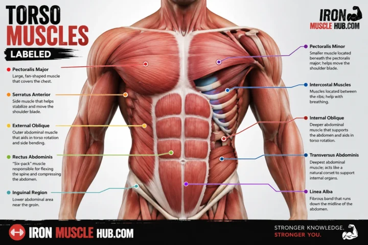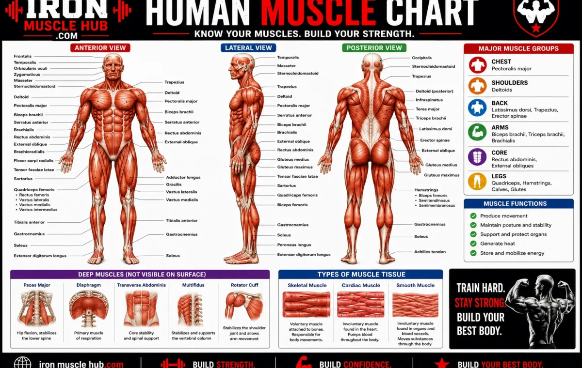
A human muscle chart is a visual guide that helps you identify the major muscles of the body, where they are located, and what movements they help produce. Most beginner muscle charts focus on skeletal muscles, the muscles that attach to bones and help you move.
- Human Muscle Chart: Quick Guide
- What Does a Human Muscle Chart Show?
- Why are there front and back views?
- Major Muscles of the Head and Neck
- Frontalis
- Masseter
- Temporalis
- Sternocleidomastoid
- Trapezius
- Major Chest and Abdominal Muscles
- Pectoralis Major
- Rectus Abdominis
- External Obliques
- Major Back Muscles
- Latissimus Dorsi
- Trapezius
- Erector Spinae
- Major Shoulder and Arm Muscles
- Deltoid
- Biceps Brachii
- Triceps Brachii
- Brachialis
- Major Hip and Leg Muscles
- Gluteus Maximus
- Quadriceps
- Hamstrings
- Gastrocnemius
- Soleus
- Tibialis Anterior
- The Three Types of Muscle Tissue
- How Skeletal Muscles Actually Move You
- A Simple Way to Learn a Muscle Chart
- Step 1: Learn the body regions
- Step 2: Learn the largest landmarks
- Step 3: Add the function
- Step 4: Test yourself without looking
- Common Mistakes When Reading a Human Muscle Chart
- Mistake 1: Assuming every chart is complete
- Mistake 2: Confusing a muscle group with one muscle
- Mistake 3: Learning only the front view
- Mistake 4: Assuming "muscle group" is a strict medical category
- A Better Way to Use a Muscle Chart for Exercise
- Where to Find Reliable Human Muscle Charts
- Internal Link Ideas
- Frequently Asked Questions
- What is a human muscle chart?
- What are the major muscles of the human body?
- How many muscles are in the human body?
- What are the three types of muscle?
- What is the difference between skeletal and smooth muscle?
- Should I learn the front or back muscle chart first?
- A Simple Muscle-Chart Study Strategy
- Key Muscle Groups to Learn Next
- Understanding the Rotator Cuff
- Understanding the Muscles of the Back
- Trapezius
- Latissimus Dorsi
- Rhomboids
- Erector Spinae
- Understanding the Quadriceps
- Why the Rectus Femoris Looks Different
- Understanding the Hamstrings
- The Gluteal Muscles
- Muscle Names Can Give You Clues
- Anatomical Directions You Should Know
- Flexion and Extension Explained Simply
- Flexion
- Extension
- Other Muscle Actions to Recognize
- Abduction
- Adduction
- Rotation
- Dorsiflexion
- Plantar Flexion
- Why One Muscle Can Have More Than One Function
- Agonists, Antagonists and Synergists
- Agonist
- Antagonist
- Synergist
- Why Charts Show Muscles in Different Colors
- Superficial vs Deep Muscles
- How to Memorize Muscle Names Faster
- Stage 1: Recognize
- Stage 2: Recall
- Stage 3: Explain
- A 7-Day Beginner Muscle Chart Study Plan
- The Best Test: Draw What You Remember
- Human Muscle Chart vs Muscle Diagram
- What a Beginner Should Memorize First
- Final Tip for Using a Human Muscle Chart
- Quick Muscle Anatomy Reference
- Head and Face Muscles
- Masseter
- Temporalis
- Orbicularis Oculi
- Orbicularis Oris
- Neck Muscles
- Sternocleidomastoid
- Trapezius
- Chest Muscles
- Pectoralis Major
- Pectoralis Minor
- Shoulder Muscles
- Deltoid
- Rotator Cuff
- Upper Arm Anatomy
- Biceps Brachii
- Triceps Brachii
- Brachialis
- Forearm Muscles
- Flexor Muscles
- Extensor Muscles
- Abdominal Muscles
- Rectus Abdominis
- External Oblique
- Internal Oblique
- Transversus Abdominis
- Back Muscle Anatomy
- Trapezius
- Rhomboid Major and Minor
- Latissimus Dorsi
- Erector Spinae
- Hip Muscles
- Gluteus Maximus
- Gluteus Medius
- Gluteus Minimus
- Iliopsoas
- Thigh Muscles
- Front of the Thigh
- Back of the Thigh
- Inner Thigh
- Lower Leg Muscles
- Gastrocnemius
- Soleus
- Tibialis Anterior
- Fibularis Muscles
- Why the Same Muscle Can Look Different on Different Charts
- How to Read a Detailed Muscle Chart
- 1. Identify the view
- 2. Find the skeleton landmarks
- 3. Identify the large muscles
- 4. Add smaller muscles
- 5. Check the labels
- Muscle Chart vs Skeleton Chart
- Why Muscle Attachments Matter
- A Practical Muscle Chart Quiz
- A Useful Memory Rule: Location Before Detail
- Human Muscle Chart: What to Learn First
- Front
- Back
- Frequently Asked Questions
- What is the easiest way to memorize a human muscle chart?
- What muscles should I learn first?
- What does anterior mean on a muscle chart?
- Why do some muscle charts show different muscles?
- Are the biceps and triceps opposites?
- How many muscles should a beginner memorize?
- Build Your Own Muscle Reference Sheet
- Final Thoughts: Make the Chart Work for You
You don’t need to memorize hundreds of muscle names at once. Start with the large, easy-to-identify muscles in the head, chest, back, arms, abdomen, hips and legs. Once those landmarks make sense, the smaller muscles become much easier to learn. For more insights read here :- how to get a lean body and lean exercises .
Quick answer: The major muscle groups commonly shown on a human muscle chart include the muscles of the head and neck, trunk, upper extremities and lower extremities.
Human Muscle Chart: Quick Guide
| Body area | Major muscles to know | Main role |
|---|---|---|
| Head and face | Frontalis, masseter, temporalis, orbicularis muscles | Facial movement and chewing |
| Neck | Sternocleidomastoid, trapezius | Head and neck movement |
| Chest | Pectoralis major | Moving the arm and shoulder |
| Abdomen | Rectus abdominis, external oblique | Trunk movement and stability |
| Back | Trapezius, latissimus dorsi, erector spinae | Shoulder and trunk movement |
| Shoulder | Deltoid, rotator cuff muscles | Moving and stabilizing the shoulder |
| Upper arm | Biceps brachii, triceps brachii, brachialis | Bending and straightening the elbow |
| Forearm | Flexor and extensor muscles | Wrist and hand movement |
| Glutes and hip | Gluteus maximus, medius and minimus | Hip movement and stability |
| Thigh | Quadriceps, hamstrings, adductors | Knee and hip movement |
| Lower leg | Gastrocnemius, soleus, tibialis anterior | Ankle and foot movement |
This is a learning chart, not a complete list of every muscle in the body. A full anatomy reference contains many more structures.
What Does a Human Muscle Chart Show?
A muscle chart normally identifies muscles by their anatomical location, name and sometimes their action.
For example, a chart may label the biceps brachii on the front of the upper arm and the triceps brachii on the back. It may also show the quadriceps at the front of the thigh and the hamstrings toward the back.
Anatomists don’t choose muscle names randomly. Names can describe a muscle’s location, shape, size, fiber direction, number of origins or action. Learning these naming patterns can make unfamiliar terms less intimidating.
Why are there front and back views?
A single front view can’t show every muscle clearly because many muscles lie underneath other muscles.
That’s why a useful human muscle chart often has:
- Anterior view: the front of the body
- Posterior view: the back of the body
- Sometimes a deep-muscle view: structures underneath superficial muscles
- Occasionally separate close-ups of the head, shoulder, arm, hand, hip or leg
OpenStax’s anatomy material, for example, uses anterior and posterior views to identify major superficial and deeper muscles.
Major Muscles of the Head and Neck
The head and neck contain muscles involved in facial expression, chewing and movement of the head and neck.
Some important names you’ll see on a chart are:
Frontalis
The frontalis is associated with movement of the forehead and is one of the muscles involved in facial expression.
Masseter
The masseter is one of the major muscles involved in chewing. It connects with the mandible and helps produce jaw movement.
Temporalis
The temporalis is another important chewing muscle located around the temporal region of the skull.
Sternocleidomastoid
This prominent neck muscle helps with movements of the head and neck. It’s often easy to spot on a labeled anatomy illustration.
Trapezius
The trapezius extends across the upper back and neck region. Although people often think of it as a back muscle, it also has an important relationship with shoulder and neck movement.
Major Chest and Abdominal Muscles
The chest and trunk contain several muscles that help move the upper body and maintain stability.
Pectoralis Major
The pectoralis major is the large muscle covering much of the front of the chest. It contributes to movements of the arm at the shoulder.
Rectus Abdominis
The rectus abdominis runs vertically along the front of the abdomen. It’s commonly shown on fitness-oriented muscle charts.
External Obliques
The external obliques are located along the sides of the abdomen. They contribute to movements and rotation of the trunk.
The abdominal muscles aren’t simply decorative structures. Skeletal muscles throughout the trunk also contribute to posture, movement and stability. Skeletal muscle contraction has roles that include movement, posture, joint stability and heat production.
Major Back Muscles
The back contains several large and important muscles, which is why posterior muscle charts can look particularly busy.
Latissimus Dorsi
The latissimus dorsi, often called the “lats,” is a large back muscle involved in movement of the upper limb.
Trapezius
The trapezius occupies a broad area around the upper back and neck and helps with movements involving the shoulder region.
Erector Spinae
The erector spinae refers to a group of muscles associated with the back and spinal posture. These muscles contribute to maintaining and moving the trunk.
A useful way to study the back is to first identify the large superficial muscles before trying to learn deeper structures.
Major Shoulder and Arm Muscles
The shoulder and arm are easier to understand if you separate them into the shoulder, upper arm and forearm.
Deltoid
The deltoid is the large, rounded muscle covering the shoulder. It contributes to movement of the arm.
Biceps Brachii
The biceps brachii is located on the front of the upper arm. It contributes to bending the elbow and other forearm movements.
Triceps Brachii
The triceps brachii is located on the back of the upper arm and is involved in straightening the elbow.
Brachialis
The brachialis lies deep to the biceps in the upper arm and contributes to elbow flexion.
The upper extremity contains muscles that move the shoulder, arm, forearm, wrist and hand. The National Cancer Institute’s anatomy training materials group these structures by their location and movement functions.
Major Hip and Leg Muscles
The lower body contains some of the largest muscle groups you’ll see on a human muscle chart.
Gluteus Maximus
The gluteus maximus is the largest of the three gluteal muscles by name convention, with the gluteus medius and gluteus minimus representing the other major gluteal muscles.
Quadriceps
The quadriceps femoris is a group of muscles at the front of the thigh. Collectively, the quadriceps help straighten the leg at the knee.
Hamstrings
The hamstrings are a group of muscles toward the back of the thigh. They work in opposition to the quadriceps during certain knee movements.
Gastrocnemius
The gastrocnemius is one of the prominent muscles of the calf.
Soleus
The soleus lies deeper in the calf and works with the gastrocnemius in movements involving the ankle.
Tibialis Anterior
The tibialis anterior is located toward the front of the lower leg. It helps lift the foot upward at the ankle, a movement called dorsiflexion.
The Three Types of Muscle Tissue
One common mistake is assuming that every muscle shown in an anatomy resource is the same type of muscle.
There are three major types of muscle tissue:
| Muscle type | Where you’ll find it | Voluntary control? | Main job |
| Skeletal | Attached to bones and other structures | Generally voluntary | Body movement, posture and stability |
| Cardiac | Heart | No | Pumps blood |
| Smooth | Walls of many internal organs and blood vessels | No | Moves and regulates materials inside the body |
A typical human muscle chart used for fitness or basic anatomy usually concentrates on skeletal muscles. Cardiac and smooth muscle are generally studied separately because they aren’t displayed as the familiar large muscles of the body’s surface.
How Skeletal Muscles Actually Move You
Skeletal muscles don’t push bones around like hydraulic pistons. They contract and pull on their attachment points, with force transferred through connective tissue and tendons to the bones.
Think about bending your elbow.
The muscles around the upper arm change their tension, producing a pull that moves the forearm around the elbow joint. Different muscles can assist, oppose or stabilize a movement.
This is why learning a muscle’s location alone isn’t enough. A useful chart should eventually help you connect three things:
Muscle → location → action
For example:
Biceps brachii → front of upper arm → helps flex the elbow
That simple pattern is much easier to remember than memorizing a list of names without context.
A Simple Way to Learn a Muscle Chart
If you’re studying anatomy for the first time, don’t try to memorize the entire body in one sitting.
Step 1: Learn the body regions
Start with:
- Head and neck
- Chest and abdomen
- Back
- Shoulder and arm
- Hip and thigh
- Lower leg
Step 2: Learn the largest landmarks
Memorize a handful of recognizable muscles in each region first.
For example:
- Chest: pectoralis major
- Shoulder: deltoid
- Upper arm: biceps and triceps
- Back: trapezius and latissimus dorsi
- Abdomen: rectus abdominis and obliques
- Thigh: quadriceps and hamstrings
- Calf: gastrocnemius and soleus
Step 3: Add the function
Don’t stop at “that’s the biceps.”
Ask:
Where is it? What joint does it influence? What movement does it help produce?
This turns passive memorization into active learning.
Step 4: Test yourself without looking
Cover the labels on a chart and try to identify each muscle.
Then reverse the exercise: look at a muscle name and point to where you think it belongs.
This is one of the simplest ways to find the areas you haven’t learned yet.
Common Mistakes When Reading a Human Muscle Chart
Mistake 1: Assuming every chart is complete
A chart showing 20 or 30 prominent muscles isn’t necessarily showing every muscle in the body. Anatomy illustrations simplify complex structures so they’re easier to read.
Mistake 2: Confusing a muscle group with one muscle
The quadriceps and hamstrings are examples of groups rather than single muscles. A chart may label the group while a more detailed anatomy resource identifies the individual muscles.
Mistake 3: Learning only the front view
The posterior view matters just as much. The back contains important muscles such as the trapezius, latissimus dorsi and gluteal muscles.
Mistake 4: Assuming “muscle group” is a strict medical category
Fitness terminology often groups muscles by body region or movement. Medical and anatomy resources may classify muscle tissue differently. Cleveland Clinic, for example, distinguishes the three muscle tissue types while also discussing common fitness-style groupings.
A Better Way to Use a Muscle Chart for Exercise
If you’re using an anatomy chart to understand training, connect each exercise to the movement, not just the muscle name.
For example:
- A pressing movement involves muscles around the chest, shoulder and arm.
- A pulling movement can involve muscles of the back and arm.
- Squatting movements involve several muscles around the hips and legs.
- Calf-raising movements emphasize muscles responsible for ankle movement.
No single exercise isolates one muscle perfectly. Your joints, connective tissues and neighboring muscles all contribute to movement.
For basic anatomy study, the most useful question is therefore not simply, “Which muscle does this exercise work?” A better question is, “Which muscles contribute to this movement, and what are their roles?”
Where to Find Reliable Human Muscle Charts
For trustworthy anatomy references, prioritize educational or medical sources rather than random social-media diagrams.
The U.S. National Cancer Institute’s SEER Training program provides anatomy material covering the head and neck, trunk, upper extremity and lower extremity muscle groups.
NCI SEER Training: Muscular System
OpenStax also provides detailed anatomy and physiology material covering skeletal muscle structure, muscle actions and the naming of skeletal muscles.
For additional patient-friendly anatomy and muscle information, MedlinePlus maintains a body-map resource covering muscles and related musculoskeletal topics.
MedlinePlus: Bones, Joints and Muscles
Internal Link Ideas
If your site publishes related fitness or anatomy content, useful internal links could include:
- Body Muscle Chart: Major Muscles Explained
- Muscle Anatomy Chart: A Beginner’s Guide
- Cardiac vs Skeletal Muscle: What’s the Difference?
These topics naturally support readers who want to move from a basic labeled chart to more detailed muscle anatomy.
Frequently Asked Questions
What is a human muscle chart?
A human muscle chart is a labeled visual reference showing muscles and their locations on the body. Most general-purpose charts focus on skeletal muscles, often using front and back views. More detailed charts can show deeper muscles, individual muscle groups, attachments or actions.
What are the major muscles of the human body?
Commonly studied major muscles include the pectoralis major, deltoid, trapezius, latissimus dorsi, biceps brachii, triceps brachii, rectus abdominis, gluteus maximus, quadriceps, hamstrings and gastrocnemius. These are only some of the body’s many muscles, not a complete anatomical list.
How many muscles are in the human body?
The exact count depends on how individual muscles and anatomical variations are defined. Cleveland Clinic describes the human body as having more than 600 muscles. A beginner chart normally displays only a selection of the most recognizable muscles rather than every structure.
What are the three types of muscle?
The three major types are skeletal, cardiac and smooth muscle. Skeletal muscle is associated mainly with voluntary movement, cardiac muscle forms the muscular tissue of the heart, and smooth muscle is found in structures such as the walls of internal organs and blood vessels.
What is the difference between skeletal and smooth muscle?
Skeletal muscle is generally under voluntary control and is responsible for much of the body’s movement. Smooth muscle is involuntary and is found in places such as the walls of internal organs and blood vessels, where it helps move or regulate materials within the body.
Should I learn the front or back muscle chart first?
Start with the front view if you’re completely new to anatomy because several major muscles are easy to recognize there, including the pectoralis major, deltoid, biceps and quadriceps. Then add the back view so you can learn muscles such as the trapezius, latissimus dorsi, triceps and hamstrings.
A Simple Muscle-Chart Study Strategy
You don’t need to become an anatomy expert to get value from a human muscle chart.
Start by learning the body’s major regions, then memorize the largest visible muscles in each region. After that, connect each muscle with its basic action. Finally, test yourself using an unlabeled chart.
The biggest improvement usually comes from studying location and function together. Once you can look at a chart and say “this is the muscle, this is where it sits, and this is the movement it helps produce,” the names stop feeling like random vocabulary.
Use reliable anatomy references for deeper study, especially if you’re learning for a course, healthcare profession or structured fitness education.
Muscle Names, Groups and Functions
If you’ve already learned the biggest muscles from a human muscle chart, the next step is understanding how those muscles fit together. A good anatomy study method isn’t just memorizing names. You want to recognize the muscle, know its general location, understand its main action, and see how it works with nearby muscles.
This second part focuses on the areas that beginners often find confusing: muscle groups, deeper muscles, anatomical directions, muscle actions, and the difference between what a chart shows and what actually happens during movement.
Key Muscle Groups to Learn Next
Once you’ve learned the major surface muscles, organize your knowledge by body region.
| Region | Muscles or groups worth learning next | What to focus on |
|---|---|---|
| Shoulder | Deltoid, rotator cuff | Shoulder movement and stability |
| Chest | Pectoralis major and minor | Arm movement |
| Upper back | Trapezius, rhomboids, latissimus dorsi | Shoulder-blade and arm movement |
| Lower back | Erector spinae | Trunk extension and posture |
| Arm | Biceps, triceps, brachialis | Elbow movement |
| Forearm | Flexors and extensors | Wrist and finger movement |
| Hip | Gluteals and hip flexors | Hip movement and stability |
| Thigh | Quadriceps, hamstrings, adductors | Knee and hip movement |
| Lower leg | Gastrocnemius, soleus, tibialis anterior | Ankle and foot movement |
The important point is that muscle groups don’t always work independently. Human movement normally involves several muscles working together.
Understanding the Rotator Cuff
The rotator cuff is a common source of confusion because it isn’t one muscle.
It’s a group of four muscles associated with the shoulder:
- Supraspinatus
- Infraspinatus
- Teres minor
- Subscapularis
These muscles contribute to movement and stabilization of the shoulder joint.
On a simple front-and-back human muscle chart, some of these muscles may be difficult to see because they occupy deeper or less obvious positions around the shoulder blade.
For beginners, remember the group as SITS:
Supraspinatus
Infraspinatus
Teres minor
Subscapularis
The acronym is useful for memorization, but don’t stop there. A more complete anatomy study should connect each name to its location and function.
Understanding the Muscles of the Back
The back is easier to study if you divide it into layers.
Trapezius
The trapezius is a large superficial muscle extending across the upper back and neck region. It contributes to movements of the shoulder blade and neck.
Latissimus Dorsi
The latissimus dorsi covers a broad portion of the back and contributes to movement of the upper limb.
Rhomboids
The rhomboid major and minor sit deeper in the upper back. They are associated with movement and positioning of the scapula.
Erector Spinae
The erector spinae refers to a group of muscles running along the back. These muscles contribute to extension and stabilization of the vertebral column.
A useful study trick is to learn the back from outside to inside. First identify the obvious surface muscles, then add deeper structures.
Understanding the Quadriceps
The word “quadriceps” means a group of four muscles.
The four muscles are:
- Rectus femoris
- Vastus lateralis
- Vastus medialis
- Vastus intermedius
They’re located at the front of the thigh and work together to extend the knee.
A labeled chart can make these muscles easier to distinguish because their positions are different even though they belong to the same group.
Why the Rectus Femoris Looks Different
The rectus femoris crosses both the hip and knee joints, unlike the three vastus muscles, which primarily cross the knee.
That distinction becomes useful when you move from beginner anatomy into more detailed study.
Understanding the Hamstrings
The hamstrings are another major muscle group.
The group generally includes:
- Biceps femoris
- Semitendinosus
- Semimembranosus
These muscles are located toward the back of the thigh and contribute to movements at the knee and hip.
A common beginner mistake is thinking “hamstring” refers to one specific muscle. It refers to a group.
The Gluteal Muscles
A human muscle chart may make the gluteal region appear to be one large muscle, but there are several muscles in this area.
The three commonly discussed gluteal muscles are:
- Gluteus maximus
- Gluteus medius
- Gluteus minimus
The gluteus maximus is the largest and most superficial of the three. The medius and minimus occupy deeper positions.
For chart study, don’t worry about memorizing every attachment immediately. First learn which muscle is superficial, where each is generally positioned, and what broad movement each contributes to.
Muscle Names Can Give You Clues
One of the easiest ways to improve your anatomy vocabulary is to understand how muscles are named.
Muscle names can describe characteristics such as:
- Location
- Shape
- Size
- Direction of fibers
- Number of origins
- Attachment points
- Action
For example, “biceps” relates to a muscle having two heads or origins, while “triceps” indicates three.
Terms such as maximus, medius and minimus indicate relative size. Directional terms can also appear in names.
Once you understand these naming patterns, unfamiliar muscle names become less intimidating.
Anatomical Directions You Should Know
Muscle charts often use anatomical terminology rather than everyday language.
Here are some important terms:
| Term | Simple meaning |
| Anterior | Toward the front |
| Posterior | Toward the back |
| Medial | Toward the body’s midline |
| Lateral | Away from the midline |
| Superior | Toward the head or upper part |
| Inferior | Toward the feet or lower part |
| Superficial | Closer to the surface |
| Deep | Farther beneath the surface |
For example, the anterior thigh means the front portion of the thigh, while the posterior thigh means the back.
Learning these terms makes almost every anatomy chart easier to read.
Flexion and Extension Explained Simply
Muscle charts sometimes include actions rather than just names.
Two of the most common actions are flexion and extension.
Flexion
Flexion generally decreases the angle between bones at a joint.
Bending your elbow is a familiar example.
Extension
Extension generally increases the angle between bones at a joint.
Straightening your elbow is an example.
The exact movement depends on the joint, so these terms shouldn’t be treated as interchangeable with everyday words such as “bend” and “straighten.”
Other Muscle Actions to Recognize
As you progress, you’ll encounter several other movement terms.
Abduction
Moving a body part away from the body’s midline.
Adduction
Moving a body part toward the body’s midline.
Rotation
Turning a body part around an axis.
Dorsiflexion
Moving the top of the foot upward toward the lower leg.
Plantar Flexion
Pointing the foot downward, such as when standing on your toes.
You don’t need to memorize all these terms in one session. Add them as you encounter them on your chart.
Why One Muscle Can Have More Than One Function
A muscle doesn’t necessarily perform one isolated movement.
Its action can change depending on:
- The joint position
- The direction of resistance
- Which other muscles are active
- Whether the muscle is shortening, lengthening or stabilizing
- The specific movement being performed
That’s why simplified charts should be treated as learning tools rather than complete descriptions of human movement.
Agonists, Antagonists and Synergists
Another useful concept is how muscles cooperate.
Agonist
An agonist is a muscle primarily responsible for producing a particular movement in a given context.
Antagonist
An antagonist performs an opposing action.
Synergist
A synergist assists another muscle in producing or controlling movement.
These relationships aren’t permanent labels. The role a muscle plays can depend on the movement being analyzed.
For beginners, think of muscles as a team rather than isolated machines.
Why Charts Show Muscles in Different Colors
Many anatomy charts use different colors to separate muscles visually.
The colors themselves generally aren’t a biological code that says one muscle is “stronger” or more important than another. They’re primarily an illustration technique that helps you distinguish neighboring structures.
Some specialized charts may use color to indicate different anatomical categories or structures, so always check the chart’s legend before interpreting the colors.
Superficial vs Deep Muscles
This distinction is especially important when looking at detailed anatomy illustrations.
A superficial muscle is closer to the body’s surface.
A deep muscle lies beneath other structures.
For example, the biceps brachii is visible on the front of the upper arm, while the brachialis is positioned deeper beneath it in that region.
This explains why a detailed anatomy chart may look different from a basic fitness poster. They’re showing different layers of the same body.
How to Memorize Muscle Names Faster
Trying to memorize a giant list can become frustrating.
Instead, use a three-stage method.
Stage 1: Recognize
Look at the chart and identify the muscle.
Stage 2: Recall
Hide the label and name the muscle yourself.
Stage 3: Explain
Without looking at the answer, describe:
where it is + what it does + which region it belongs to.
For example:
“The deltoid is the rounded shoulder muscle and contributes to movement of the arm.”
That is much stronger learning than simply repeating the word “deltoid.”
A 7-Day Beginner Muscle Chart Study Plan
If you’re starting from zero, you can split your study into manageable sessions.
Day 1: Head, neck and chest
Day 2: Abdomen and front of the shoulder
Day 3: Upper arm and forearm
Day 4: Upper back and lower back
Day 5: Glutes, hip and front of thigh
Day 6: Hamstrings, calf and lower leg
Day 7: Full-body review using an unlabeled chart
On each day, focus on recognition first. Don’t move on simply because you’ve read the names once.
The Best Test: Draw What You Remember
You don’t need artistic skills for this exercise.
Draw a very simple human outline and divide it into major regions. Then try to place the muscles you’ve learned.
For example, mark:
- Chest
- Shoulders
- Upper arms
- Abdomen
- Back
- Glutes
- Thighs
- Calves
Then add the muscle names from memory.
The mistakes are useful. They show you exactly which areas need another review.
Human Muscle Chart vs Muscle Diagram
These terms are often used interchangeably, but they can describe slightly different resources.
A muscle chart is usually designed as a reference, with labels arranged around an illustration.
A muscle diagram may focus more heavily on explaining relationships, structures or specific anatomical details.
Neither format is automatically better. Choose the one that matches your goal.
If you’re learning major muscles for the first time, a clean labeled chart is usually easier. If you’re studying anatomy in greater depth, layered diagrams can provide more information.
What a Beginner Should Memorize First
Don’t try to learn everything shown in a detailed anatomy textbook.
Start with these landmarks:
Head and neck
- Frontalis
- Masseter
- Sternocleidomastoid
Chest and abdomen
- Pectoralis major
- Rectus abdominis
- External oblique
Back
- Trapezius
- Latissimus dorsi
- Erector spinae
Shoulder and arm
- Deltoid
- Biceps brachii
- Triceps brachii
Hip and leg
- Gluteus maximus
- Quadriceps
- Hamstrings
- Gastrocnemius
- Soleus
- Tibialis anterior
Once these landmarks are familiar, adding smaller muscles becomes much easier.
Final Tip for Using a Human Muscle Chart
A good chart shouldn’t be something you glance at once and forget.
Use it actively. Point to a muscle, name it, locate it on your own body, describe its broad function, and then test yourself without looking.
The goal isn’t to memorize an impressive list of Latin-sounding names. The goal is to build a mental map of the body.
Once that map is in place, anatomy becomes much easier to understand because every new muscle has somewhere to fit.
A human muscle chart becomes much easier to understand when you stop treating it as one giant list of names. Instead, divide the body into regions and learn the muscles in each area together. This approach helps you remember not only what a muscle is called, but also where it sits and what movement it helps produce.
This part takes a closer look at the major muscle groups from head to toe. It also explains common anatomy terms and gives you a practical way to use a chart for studying, exercise education, or general body awareness.
Quick Muscle Anatomy Reference
| Body region | Key muscles | Main movement or role |
|---|---|---|
| Face | Masseter, temporalis, orbicularis muscles | Chewing and facial movement |
| Neck | Sternocleidomastoid, trapezius | Head and neck movement |
| Chest | Pectoralis major, pectoralis minor | Shoulder and arm movement |
| Shoulder | Deltoid, rotator cuff | Arm movement and shoulder stability |
| Upper back | Trapezius, rhomboids | Scapular movement and positioning |
| Mid/lower back | Latissimus dorsi, erector spinae | Arm and trunk movement |
| Abdomen | Rectus abdominis, obliques | Trunk movement and stability |
| Upper arm | Biceps brachii, triceps brachii, brachialis | Elbow movement |
| Forearm | Flexor and extensor groups | Wrist and hand movement |
| Hip | Gluteal muscles, iliopsoas | Hip movement and stability |
| Thigh | Quadriceps, hamstrings, adductors | Knee and hip movement |
| Lower leg | Gastrocnemius, soleus, tibialis anterior | Ankle and foot movement |
This is a simplified study guide. It isn’t a complete anatomical inventory.
Head and Face Muscles
A detailed human muscle chart may contain numerous muscles in the face because facial expression and jaw movement require many small structures.
Masseter
The masseter is one of the principal muscles involved in chewing. It’s located along the side of the jaw and can often be felt when you clench your teeth.
Temporalis
The temporalis occupies the side of the skull around the temple and also contributes to chewing.
Orbicularis Oculi
This muscle surrounds the eye and contributes to movements of the eyelids.
Orbicularis Oris
This circular muscle around the mouth contributes to movements of the lips.
You don’t need to memorize every facial muscle at first. Learn the jaw and major facial landmarks, then add smaller muscles as your anatomy knowledge develops.
Neck Muscles
The neck contains muscles responsible for positioning and moving the head, neck and nearby structures.
Sternocleidomastoid
The sternocleidomastoid is one of the easiest neck muscles to recognize on an anatomy diagram. It runs from the sternum and clavicle toward the skull.
Its actions include helping move and rotate the head and neck.
Trapezius
Although often categorized as a back muscle, the trapezius extends upward into the neck and works with the shoulder region.
This is a good example of why anatomy charts shouldn’t always be divided into rigid boxes. Muscles can cross or influence several regions.
Chest Muscles
The chest is dominated visually by the pectoral muscles.
Pectoralis Major
The pectoralis major is the large, superficial muscle covering much of the front of the chest. It contributes to movements of the upper limb at the shoulder.
Pectoralis Minor
The pectoralis minor sits deeper than the pectoralis major and has connections with the scapula and ribs.
On a simple human muscle chart, the pectoralis minor may not be visible because the larger pectoralis major covers it.
This is an important lesson:
If you can’t see a muscle on a surface chart, it doesn’t mean the muscle isn’t there.
It may simply be underneath another structure.
Shoulder Muscles
The shoulder is complex because it must provide both movement and stability.
Deltoid
The deltoid forms the rounded contour of the shoulder.
It’s divided anatomically into different portions, often described as anterior, middle and posterior fibers.
These portions can contribute differently to shoulder movements.
Rotator Cuff
The rotator cuff consists of four muscles:
- Supraspinatus
- Infraspinatus
- Teres minor
- Subscapularis
Together, they contribute to shoulder stability and movement.
A beginner chart may label the group or individual muscles depending on its level of detail.
Upper Arm Anatomy
The upper arm is comparatively simple to learn.
Biceps Brachii
The biceps brachii is on the anterior side of the upper arm.
Its actions include elbow flexion and forearm supination.
Triceps Brachii
The triceps brachii is positioned on the posterior side of the upper arm.
It is the major muscle responsible for extending the elbow.
Brachialis
The brachialis lies deep to the biceps brachii in the upper arm.
It contributes strongly to elbow flexion.
A useful visual memory trick is:
Front of upper arm = biceps region
Back of upper arm = triceps region
This isn’t the whole story, but it’s a useful starting point for a beginner.
Forearm Muscles
The forearm contains many muscles because the wrist, hand and fingers require precise movements.
Rather than memorizing every forearm muscle immediately, begin with two broad categories:
Flexor Muscles
Many muscles on the anterior side of the forearm contribute to flexion of the wrist and fingers.
Extensor Muscles
Many muscles on the posterior side contribute to extension of the wrist and fingers.
Once you’ve learned these categories, you can gradually study individual muscles such as flexor carpi radialis, flexor carpi ulnaris and extensor digitorum.
Abdominal Muscles
A fitness-style muscle chart often makes the abdomen look simpler than it actually is.
Rectus Abdominis
The rectus abdominis runs vertically along the front of the abdomen.
It’s associated with flexion of the trunk and contributes to abdominal wall function.
External Oblique
The external oblique occupies the side of the abdomen.
It contributes to trunk rotation and other trunk movements.
Internal Oblique
The internal oblique lies deeper than the external oblique and contributes to movements of the trunk.
Transversus Abdominis
The transversus abdominis is a deeper abdominal muscle with fibers running generally horizontally.
It contributes to the abdominal wall and trunk stability.
A surface chart may show only the rectus abdominis and external obliques. A more detailed anatomy chart can reveal the deeper layers.
Back Muscle Anatomy
Turn the human body around and the anatomy becomes noticeably different.
Trapezius
The trapezius spans a broad region of the upper back.
Its fibers are commonly discussed in upper, middle and lower portions because their orientations and functions differ.
Rhomboid Major and Minor
These muscles sit between the spine and scapula and contribute to scapular movement and positioning.
Latissimus Dorsi
The latissimus dorsi is a large muscle extending across much of the lower and lateral back.
It contributes to movement of the upper limb.
Erector Spinae
The erector spinae is a group of muscles associated with the back of the trunk and vertebral column.
They help maintain posture and contribute to spinal and trunk movement.
Hip Muscles
The hip region contains muscles that control the position and movement of the thigh and pelvis.
Gluteus Maximus
The gluteus maximus is the largest and most superficial of the three major gluteal muscles.
It contributes to hip extension and other movements.
Gluteus Medius
The gluteus medius sits partly underneath the gluteus maximus and contributes to movement and stabilization of the hip.
Gluteus Minimus
The gluteus minimus is deeper than the gluteus medius and also contributes to hip movement.
Iliopsoas
The iliopsoas refers to a functional muscle group that includes the psoas major and iliacus.
It’s an important hip flexor.
Thigh Muscles
The thigh is best learned by dividing it into major compartments.
Front of the Thigh
The quadriceps group occupies much of the anterior thigh.
Its four muscles are:
- Rectus femoris
- Vastus lateralis
- Vastus medialis
- Vastus intermedius
Their combined action includes extension of the knee.
Back of the Thigh
The hamstring group includes:
- Biceps femoris
- Semitendinosus
- Semimembranosus
These muscles contribute to knee flexion and hip movement.
Inner Thigh
The adductor muscles are associated with bringing the thigh toward the body’s midline.
This region contains several individual muscles, so a detailed chart will show considerably more labels than a simplified fitness diagram.
Lower Leg Muscles
The lower leg also becomes easier when separated into regions.
Gastrocnemius
The gastrocnemius is the prominent superficial calf muscle.
It crosses the knee and ankle joints and contributes to movements at both.
Soleus
The soleus lies deep to the gastrocnemius and contributes to plantar flexion of the ankle.
Tibialis Anterior
The tibialis anterior is located along the front of the lower leg.
It contributes to dorsiflexion of the ankle.
Fibularis Muscles
The fibularis, or peroneal, muscles are located on the lateral side of the lower leg and contribute to movements of the foot.
Why the Same Muscle Can Look Different on Different Charts
If you’ve compared two anatomy illustrations and wondered why they don’t look identical, that’s normal.
Charts can differ because they may show:
- Different anatomical layers
- Different viewing angles
- Different artistic conventions
- Superficial versus deep muscles
- Different levels of detail
- Different labeling systems
A beginner fitness chart and a medical anatomy atlas aren’t designed for exactly the same purpose.
Don’t assume one must be wrong simply because the illustrations look different.
How to Read a Detailed Muscle Chart
When you first open a complicated chart, don’t try to read every label.
Use this order instead:
1. Identify the view
Ask whether you’re looking at the anterior, posterior, lateral or another view.
2. Find the skeleton landmarks
Look for obvious structures such as the skull, spine, ribs, pelvis and major joints.
3. Identify the large muscles
Find the muscles that occupy the most visible space.
4. Add smaller muscles
Only after you understand the large structures should you learn the smaller ones.
5. Check the labels
Trace each label carefully. Lines can cross over neighboring muscles in crowded illustrations.
This method reduces the chance of memorizing a label incorrectly.
Muscle Chart vs Skeleton Chart
These two charts are related, but they answer different questions.
| Chart | Mainly shows | Best for |
| Muscle chart | Muscles and their relationships | Muscle identification |
| Skeleton chart | Bones and joints | Bone identification |
| Musculoskeletal chart | Bones plus muscles | Understanding movement |
| Deep muscle chart | Muscles underneath superficial layers | Detailed anatomy |
If you’re trying to understand why a muscle moves a particular joint, looking at the skeleton alongside the muscle chart can be especially helpful.
Why Muscle Attachments Matter
Muscles create movement because they connect to anatomical structures through connective tissues, including tendons.
A basic chart may not show these attachment points clearly.
In more advanced anatomy, you’ll encounter terms such as:
- Origin
- Insertion
- Tendon
- Fascia
- Aponeurosis
Understanding attachments helps explain why a muscle produces a particular movement.
You don’t need this level of detail for a basic muscle-name quiz, but it becomes increasingly useful in formal anatomy study.
A Practical Muscle Chart Quiz
Try answering these without looking at a chart:
- Which muscle sits on the front of the upper arm?
- Which major muscle covers the shoulder?
- What group occupies the front of the thigh?
- Which group is found at the back of the thigh?
- Which large muscle occupies much of the upper back?
- What are the four rotator cuff muscles?
- Which muscle is prominent in the calf?
- Which muscle group is located along the front of the thigh?
- What are the three major gluteal muscles?
- What is the difference between an anterior and posterior view?
If you miss several answers, don’t treat that as failure. Those are simply the areas that need another review.
A Useful Memory Rule: Location Before Detail
One of the most effective ways to learn anatomy is to build from broad to specific.
Think:
Region → muscle group → individual muscle → action
For example:
Thigh → quadriceps → vastus lateralis → knee extension
You can later add attachments, nerve supply and other details.
Trying to learn all of those facts simultaneously can make a beginner’s chart feel unnecessarily complicated.
Human Muscle Chart: What to Learn First
If you’re overwhelmed by a large anatomy poster, narrow your first study list to these landmarks:
Front
- Pectoralis major
- Deltoid
- Biceps brachii
- Rectus abdominis
- External oblique
- Quadriceps
- Tibialis anterior
Back
- Trapezius
- Triceps brachii
- Latissimus dorsi
- Erector spinae
- Gluteus maximus
- Hamstrings
- Gastrocnemius
After these become familiar, add the rotator cuff, deeper abdominal muscles, smaller forearm muscles, individual quadriceps and hamstring muscles, and other structures.
Frequently Asked Questions
What is the easiest way to memorize a human muscle chart?
Start with body regions rather than individual muscles. Learn the major muscles of the chest, back, shoulder, arms, abdomen, hips and legs first. Then connect each muscle to its basic action. Finally, use an unlabeled chart to test your memory.
What muscles should I learn first?
Start with the large and easily recognizable muscles: pectoralis major, deltoid, biceps brachii, triceps brachii, trapezius, latissimus dorsi, rectus abdominis, gluteus maximus, quadriceps, hamstrings and gastrocnemius. These give you useful landmarks before you move into smaller muscles.
What does anterior mean on a muscle chart?
Anterior means toward the front of the body. A chart labeled “anterior view” is therefore showing the body from the front. Posterior means toward the back, so a posterior muscle chart shows the body from behind.
Why do some muscle charts show different muscles?
Different charts may show different layers, views or levels of detail. A basic chart usually emphasizes large superficial muscles, while a detailed anatomy illustration may remove or separate superficial structures to reveal deeper muscles.
Are the biceps and triceps opposites?
They generally perform opposing actions at the elbow. The biceps brachii contributes to elbow flexion, while the triceps brachii contributes to elbow extension. During real movement, however, muscles work as part of coordinated systems rather than operating as simple on/off switches.
How many muscles should a beginner memorize?
There’s no universal number you need to memorize. A better goal is to first recognize the major muscles by region and understand their broad functions. Once those landmarks are familiar, you can add smaller muscles according to your course, profession or personal interest.
Build Your Own Muscle Reference Sheet
A useful exercise is to create a one-page study sheet with six sections:
Head & Neck → Chest → Back → Arms → Core → Legs
Under each heading, write five to ten muscles you want to remember.
For every muscle, add three pieces of information:
Name | Location | Main action
For example:
Deltoid | Shoulder | Moves the arm at the shoulder
This format keeps your notes compact and forces you to connect the name with useful information.
Final Thoughts: Make the Chart Work for You
A human muscle chart is most useful when it becomes a map rather than a poster full of unfamiliar labels.
Start with the major regions. Learn the large muscles first. Then add smaller structures, deeper layers and muscle actions as your knowledge improves. Front and back views should be studied together because many important muscles aren’t visible from one side.
Most importantly, don’t rush. Anatomy is easier to remember when you repeatedly identify a muscle, locate it, and connect it with its movement. A few minutes of active recall can be more useful than simply staring at a complicated chart.