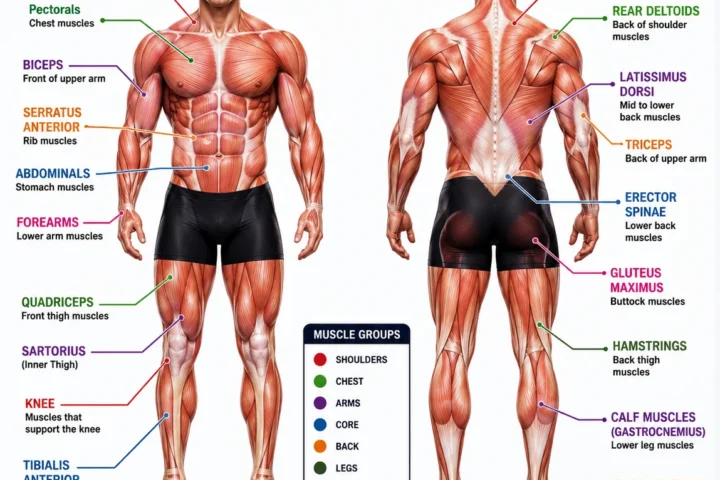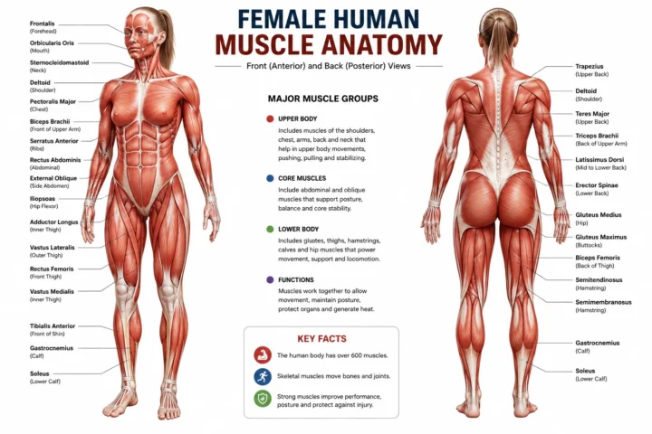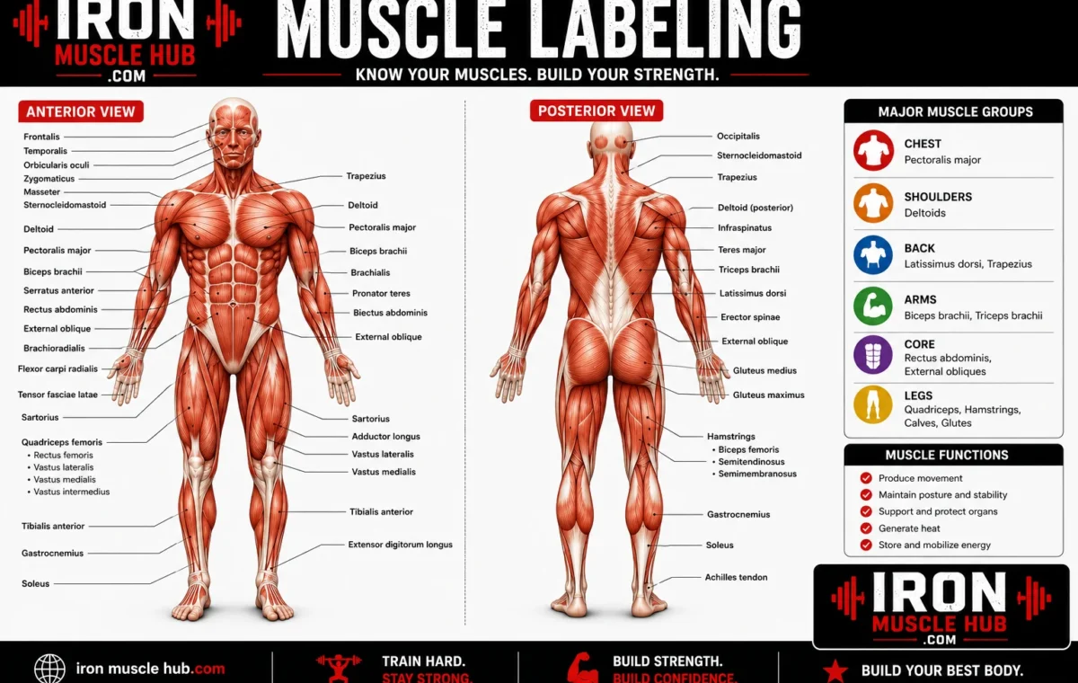
Muscle labeling means identifying muscles on an anatomy diagram and matching each muscle with its correct name and location. For beginners, the easiest approach is to learn the body region first, then recognize a muscle by its position, shape, and function. You don’t need to memorize hundreds of names at once.
- Quick Muscle Labeling Guide
- What Is Muscle Labeling?
- The Major Muscles You Should Know First
- 1. Deltoid
- 2. Biceps Brachii
- 3. Triceps Brachii
- 4. Pectoralis Major
- 5. Rectus Abdominis
- 6. Trapezius
- 7. Latissimus Dorsi
- 8. Gluteus Maximus
- 9. Quadriceps
- 10. Hamstrings
- 11. Gastrocnemius
- 12. Tibialis Anterior
- How to Label a Muscle Diagram Step by Step
- Step 1: Identify the view
- Step 2: Identify the body region
- Step 3: Find the obvious landmarks
- Step 4: Follow the label line carefully
- Step 5: Check the muscle's shape
- Step 6: Check neighboring structures
- Step 7: Say the answer aloud
- A Simple Muscle Labeling Study Method
- Round 1: Learn
- Round 2: Cover
- Round 3: Check
- Round 4: Repeat
- Why Muscle Names Can Help You Label Faster
- Common Muscle Labeling Mistakes
- Mixing up anterior and posterior
- Confusing biceps and triceps
- Treating a muscle group as one muscle
- Guessing from the label's position
- Trying to learn everything at once
- Memorizing spelling without location
- A Worked Muscle Labeling Example
- Where to Find Reliable Muscle Diagrams
- Internal Link Ideas
- Frequently Asked Questions
- What is muscle labeling?
- What are the major muscles to learn for muscle labeling?
- How can I memorize muscles quickly?
- What is the difference between an anterior and posterior muscle diagram?
- Why are muscle names so complicated?
- Is a muscle labeling worksheet useful for studying?
- Learn Muscle Labeling by Thinking, Not Just Memorizing
- Muscle Labeling by Body Region
- Facial Muscle Labeling
- Masseter
- Temporalis
- Neck Muscle Labeling
- Sternocleidomastoid
- Shoulder and Chest Labeling
- Deltoid
- Pectoralis Major
- Serratus Anterior
- Back Muscle Labeling
- Trapezius
- Latissimus Dorsi
- Rhomboid Muscles
- Arm and Forearm Muscle Labeling
- Biceps Brachii
- Triceps Brachii
- Brachialis
- Forearm Muscle Labeling
- A Useful Forearm Strategy
- Abdominal Muscle Labeling
- Rectus Abdominis
- External Oblique
- Internal Oblique
- Hip and Gluteal Muscle Labeling
- Gluteus Maximus
- Gluteus Medius
- Gluteus Minimus
- Thigh Muscle Labeling
- Quadriceps
- Sartorius
- Posterior Thigh Labeling
- Hamstrings
- Lower-Leg Muscle Labeling
- Tibialis Anterior
- Gastrocnemius
- Soleus
- How to Read a Difficult Muscle Diagram
- 1. What view am I seeing?
- 2. What body region is shown?
- 3. What structures surround it?
- 4. Is the muscle superficial or deep?
- 5. What does the name tell me?
- Muscle Labeling Memory Tricks
- A 10-Minute Muscle Labeling Practice Routine
- Practice Muscle Labeling Questions
- Question 1
- Question 2
- Question 3
- Question 4
- Question 5
- Question 6
- Question 7
- Question 8
- Question 9
- Question 10
- The Biggest Muscle Labeling Mistake to Avoid
- How to Build Your Own Muscle Labeling Chart
- Muscle Labeling: What to Learn Next
- Final Study Tip
- Muscle Labeling: Front-View Practice
- Head and Neck
- Shoulder and Chest
- Upper Arm
- Abdomen
- Muscle Labeling: Back-View Practice
- Upper Back
- Back of the Arm
- Gluteal Region
- Back of the Thigh
- Calf
- Advanced Muscle Labeling: Muscles You May Meet Next
- Vastus Lateralis
- Vastus Medialis
- Rectus Femoris
- Biceps Femoris
- Muscle Names Can Give You Clues
- A Common Exam Trap: Similar Names
- Biceps Brachii vs. Biceps Femoris
- Gluteus Maximus vs. Gluteus Medius
- Gastrocnemius vs. Soleus
- How to Practice With a Blank Diagram
- First Pass
- Second Pass
- Third Pass
- Fourth Pass
- Try This Mini Muscle Labeling Quiz
- 1. Which muscle covers the rounded shoulder?
- 2. Which muscle is found on the front of the upper arm?
- 3. Which muscle is found on the back of the upper arm?
- 4. Which muscle occupies the center of the front abdomen?
- 5. Which muscle is prominent across the upper back?
- 6. Which major muscle occupies the front of the thigh as part of the quadriceps group?
- 7. Which muscle group occupies the posterior thigh?
- 8. Which large muscle forms much of the buttock?
- 9. Which prominent muscle is found on the posterior lower leg?
- 10. Which major muscle is located toward the front of the lower leg?
- Quiz Answers
- The “Three Clues” Method
- Clue 1: Direction
- Clue 2: Region
- Clue 3: Shape
- Don't Ignore the Label Line
- What to Do If a Diagram Looks Different
- A 15-Minute Final Review
- Your Muscle Labeling Master Checklist
- What Comes After Muscle Labeling?
- Final Thoughts
A good muscle-labeling routine starts with large, recognizable muscles such as the deltoid, biceps brachii, triceps brachii, pectoralis major, rectus abdominis, gluteus maximus, quadriceps, hamstrings, and gastrocnemius. Once those landmarks become familiar, smaller muscles are much easier to place. For more insights read here :- lean build and how to build muscle .
Quick answer: To label muscles correctly, identify whether you’re looking at the front (anterior) or back (posterior) of the body, locate the body region, use surrounding landmarks, and then match the muscle’s position and shape with its name.
Quick Muscle Labeling Guide
| Body region | Major muscles to learn first | Easy location clue |
|---|---|---|
| Head | Masseter, temporalis | Jaw and side of skull |
| Neck | Sternocleidomastoid | Prominent muscle along the side of the neck |
| Shoulder | Deltoid | Rounded shoulder cap |
| Chest | Pectoralis major | Large muscle across the upper chest |
| Upper arm | Biceps brachii, triceps brachii | Front and back of upper arm |
| Abdomen | Rectus abdominis, external oblique | Front and sides of the abdomen |
| Back | Trapezius, latissimus dorsi | Upper back and broad side of back |
| Buttocks | Gluteus maximus | Large superficial muscle of the buttock |
| Thigh | Quadriceps, hamstrings | Front and back of thigh |
| Lower leg | Tibialis anterior, gastrocnemius, soleus | Front and back of lower leg |
These are useful starting points rather than a complete list. The human muscular system contains many more muscles, and anatomy courses may require considerably more detailed labeling.
What Is Muscle Labeling?
Muscle labeling is an anatomy study skill in which you identify muscles shown on a diagram, model, worksheet, or other visual representation.
A typical exercise might show an anterior view of the body with arrows pointing toward several muscles. Your task is to supply names such as deltoid, biceps brachii, or rectus abdominis.
A posterior diagram works the same way, but you’ll be looking at the back of the body. This distinction matters because a muscle that is easy to recognize from the front may not be visible from the same angle on the back.
Muscle diagrams can also show superficial muscles near the body’s surface or reveal deeper structures underneath them. OpenStax’s anatomy material, for example, uses anterior and posterior views to distinguish superficial and deeper muscles.
The Major Muscles You Should Know First
Trying to memorize every anatomical name immediately can make muscle labeling unnecessarily difficult. Start with the large landmarks.
1. Deltoid
The deltoid forms the rounded contour of the shoulder. On a basic anterior or posterior body diagram, it’s usually one of the easiest muscles to identify.
Its name is related to its triangular shape, which is one example of how anatomical muscle names can describe a muscle’s characteristics.
2. Biceps Brachii
The biceps brachii is on the front of the upper arm.
The word bi is useful here because it relates to two origins. Learning the meaning behind anatomical names can give you an extra memory cue instead of forcing you to memorize the name as an isolated word.
3. Triceps Brachii
The triceps brachii is on the back of the upper arm.
A simple way to remember the pair is:
- Biceps = front of upper arm
- Triceps = back of upper arm
They provide a useful landmark pair when you’re working through an arm-labeling exercise.
4. Pectoralis Major
The pectoralis major is the large, superficial muscle covering much of the upper front chest.
On an anterior diagram, look below the neck and medial to the shoulder. Its broad position makes it a useful landmark for orienting yourself.
5. Rectus Abdominis
The rectus abdominis runs vertically along the front of the abdomen.
The word rectus relates to a straight arrangement, which gives you another clue when remembering the name. OpenStax explains that muscle names can describe fiber direction, shape, location, size, attachments, or action.
6. Trapezius
The trapezius is a large muscle covering much of the upper back and extending toward the neck and shoulders.
It’s particularly useful on a posterior muscle diagram because its broad shape helps establish where the upper back begins.
7. Latissimus Dorsi
The latissimus dorsi, often called the “lats,” occupies a large area of the back.
It can be easier to identify by its location and broad shape than by trying to memorize the Latin name first.
8. Gluteus Maximus
The gluteus maximus is the large superficial muscle of the buttock region.
The gluteal muscles provide a useful example of anatomical naming based partly on size: maximus, medius, and minimus distinguish relative size within the group.
9. Quadriceps
The quadriceps is a group of four muscles on the front of the thigh.
That’s important for labeling: “quadriceps” refers to a muscle group rather than one individual muscle. If your course requires individual labels, you’ll need to learn the specific muscles within the group as well.
10. Hamstrings
The hamstrings are a group of muscles on the posterior thigh.
When looking at a back-view diagram, first locate the buttocks and knee. The hamstring region lies between them on the back of the thigh.
11. Gastrocnemius
The gastrocnemius is the prominent superficial muscle of the back of the lower leg.
On a posterior diagram, look below the knee. Its large calf position makes it a useful landmark.
12. Tibialis Anterior
The tibialis anterior is found on the front of the lower leg.
The name provides a location clue: anterior means toward the front, while tibialis connects the name with the tibia. Learning these terminology clues can make labeling easier.
How to Label a Muscle Diagram Step by Step
If you’re given a blank muscle diagram, don’t start guessing individual names immediately. Use a consistent process.
Step 1: Identify the view
First ask whether you’re looking at:
- Anterior: front of the body
- Posterior: back of the body
- Lateral: side of the body
This single step can eliminate many wrong answers.
Step 2: Identify the body region
Determine whether the arrow points toward the head, neck, shoulder, arm, chest, abdomen, back, thigh, or lower leg.
You can then narrow the possible answers considerably.
Step 3: Find the obvious landmarks
Start with large muscles that are easy to recognize.
For example, on an anterior diagram, you might first locate the deltoid, pectoralis major, biceps brachii, and rectus abdominis.
On a posterior diagram, look for the trapezius, latissimus dorsi, triceps brachii, gluteus maximus, and gastrocnemius.
Step 4: Follow the label line carefully
This is a surprisingly common source of mistakes.
Don’t identify a muscle simply because the text box appears beside it. Follow the line or pointer all the way to its endpoint. The line may cross another muscle before reaching the intended structure.
Step 5: Check the muscle’s shape
Shape can provide an important clue. Some muscle names themselves describe shape, size, or fiber direction.
Step 6: Check neighboring structures
If you’re unsure, look at what surrounds the muscle.
For example, a structure on the front of the upper arm is more likely to be the biceps brachii or brachialis than the triceps brachii, which is located posteriorly.
Step 7: Say the answer aloud
After writing a label, say it aloud and point to the muscle.
This connects three pieces of information:
name → location → visual shape
That is more useful for recall than repeatedly reading a list of names.
A Simple Muscle Labeling Study Method
One of the most useful ways to study is to work from a labeled diagram to an unlabeled diagram.
Round 1: Learn
Study a labeled diagram for a few minutes. Concentrate on where each major muscle sits rather than trying to memorize spelling immediately.
Round 2: Cover
Hide the labels or switch to an unlabeled version.
Try to name the muscles without looking at the answer key.
Round 3: Check
Mark the answers you missed.
Don’t simply reread the correct names. Return to the diagram and identify exactly where those muscles are located.
Round 4: Repeat
Try the same diagram again later.
A useful variation is to study the body region in sections:
- Head and neck
- Shoulder and upper arm
- Chest and abdomen
- Back
- Hip and thigh
- Lower leg
Regional study is also recommended by anatomy-learning resources because it reduces the size of the problem you’re trying to memorize at one time.
Why Muscle Names Can Help You Label Faster
Anatomical names aren’t always random words. Many contain clues about the muscle.
| Name clue | What it can tell you | Example |
| maximus | Larger | Gluteus maximus |
| medius | Medium | Gluteus medius |
| minimus | Smaller | Gluteus minimus |
| bi- | Two | Biceps |
| tri- | Three | Triceps |
| rectus | Straight | Rectus abdominis |
| longus | Long | Various muscles with “longus” in the name |
| brevis | Short | Various muscles with “brevis” in the name |
| anterior | Front | Tibialis anterior |
| lateralis | Toward the side | Various muscles with “lateralis” in the name |
OpenStax explains that skeletal muscles can be named according to characteristics including location, shape, relative size, number of origins, attachments, and action.
This means learning basic terminology can improve your ability to work out an unfamiliar label instead of relying entirely on memory.
Common Muscle Labeling Mistakes
Mixing up anterior and posterior
Always establish the view before naming anything.
Fix: Write “front” or “back” at the top of your worksheet if necessary.
Confusing biceps and triceps
Remember their basic positions:
Biceps = front of upper arm.
Triceps = back of upper arm.
Treating a muscle group as one muscle
Terms such as quadriceps refer to groups. If your test asks for individual muscles, read the instructions carefully.
Guessing from the label’s position
The written label may be far away from the actual muscle.
Fix: Trace the pointer to its endpoint.
Trying to learn everything at once
A long list of muscle names can quickly become confusing.
Fix: Learn one region at a time and repeatedly test yourself without the labels.
Memorizing spelling without location
Knowing how to spell sternocleidomastoid doesn’t help much if you can’t identify where it is.
Fix: Learn every muscle as a three-part package: name + location + visual landmark.
A Worked Muscle Labeling Example
Imagine a worksheet shows the front of the human body with an arrow pointing to the rounded muscle covering the shoulder.
You could reason through it like this:
- The diagram is an anterior view.
- The arrow points to the shoulder region.
- The muscle forms a rounded cap over the shoulder.
- The major muscle matching those clues is the deltoid.
- Write deltoid as the label.
Now imagine another arrow points to the front of the upper arm.
The region is the same, but the muscle isn’t the deltoid. The major muscle on the front of the upper arm is the biceps brachii.
This approach is better than memorizing “arrow 4 = biceps.” You’re learning the reasoning that lets you identify the muscle even when the diagram changes.
Where to Find Reliable Muscle Diagrams
For study, use sources that identify their anatomy references and licensing clearly.
OpenStax Anatomy & Physiology: Naming Skeletal Muscles provides explanations of muscle names and a labeled anterior/posterior muscular-system figure.
Wikimedia Commons: OpenStax Muscle Diagram hosts the OpenStax anterior and posterior muscle diagram under a CC BY 4.0 license. If you reuse it, follow the attribution requirements.
Kenhub: Muscle Diagrams and Quizzes offers labeled and unlabeled diagrams and quiz-based study material.
For general anatomy background, Cleveland Clinic explains that skeletal muscles attach to bones through tendons and contribute to movement, posture, joint stability, and heat production.
Internal Link Ideas
For a site covering fitness and beginner-friendly anatomy, consider linking this article to:
- Body Muscle Chart: A Beginner’s Guide to Major Muscles
- Muscle Anatomy Chart: Names, Locations, and Functions
- Body Diagram of Muscles: Front and Back Views Explained
These articles can form a useful topic cluster around muscle anatomy and help readers move from basic labeling to more detailed study.
Frequently Asked Questions
What is muscle labeling?
Muscle labeling is the process of identifying muscles on an anatomical diagram or model. A student usually follows a pointer to a particular structure and supplies its correct anatomical name. The easiest way to learn is to combine the muscle’s name with its location, shape, and surrounding landmarks rather than memorizing names in isolation.
What are the major muscles to learn for muscle labeling?
Good starting points include the deltoid, pectoralis major, biceps brachii, triceps brachii, rectus abdominis, trapezius, latissimus dorsi, gluteus maximus, quadriceps, hamstrings, gastrocnemius, and tibialis anterior. The exact list depends on the course or worksheet you’re studying.
How can I memorize muscles quickly?
Study muscles by body region rather than trying to memorize the entire muscular system at once. Start with a labeled diagram, then cover the labels and test yourself using an unlabeled version. Repeating the recall process is more useful than simply rereading the same list.
What is the difference between an anterior and posterior muscle diagram?
An anterior diagram shows the front of the body, while a posterior diagram shows the back. The same person can therefore look completely different from each view because different muscles are visible. Always identify the view before attempting to label the structures.
Why are muscle names so complicated?
Many anatomical muscle names contain information about location, shape, size, direction, attachments, or action. For example, words such as maximus, minimus, rectus, anterior, and prefixes such as bi- can provide clues about the structure being described.
Is a muscle labeling worksheet useful for studying?
Yes. A worksheet turns passive reading into active recall. The most useful approach is to study a labeled diagram first, attempt an unlabeled worksheet without looking at the answers, and then review only the structures you missed. This makes the exercise a test of recognition rather than simple copying.
Learn Muscle Labeling by Thinking, Not Just Memorizing
Muscle labeling becomes much easier once you stop treating every anatomical name as a separate fact.
Start with the body’s major regions. Learn the obvious landmarks. Decide whether you’re looking at the front or back. Then use location, shape, neighboring muscles, and the meaning of the name to narrow down the answer.
A strong study session doesn’t require dozens of pages of notes. A labeled diagram, an unlabeled version, and repeated self-testing can give you a much clearer picture of where the major muscles actually sit.
For beginners, focus on accuracy before speed. Once the major landmarks become familiar, more detailed muscle anatomy has somewhere to fit.
Memory Tricks, and Practice
If you’ve learned the major muscles from the first part, the next step is to make muscle labeling more precise. Instead of only recognizing large muscles such as the biceps or quadriceps, you can start identifying smaller muscles, understanding muscle groups, and using anatomical terminology to work out unfamiliar labels.
The key is not to memorize a giant list in one sitting. Break the body into regions and learn how neighboring muscles relate to one another. This makes an unlabeled diagram much easier to solve.
Muscle Labeling by Body Region
A useful way to organize your study is from the top of the body downward.
| Region | Muscles worth learning | What to look for |
|---|---|---|
| Face | Masseter, temporalis | Jaw and side of skull |
| Neck | Sternocleidomastoid | Long muscle on side of neck |
| Shoulder | Deltoid | Rounded shoulder |
| Chest | Pectoralis major | Broad upper-chest muscle |
| Upper back | Trapezius | Large upper-back region |
| Side of back | Latissimus dorsi | Broad muscle below the shoulder |
| Upper arm | Biceps brachii, triceps brachii | Front vs. back of arm |
| Forearm | Flexor and extensor groups | Front and back of forearm |
| Abdomen | Rectus abdominis, external oblique | Center and sides of abdomen |
| Hip | Gluteus maximus, gluteus medius | Buttock and upper hip |
| Thigh | Quadriceps, sartorius, hamstrings | Front and back of thigh |
| Lower leg | Tibialis anterior, gastrocnemius, soleus | Front and calf region |
The exact muscles required will depend on your anatomy course, worksheet, or exam.
Facial Muscle Labeling
Facial diagrams can look intimidating because several muscles are packed into a relatively small area.
Masseter
The masseter is associated with the jaw and is one of the main muscles involved in chewing.
On a side-view diagram of the head, look around the cheek and jaw area.
Temporalis
The temporalis sits on the side of the skull in the temporal region.
A useful distinction is:
- Temporalis: side of skull
- Masseter: jaw region
If a diagram has arrows pointing to both structures, their positions provide a strong clue.
Neck Muscle Labeling
Sternocleidomastoid
The sternocleidomastoid runs along the side of the neck.
Its long name looks difficult, but breaking it into parts can help:
sterno + cleido + mastoid
These terms relate to anatomical attachment locations. Learning the meaning of a name can therefore give you additional clues instead of making you depend entirely on visual memorization.
When labeling a neck diagram, first find the large, prominent muscle running diagonally along the side of the neck.
Shoulder and Chest Labeling
Deltoid
The deltoid creates much of the rounded appearance of the shoulder.
A simple visual cue is:
Shoulder cap = deltoid
Pectoralis Major
The pectoralis major covers a large portion of the upper front chest.
If you’re working from an anterior diagram, identify the shoulder first and then move inward toward the chest. The broad muscle in that region is an important landmark.
Serratus Anterior
The serratus anterior is located along the side of the chest.
It can be more difficult to identify than the pectoralis major because it has a different shape and position.
If your worksheet shows several smaller projections along the ribs, don’t automatically label them as the pectoralis. Check the muscle’s location and pattern carefully.
Back Muscle Labeling
Posterior diagrams become much easier when you recognize the major landmarks.
Trapezius
The trapezius occupies a large area of the upper back and extends toward the neck and shoulders.
On a posterior diagram, it’s one of the first muscles you should try to identify.
Latissimus Dorsi
The latissimus dorsi occupies a broad region of the back below the upper trapezius area.
A simple study cue is:
Trapezius = upper back
Latissimus dorsi = broad lower/lateral back
The two aren’t identical in shape or location, so learning them as neighboring landmarks can help.
Rhomboid Muscles
The rhomboid major and rhomboid minor are located in the upper back, deep to the trapezius.
This creates an important labeling lesson: not every muscle will be clearly visible on a basic superficial diagram.
If your worksheet includes deeper muscles, you may need to understand which superficial structures have been removed or made transparent in the illustration.
Arm and Forearm Muscle Labeling
The upper arm is a great place to practice positional reasoning.
Biceps Brachii
The biceps brachii is located on the anterior upper arm.
Remember:
Biceps → front of upper arm
Triceps Brachii
The triceps brachii is located on the posterior upper arm.
Remember:
Triceps → back of upper arm
This pair is one of the simplest ways to practice anterior-versus-posterior reasoning.
Brachialis
The brachialis lies deep to the biceps brachii and contributes to movement at the elbow.
If your diagram exposes deeper structures, don’t assume every muscle in the front of the upper arm is the biceps.
The diagram’s depth and labeling style matter.
Forearm Muscle Labeling
Forearm anatomy becomes significantly more detailed than upper-arm anatomy.
Instead of immediately trying to memorize every individual muscle, start by understanding the major groups.
A beginner may encounter:
- Wrist and finger flexor muscles
- Wrist and finger extensor muscles
- Muscles associated with rotation of the forearm
The exact structures shown depend on how detailed the diagram is.
A Useful Forearm Strategy
When an arrow points to the forearm, ask:
- Is the diagram showing the front or back?
- Is the palm facing toward or away from you?
- Is the muscle superficial or deep?
- Is the arrow pointing to one muscle or a group?
These questions can prevent several common labeling mistakes.
Abdominal Muscle Labeling
The abdomen contains several muscles that are commonly included in introductory diagrams.
Rectus Abdominis
The rectus abdominis is positioned vertically along the front of the abdomen.
It’s usually one of the easiest abdominal muscles to recognize.
External Oblique
The external oblique is positioned more laterally on the abdomen.
A useful visual distinction is:
Rectus abdominis = central front
External oblique = sides
Internal Oblique
The internal oblique lies deeper than the external oblique.
If you’re using a basic superficial diagram, it may not be shown.
This is another reason to examine the diagram’s level of detail before attempting to label every structure.
Hip and Gluteal Muscle Labeling
Gluteus Maximus
The gluteus maximus is the large superficial muscle of the buttock.
It’s generally easy to recognize on a posterior diagram.
Gluteus Medius
The gluteus medius sits higher and more laterally around the upper hip region.
If both gluteus maximus and gluteus medius appear on your diagram, compare their positions rather than relying only on the names.
Gluteus Minimus
The gluteus minimus is deeper than the gluteus medius.
A superficial diagram may not show it clearly.
This is where understanding anatomical depth becomes important.
Thigh Muscle Labeling
The thigh contains several major muscle groups.
Quadriceps
The quadriceps are located on the anterior thigh.
The name indicates a group containing four muscles.
The four individual muscles are:
- Rectus femoris
- Vastus lateralis
- Vastus medialis
- Vastus intermedius
The vastus intermedius is deep to the rectus femoris, so it isn’t always visible on a basic superficial diagram.
Sartorius
The sartorius is a long, narrow muscle that crosses the thigh diagonally.
Its unusual path makes it a useful landmark when studying an anterior thigh diagram.
If you see a long diagonal muscle crossing the front of the thigh, consider sartorius rather than automatically choosing one of the quadriceps muscles.
Posterior Thigh Labeling
Hamstrings
The hamstrings are a group of muscles on the posterior thigh.
Major muscles in the group include:
- Biceps femoris
- Semitendinosus
- Semimembranosus
For a beginner diagram, you may first need to recognize the overall hamstring region before learning each individual structure.
A more advanced diagram may require the individual names.
Lower-Leg Muscle Labeling
The lower leg is another area where front-versus-back positioning is extremely useful.
Tibialis Anterior
The tibialis anterior is located toward the front of the lower leg.
If you’re looking at an anterior view and an arrow points alongside the shin, this muscle is an important possibility.
Gastrocnemius
The gastrocnemius is the prominent superficial calf muscle.
On a posterior diagram, look below the knee and identify the large calf structure.
Soleus
The soleus lies deep to the gastrocnemius in much of the lower leg.
If a diagram shows deeper anatomy, you may need to distinguish it from the gastrocnemius.
How to Read a Difficult Muscle Diagram
Some anatomy diagrams are much harder than simple front-and-back charts.
When the image looks confusing, use this five-question method.
1. What view am I seeing?
Determine whether it’s:
- Anterior
- Posterior
- Lateral
- A sectional or deeper view
2. What body region is shown?
Don’t try to identify the muscle before establishing the region.
3. What structures surround it?
Neighboring anatomy is often the best clue.
4. Is the muscle superficial or deep?
A muscle may be hidden underneath another structure in a basic view.
5. What does the name tell me?
Words such as anterior, posterior, maximus, medius, minimus, longus, brevis, major, and minor can provide useful clues.
Muscle Labeling Memory Tricks
You don’t need a complicated memory system.
Use short associations that connect a name with a location.
| Muscle | Memory clue |
| Deltoid | Shoulder cap |
| Biceps brachii | Front upper arm |
| Triceps brachii | Back upper arm |
| Pectoralis major | Large chest muscle |
| Rectus abdominis | Center abdomen |
| External oblique | Side abdomen |
| Trapezius | Upper back |
| Latissimus dorsi | Broad back |
| Gluteus maximus | Large buttock muscle |
| Sartorius | Diagonal thigh |
| Quadriceps | Front thigh group |
| Hamstrings | Back thigh group |
| Tibialis anterior | Front lower leg |
| Gastrocnemius | Large calf muscle |
The goal isn’t to replace anatomical knowledge with silly shortcuts. Memory cues are simply a bridge that helps you recall the correct structure.
A 10-Minute Muscle Labeling Practice Routine
If you only have a short study session, try this routine.
Minute 1–2: Look at a labeled diagram.
Minute 3–4: Cover the labels and identify the major muscles.
Minute 5: Write down every muscle you couldn’t remember.
Minute 6–7: Study only those missed structures.
Minute 8–9: Repeat the unlabeled test.
Minute 10: Say the names aloud while pointing to their locations.
The important part is the recall test. Looking at a labeled diagram feels productive, but actively trying to retrieve the answer gives you a clearer indication of what you actually know.
Practice Muscle Labeling Questions
Try answering these without looking at the table above.
Question 1
Which major muscle forms the rounded cap of the shoulder?
Answer: Deltoid.
Question 2
Which major muscle is found on the front of the upper arm?
Answer: Biceps brachii.
Question 3
Which major muscle is found on the back of the upper arm?
Answer: Triceps brachii.
Question 4
Which large muscle covers much of the upper chest?
Answer: Pectoralis major.
Question 5
Which major muscle occupies the upper back and extends toward the shoulders and neck?
Answer: Trapezius.
Question 6
Which major muscle is located on the front of the abdomen?
Answer: Rectus abdominis.
Question 7
Which major muscle group occupies the front of the thigh?
Answer: Quadriceps.
Question 8
Which major muscle group occupies the back of the thigh?
Answer: Hamstrings.
Question 9
Which prominent superficial muscle is found on the back of the lower leg?
Answer: Gastrocnemius.
Question 10
Which major muscle is found toward the front of the lower leg?
Answer: Tibialis anterior.
The Biggest Muscle Labeling Mistake to Avoid
The biggest mistake isn’t forgetting a difficult Latin name.
It’s guessing before establishing where the muscle is.
Suppose you see an arrow pointing somewhere around the upper arm. If you immediately guess “biceps,” you have skipped several useful clues.
Instead:
View → region → position → neighboring structures → muscle name
That sequence gives you a repeatable way to solve unfamiliar diagrams.
It also helps when your teacher uses a diagram that looks different from the one you studied.
How to Build Your Own Muscle Labeling Chart
You can make a simple study chart with four columns:
| Muscle | Body region | Front/back | Recognition clue |
| Deltoid | Shoulder | Both views | Rounded shoulder |
| Biceps brachii | Upper arm | Front | Front of arm |
| Triceps brachii | Upper arm | Back | Back of arm |
| Rectus abdominis | Abdomen | Front | Vertical central muscle |
| Trapezius | Upper back | Back | Broad upper-back muscle |
| Gluteus maximus | Buttocks | Back | Large superficial buttock muscle |
| Quadriceps | Thigh | Front | Front thigh group |
| Hamstrings | Thigh | Back | Back thigh group |
| Gastrocnemius | Lower leg | Back | Large calf muscle |
Then turn the chart into a quiz by covering the second and third columns.
That forces you to recall the information rather than simply recognize it.
Muscle Labeling: What to Learn Next
Once you can confidently identify the major superficial muscles, move toward individual muscles within groups.
A sensible progression is:
- Major muscles
- Muscle groups
- Individual muscles within each group
- Superficial versus deep muscles
- Muscle attachments
- Muscle actions
- More detailed regional anatomy
You don’t need to jump directly from a beginner diagram to a highly detailed anatomical atlas.
Build the map gradually.
Final Study Tip
If you’re preparing for a quiz, don’t spend your entire study session staring at the answer sheet.
Use the diagram as a test.
Point to a structure, name it, explain where it is, and then check your answer. If you get it wrong, immediately reconnect the name with its location.
That simple cycle — look, recall, check, repeat — turns muscle labeling from a memorization task into a recognition skill.
The more often you practice with unlabeled diagrams, the easier it becomes to identify muscles even when the artwork, angle, or labeling style changes.
Final Review
If you’ve worked through Parts 1 and 2, you’re ready to take muscle labeling one step further. This section focuses on practice, harder-to-identify muscles, common exam traps, and a simple review system you can use before a quiz.
The biggest improvement usually comes from moving away from simply looking at labeled pictures. Instead, test yourself with an unlabeled diagram and explain why you think a particular muscle belongs in each position.
Muscle Labeling: Front-View Practice
An anterior, or front, view commonly gives you several recognizable landmarks.
Start at the top and work downward.
Head and Neck
Look for:
- Temporalis around the side of the skull
- Masseter around the jaw
- Sternocleidomastoid along the side of the neck
Don’t rush through this region. The face and neck contain smaller structures that can be difficult to distinguish on simplified diagrams.
Shoulder and Chest
The deltoid forms the rounded shoulder area.
Below and toward the center of the upper torso, the pectoralis major is a major landmark.
If a diagram includes deeper or smaller muscles, use the surrounding structures to determine exactly where the arrow ends.
Upper Arm
The front of the upper arm is associated with the biceps brachii.
A deeper diagram may also show the brachialis, so don’t automatically label every anterior upper-arm structure as biceps.
Abdomen
The central abdominal region contains the rectus abdominis.
Toward the sides, you’ll encounter the external oblique.
This gives you an easy positional rule:
Center = rectus abdominis
Sides = external oblique
Muscle Labeling: Back-View Practice
Posterior diagrams require you to recognize a different set of major landmarks.
Upper Back
The trapezius is a major superficial landmark around the upper back and neck.
Moving lower and outward, the latissimus dorsi covers a broad portion of the back.
Back of the Arm
The triceps brachii is the major muscle to recognize on the posterior upper arm.
If you know the front/back relationship between biceps and triceps, this becomes much easier.
Gluteal Region
The gluteus maximus is the large superficial muscle of the buttock.
Depending on the diagram, smaller or deeper gluteal muscles may also be visible.
Back of the Thigh
The hamstrings occupy the posterior thigh.
A more detailed diagram can separate the individual muscles within this group, so learn the level of detail required by your course.
Calf
The gastrocnemius is the large superficial calf muscle.
The soleus lies deeper and may appear on diagrams that show deeper structures.
Advanced Muscle Labeling: Muscles You May Meet Next
Once the major landmarks are familiar, you can expand your vocabulary.
Vastus Lateralis
The vastus lateralis is one of the quadriceps muscles and is positioned toward the lateral side of the thigh.
The word lateralis gives you a useful location clue: lateral structures are toward the side.
Vastus Medialis
The vastus medialis is another quadriceps muscle, positioned more medially.
A simple contrast is:
Vastus lateralis → lateral side
Vastus medialis → medial side
Rectus Femoris
The rectus femoris is one of the quadriceps muscles and runs along the anterior thigh.
Because several quadriceps muscles occupy the same general area, position and depth become increasingly important in advanced diagrams.
Biceps Femoris
The biceps femoris is one of the hamstring muscles.
Don’t confuse it with the biceps brachii.
The names share “biceps,” but they belong to different body regions.
Biceps brachii → upper arm
Biceps femoris → thigh
This is a common source of confusion for beginners.
Muscle Names Can Give You Clues
Anatomical terminology becomes increasingly useful as the diagrams become more detailed.
Consider these examples:
| Term | Useful clue |
|---|---|
| Anterior | Front |
| Posterior | Back |
| Medial | Toward the body’s midline |
| Lateral | Away from the midline |
| Major | Larger |
| Minor | Smaller |
| Maximus | Largest |
| Medius | Middle |
| Minimus | Smallest |
| Longus | Long |
| Brevis | Short |
| Bi- | Two |
| Tri- | Three |
These clues don’t always give you the complete answer, but they can help narrow your choices.
A Common Exam Trap: Similar Names
Some muscle names look or sound similar.
Biceps Brachii vs. Biceps Femoris
Biceps brachii is in the upper arm.
Biceps femoris is in the posterior thigh.
The second part of the name helps distinguish the region.
Gluteus Maximus vs. Gluteus Medius
Both are in the gluteal region.
The names indicate their relative size and position within the group.
Gastrocnemius vs. Soleus
Both are associated with the calf region.
The gastrocnemius is the prominent superficial calf muscle, while the soleus is deeper.
How to Practice With a Blank Diagram
Here’s a simple method that works well for beginners.
First Pass
Identify only the muscles you know immediately.
Don’t guess.
Second Pass
Return to the blank labels you skipped.
Use location and neighboring structures to narrow down the possibilities.
Third Pass
Check your answers against a reliable labeled reference.
Circle the muscles you missed.
Fourth Pass
Cover the answers again and repeat only the difficult areas.
This is much more useful than repeatedly copying the same answer list.
Try This Mini Muscle Labeling Quiz
Don’t look at the answers until you’ve tried all ten.
1. Which muscle covers the rounded shoulder?
2. Which muscle is found on the front of the upper arm?
3. Which muscle is found on the back of the upper arm?
4. Which muscle occupies the center of the front abdomen?
5. Which muscle is prominent across the upper back?
6. Which major muscle occupies the front of the thigh as part of the quadriceps group?
7. Which muscle group occupies the posterior thigh?
8. Which large muscle forms much of the buttock?
9. Which prominent muscle is found on the posterior lower leg?
10. Which major muscle is located toward the front of the lower leg?
Quiz Answers
1. Deltoid
2. Biceps brachii
3. Triceps brachii
4. Rectus abdominis
5. Trapezius
6. Rectus femoris (one of the quadriceps muscles)
7. Hamstrings
8. Gluteus maximus
9. Gastrocnemius
10. Tibialis anterior
If you missed several, don’t worry. Go back to the diagram and physically point to each answer.
The “Three Clues” Method
When you’re stuck on a label, use three clues:
Clue 1: Direction
Is the structure anterior, posterior, medial, or lateral?
Clue 2: Region
Is it in the shoulder, chest, arm, abdomen, thigh, or lower leg?
Clue 3: Shape
Is it broad, rounded, narrow, diagonal, or located between recognizable landmarks?
Combining these clues can often narrow down the answer without guessing.
Don’t Ignore the Label Line
One of the easiest mistakes to make is looking at where the label text sits rather than where the arrow actually points.
A label might be printed outside the body, with a line extending across several structures.
Always follow the line to its endpoint.
The endpoint is what matters.
This single habit can improve your accuracy on crowded anatomy diagrams.
What to Do If a Diagram Looks Different
Your textbook, teacher, website, or exam may use different illustrations.
Don’t memorize the exact position of a label on one picture.
Instead, memorize the actual anatomical relationship.
For example, don’t think:
“The deltoid is the muscle beside this particular arrow.”
Think:
“The deltoid is the rounded muscle covering the shoulder.”
That knowledge transfers to different diagrams.
A 15-Minute Final Review
Before a basic muscle-labeling test, try this routine.
Minutes 1–3: Review a labeled front-view diagram.
Minutes 4–6: Review a labeled back-view diagram.
Minutes 7–10: Complete an unlabeled diagram from memory.
Minutes 11–13: Check your mistakes.
Minutes 14–15: Repeat only the muscles you missed.
This keeps your study session focused on active recall rather than passive reading.
Your Muscle Labeling Master Checklist
Before moving to more advanced anatomy, see if you can identify:
- Masseter
- Temporalis
- Sternocleidomastoid
- Deltoid
- Pectoralis major
- Trapezius
- Latissimus dorsi
- Biceps brachii
- Triceps brachii
- Rectus abdominis
- External oblique
- Gluteus maximus
- Gluteus medius
- Quadriceps
- Rectus femoris
- Vastus lateralis
- Vastus medialis
- Hamstrings
- Tibialis anterior
- Gastrocnemius
- Soleus
You don’t have to master every structure immediately. Use this as a progress check.
What Comes After Muscle Labeling?
Once you’re comfortable identifying muscles, the next stage is understanding what those muscles actually do.
You can begin studying:
- Muscle origins
- Muscle insertions
- Muscle actions
- Agonists and antagonists
- Muscle groups
- Superficial and deep muscles
- Major joints and their movements
- Basic muscle terminology
This turns a static labeling exercise into a better understanding of human movement.
Final Thoughts
Muscle labeling is easiest when you learn relationships instead of isolated names.
Start with the big landmarks. Learn the difference between front and back views. Break the body into regions. Use anatomical terminology as a clue, and always follow the label line to its endpoint.
When you get an answer wrong, don’t simply memorize the correction. Ask why the correct muscle belongs there.
That small change can make studying much more effective.
With regular practice, you’ll begin to recognize muscles almost automatically. What first looks like a confusing collection of shapes gradually becomes a map of familiar anatomical landmarks.