A muscle anatomy chart is a visual guide that shows where important muscles are located and helps you understand their basic functions. For beginners, the easiest place to start is with the major skeletal muscles of the head, neck, shoulders, chest, arms, back, abdomen, hips, and legs.
- Muscle Anatomy Chart at a Glance
- What Is a Muscle Anatomy Chart?
- Major Muscles on the Front of the Body
- 1. Deltoids
- 2. Pectoralis Major
- 3. Biceps Brachii
- 4. Rectus Abdominis
- 5. External Obliques
- 6. Quadriceps
- Major Muscles on the Back of the Body
- 1. Trapezius
- 2. Latissimus Dorsi
- 3. Triceps Brachii
- 4. Gluteus Maximus
- 5. Hamstrings
- 6. Gastrocnemius
- Muscle Groups You Should Learn First
- How Muscles Work With Bones
- The Three Main Types of Muscle Tissue
- Anterior vs. Posterior Muscle Anatomy
- How to Read a Muscle Anatomy Chart
- Step 1: Identify the body region
- Step 2: Learn the muscle name
- Step 3: Learn one basic function
- Step 4: Test yourself without the chart
- A Useful Muscle-Chart Study Trick
- Common Mistakes Beginners Make
- Trying to memorize every muscle immediately
- Confusing a muscle with a muscle group
- Studying only the front view
- Assuming every visible muscle is superficial
- Treating an anatomy chart as a diagnostic tool
- Related Resources to Add on Iron Muscle Hub
- Frequently Asked Questions
- What are the major muscles in the human body?
- How many muscles are in the human body?
- What are the three types of muscle?
- What is the difference between anterior and posterior muscle charts?
- What is the easiest way to memorize muscle anatomy?
- Are muscle anatomy charts useful for workouts?
- Build Your Muscle Anatomy Knowledge One Region at a Time
- Quick Muscle Group Guide
- Upper-Body Muscle Anatomy
- Deltoid
- Pectoralis Major
- Trapezius
- Latissimus Dorsi
- Biceps Brachii
- Triceps Brachii
- Core Muscle Anatomy
- Rectus Abdominis
- External Obliques
- Internal Obliques
- Transversus Abdominis
- Back Muscle Anatomy
- Erector Spinae
- Rhomboids
- Multifidus
- Hip and Glute Muscle Anatomy
- Gluteus Maximus
- Gluteus Medius
- Gluteus Minimus
- Thigh Muscle Anatomy
- Quadriceps
- Hamstrings
- Adductors
- Lower-Leg Muscle Anatomy
- Gastrocnemius
- Soleus
- Tibialis Anterior
- How to Match a Muscle to Its Function
- A Simple 7-Day Muscle Anatomy Study Plan
- Day 1: Shoulders and arms
- Day 2: Chest and abdomen
- Day 3: Back
- Day 4: Hips and glutes
- Day 5: Thighs
- Day 6: Lower legs
- Day 7: Test yourself
- A Better Way to Use a Muscle Anatomy Chart
- Why Some Charts Look Different
- Superficial vs. Deep Muscles
- Muscle Names Can Give You Clues
- What a Muscle Anatomy Chart Can and Can't Tell You
- Final Takeaway
- Muscle Anatomy Chart: The Complete Beginner Map
- Face and Neck Muscle Anatomy
- Masseter
- Temporalis
- Sternocleidomastoid
- Shoulder and Chest Anatomy
- Deltoid
- Pectoralis Major
- Rotator Cuff Muscles
- Upper-Arm Muscle Anatomy
- Biceps Brachii
- Triceps Brachii
- Forearm Muscle Anatomy
- Flexor Muscles
- Extensor Muscles
- Abdominal Muscle Anatomy
- Rectus Abdominis
- External Oblique
- Internal Oblique
- Transversus Abdominis
- Back Muscle Anatomy
- Trapezius
- Latissimus Dorsi
- Rhomboids
- Erector Spinae
- Glute and Hip Anatomy
- Gluteus Maximus
- Gluteus Medius
- Gluteus Minimus
- Quadriceps Anatomy
- A Simple Visual Trick
- Hamstring Anatomy
- Calf Muscle Anatomy
- Gastrocnemius
- Soleus
- Tibialis Anterior
- How to Memorize a Muscle Anatomy Chart Faster
- Use the Body-Region Method
- Use Front-and-Back Pairs
- Add Function After Location
- What the Layers of a Skeletal Muscle Chart Mean
- Muscle Anatomy and Exercise
- A 10-Minute Muscle Chart Review
- Minute 1–2: Scan the chart
- Minute 3–5: Pick five muscles
- Minute 6–7: Add one function
- Minute 8–9: Cover the labels
- Minute 10: Correct yourself
- The Most Important Muscle Names to Know
- Frequently Asked Questions
- What is the easiest way to learn a muscle anatomy chart?
- What are the most important muscles to learn first?
- Why does my muscle anatomy chart look different from another chart?
- Are the quadriceps one muscle or four muscles?
- Are the calf muscles one muscle?
- How many muscle types are there?
- Final Thoughts on Muscle Anatomy Charts
You don’t need to memorize hundreds of anatomical names at once. Learning the major muscle groups first gives you a useful foundation for understanding exercise, movement, posture, and basic human anatomy. This guide breaks the body down into simple sections so you can learn one area at a time. For more insights read here :- lean build and lean muscle .
Quick answer: The major muscles most beginners should recognize include the deltoids, pectorals, biceps, triceps, trapezius, latissimus dorsi, abdominals, gluteals, quadriceps, hamstrings, and calf muscles.
Muscle Anatomy Chart at a Glance
| Body area | Major muscles to know | Basic role |
|---|---|---|
| Head and face | Masseter, facial muscles | Chewing and facial movement |
| Neck | Sternocleidomastoid and other neck muscles | Head and neck movement |
| Shoulders | Deltoids | Moving and stabilizing the shoulder |
| Chest | Pectoralis major | Moving the upper arm |
| Upper back | Trapezius, rhomboids | Shoulder-blade and upper-back movement |
| Back | Latissimus dorsi, erector spinae | Arm movement and spinal support |
| Upper arms | Biceps, triceps | Bending and straightening the elbow |
| Abdomen | Rectus abdominis, obliques | Trunk movement and stability |
| Hips and buttocks | Gluteus maximus, medius | Hip movement and stability |
| Front thighs | Quadriceps | Straightening the knee |
| Back thighs | Hamstrings | Bending the knee and moving the hip |
| Lower legs | Gastrocnemius, soleus | Ankle and foot movement |
This is a learning chart rather than a complete anatomical inventory. The human body contains more than 600 muscles, and many smaller muscles aren’t normally included on beginner-friendly charts.
What Is a Muscle Anatomy Chart?
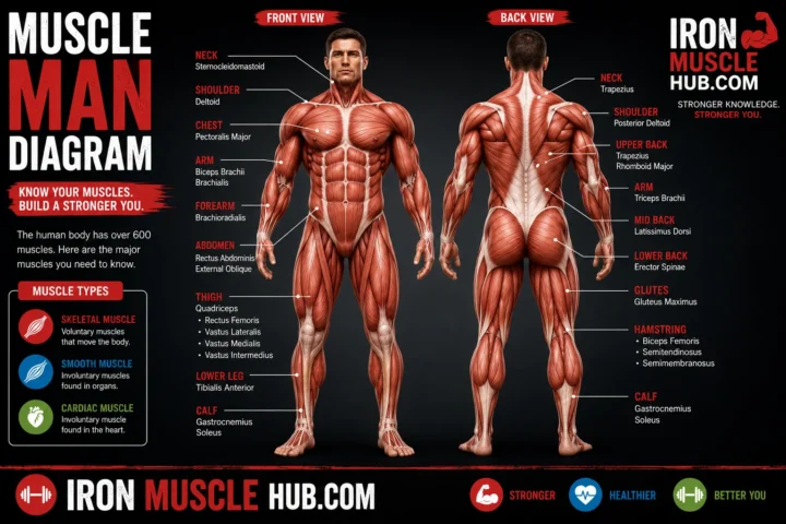
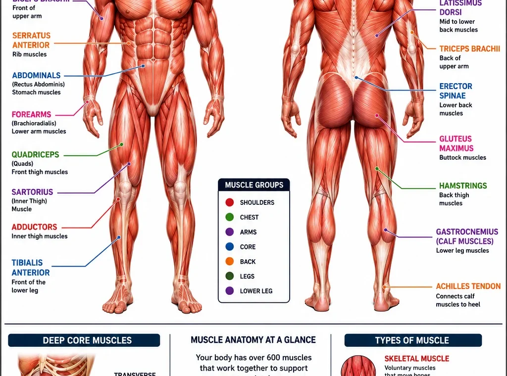
A muscle anatomy chart is an illustrated reference that identifies muscles by their location, name, and sometimes their action or attachment points.
Most basic charts concentrate on skeletal muscles, because these are the muscles associated with conscious movement and the musculoskeletal system. Skeletal muscles attach to bones through tendons and work with joints to produce movement.
An anatomy chart may show the body from the anterior view, meaning the front, and the posterior view, meaning the back. Looking at both views is important because many large muscles are easier to understand from the rear of the body.
Major Muscles on the Front of the Body
A front-view muscle chart usually makes the chest, shoulders, arms, abdomen, and thighs easiest to identify.
1. Deltoids
The deltoid is the large muscle covering the shoulder. Its position makes it one of the easiest muscles to recognize on a front or side-view anatomy chart.
The deltoid helps produce movements of the upper arm around the shoulder joint.
2. Pectoralis Major
The pectoralis major is the large muscle covering much of the front of the chest.
It contributes to movements of the upper arm, making it an important muscle to recognize when studying chest and upper-body anatomy.
3. Biceps Brachii
The biceps brachii sits on the front of the upper arm.
A simple way to remember it is “biceps = front of upper arm.” It helps flex the elbow and contributes to rotation of the forearm.
4. Rectus Abdominis
The rectus abdominis is located along the front of the abdomen.
It helps flex the trunk and is one of the muscles commonly associated with the abdominal region. A muscle chart may show it as a long paired muscle running vertically down the front of the abdomen.
5. External Obliques
The external obliques sit along the sides of the abdomen.
They help with movements and stabilization of the trunk. On a front-view chart, look beside the rectus abdominis rather than directly down the center of the stomach.
6. Quadriceps
The quadriceps are a group of muscles at the front of the thigh.
The group includes four muscles, and together they play a major role in extending the knee.
Major Muscles on the Back of the Body
A posterior muscle chart reveals several important muscles that aren’t fully visible from the front.
1. Trapezius
The trapezius is a broad muscle across the upper back and neck region.
It contributes to movement and positioning of the shoulder blades and helps with movements involving the neck and shoulders.
2. Latissimus Dorsi
The latissimus dorsi, often called the “lats,” is a large muscle covering part of the back.
It contributes to movements of the upper arm. On a back-view chart, it is located below the trapezius and extends toward the sides of the back.
3. Triceps Brachii
The triceps brachii is on the back of the upper arm.
If the biceps are the major muscle on the front of the upper arm, the triceps are the major muscle group on the back. The triceps help extend the elbow.
4. Gluteus Maximus
The gluteus maximus is the largest of the three gluteal muscles.
It is located in the buttock region and plays an important role in hip movement. The gluteal region also contains the gluteus medius and gluteus minimus.
5. Hamstrings
The hamstrings are a group of muscles located at the back of the thigh.
They work across the hip and knee and contribute to movements such as bending the knee and extending the hip.
6. Gastrocnemius
The gastrocnemius is one of the prominent muscles of the calf.
It crosses the knee and ankle and contributes to movement at the ankle. It is particularly easy to spot on a posterior lower-leg diagram.
Muscle Groups You Should Learn First
Trying to memorize every muscle can make anatomy unnecessarily difficult. Start with these major groups:
- Shoulders: deltoids
- Chest: pectorals
- Upper back: trapezius and rhomboid region
- Back: latissimus dorsi and erector spinae
- Front upper arm: biceps
- Back upper arm: triceps
- Abdomen: rectus abdominis and obliques
- Buttocks: gluteal muscles
- Front thigh: quadriceps
- Back thigh: hamstrings
- Calf: gastrocnemius and soleus
This approach gives you a practical mental map before you move on to smaller or deeper muscles.
How Muscles Work With Bones
Muscles don’t move bones by pushing them. Instead, skeletal muscles contract and pull on structures through their attachments, including tendons.
When a skeletal muscle contracts, it can create movement at a joint. Muscles, bones, joints, and connective tissues therefore work together as part of the musculoskeletal system.
For example, bending your elbow involves coordinated action from muscles around the upper arm and forearm rather than one muscle working completely by itself.
This is one reason an anatomy chart is more useful when you study location and function together.
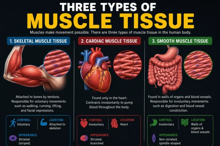
The Three Main Types of Muscle Tissue
One common beginner mistake is assuming every muscle in the body is a skeletal muscle. Human muscle tissue is generally divided into three major types.
| Type | Where you’ll find it | Main characteristic |
| Skeletal muscle | Attached to the skeleton | Primarily responsible for voluntary movement |
| Cardiac muscle | Heart | Contracts involuntarily to pump blood |
| Smooth muscle | Walls of various organs and vessels | Controls involuntary internal movements |
Skeletal muscle is the type most commonly shown on fitness-oriented muscle anatomy charts. Cardiac muscle forms the muscular tissue of the heart, while smooth muscle is found in structures such as the digestive tract and blood vessels.
Anterior vs. Posterior Muscle Anatomy
If you’re learning from a labeled chart, you’ll often see two terms:
Anterior means the front side of the body.
Posterior means the back side of the body.
A simple study method is to learn five or six muscles from the anterior view and then learn five or six from the posterior view.
For example:
Anterior: deltoids → pectorals → biceps → abdominals → quadriceps
Posterior: trapezius → latissimus dorsi → triceps → gluteals → hamstrings → calves
Once those locations become familiar, adding smaller muscles becomes much easier.
How to Read a Muscle Anatomy Chart
Use this four-step method rather than trying to memorize a picture.
Step 1: Identify the body region
Start with the head, neck, shoulder, chest, arm, abdomen, back, hip, or leg.
Step 2: Learn the muscle name
Say the name while pointing to the muscle. Repetition helps connect the word with its physical location.
Step 3: Learn one basic function
Don’t try to memorize every attachment and action immediately. Start with something simple, such as “quadriceps help straighten the knee.”
Step 4: Test yourself without the chart
Cover the labels and try to identify the muscles yourself. Then check your answers.
This simple recall exercise is often more useful than repeatedly staring at the labeled diagram.
A Useful Muscle-Chart Study Trick
Try learning muscles in opposing pairs or groups.
For example:
- Biceps: front of upper arm
- Triceps: back of upper arm
- Quadriceps: front of thigh
- Hamstrings: back of thigh
This creates a mental map instead of a random collection of anatomical vocabulary.
You can also connect each muscle to a movement. For instance, think about the quadriceps when straightening the knee and the calf muscles when moving the ankle.
Common Mistakes Beginners Make
Trying to memorize every muscle immediately
There are hundreds of muscles, so attempting to learn everything at once can quickly become overwhelming. Start with the major groups and build from there.
Confusing a muscle with a muscle group
Some names describe groups rather than one individual muscle. Quadriceps and hamstrings, for example, refer to groups of muscles.
Studying only the front view
Many important muscles are easier to understand from the back. Use both anterior and posterior views.
Assuming every visible muscle is superficial
An anatomy chart may simplify the body for educational purposes. Some muscles are deep beneath other muscles and won’t appear clearly on a basic surface-level diagram.
Treating an anatomy chart as a diagnostic tool
A chart can help you learn anatomy, but it can’t tell you what’s causing pain or injury. Persistent pain, weakness, or difficulty moving deserves appropriate medical evaluation. Cleveland Clinic recommends seeking professional care for ongoing muscle symptoms.
Related Resources to Add on Iron Muscle Hub
For a useful internal-link structure, consider linking this guide to:
- What Are Muscles? A Beginner’s Guide to Muscle Function
- Three Types of Muscle Tissue Explained
- Cardiac vs. Skeletal Muscle: Key Differences
- How Muscles Work During Exercise
These topics naturally support readers who want to move from basic anatomy into muscle function and fitness.
For deeper medical anatomy research, reputable references include Cleveland Clinic’s muscle anatomy guide and NCBI’s Skeletal Muscle anatomy reference.
Frequently Asked Questions
What are the major muscles in the human body?
Major muscles and muscle groups include the deltoids, pectorals, biceps, triceps, trapezius, latissimus dorsi, abdominal muscles, gluteal muscles, quadriceps, hamstrings, and calf muscles. These aren’t all the muscles in the body, but they’re useful starting points for learning basic human anatomy.
How many muscles are in the human body?
Sources commonly describe the human body as having more than 600 muscles, although exact counts can vary depending on how individual muscles and muscle groups are classified. Cleveland Clinic and the National Institute of Arthritis and Musculoskeletal and Skin Diseases both use figures above 600.
What are the three types of muscle?
The three major types are skeletal, cardiac, and smooth muscle. Skeletal muscle is associated with movement of the skeleton, cardiac muscle makes up the heart, and smooth muscle performs involuntary movements in various internal structures.
What is the difference between anterior and posterior muscle charts?
An anterior muscle chart shows the front of the body, while a posterior muscle chart shows the back. Using both views gives you a more complete picture of where major muscles are located.
What is the easiest way to memorize muscle anatomy?
Start with major regions instead of individual muscles. Learn the shoulders, chest, arms, abdomen, back, hips, thighs, and calves, then connect each major muscle with its location and one basic movement. Testing yourself without looking at the labels can reinforce what you’ve learned.
Are muscle anatomy charts useful for workouts?
Yes. A basic anatomy chart can help you understand which muscles an exercise is intended to involve. However, anatomy alone doesn’t determine the best exercise program, and a diagram shouldn’t be used to diagnose an injury or replace professional medical guidance.
Build Your Muscle Anatomy Knowledge One Region at a Time
A good muscle anatomy chart doesn’t need to be complicated. Start by recognizing the major muscles from the front and back, then connect each one with its basic location and function.
The most useful first landmarks are the deltoids, pectorals, biceps, triceps, trapezius, latissimus dorsi, abdominals, glutes, quadriceps, hamstrings, and calf muscles. Once those become familiar, you can gradually add smaller and deeper muscles.
Think of anatomy as a map rather than a memorization test. Learn one body region at a time, quiz yourself regularly, and use reputable medical references when you need more detail. That approach makes muscle anatomy much easier to understand and remember.
Muscle Anatomy Chart: Parts 2 — Muscle Groups, Functions, and Study Guide
A muscle anatomy chart becomes much easier to understand when you stop treating it as a collection of unfamiliar names and start grouping muscles by body region. The major skeletal muscles work with bones, joints, tendons and other tissues to produce movement and help maintain posture.
In this second part, we’ll go deeper into the major muscle groups, explain what they generally do, and show you a simple way to study a labeled muscle diagram without trying to memorize everything at once.
Quick Muscle Group Guide
| Region | Important muscles | What to remember |
|---|---|---|
| Neck | Sternocleidomastoid | Head and neck movement |
| Shoulder | Deltoid | Major shoulder muscle |
| Chest | Pectoralis major | Upper-arm movement |
| Upper back | Trapezius, rhomboids | Shoulder-blade movement |
| Mid/lower back | Latissimus dorsi, erector spinae | Arm and spinal movement/support |
| Upper arm | Biceps, triceps | Elbow movement |
| Abdomen | Rectus abdominis, obliques | Trunk movement and stability |
| Hip | Gluteal muscles | Hip movement and stability |
| Front thigh | Quadriceps | Knee extension |
| Back thigh | Hamstrings | Hip and knee movement |
| Calf | Gastrocnemius, soleus | Ankle movement |
The table is a simplified learning guide, not a complete anatomical list. A full anatomy reference contains many additional superficial and deep muscles.
Upper-Body Muscle Anatomy
Deltoid
The deltoid covers the shoulder joint and has a distinctive rounded appearance.
On a muscle anatomy chart, look for it at the top of the upper arm where the arm meets the shoulder. The deltoid is commonly divided into anterior, middle and posterior portions, which contribute to different shoulder movements.
Pectoralis Major
The pectoralis major is the large superficial muscle on the front of the chest.
Its main actions involve movement of the upper arm at the shoulder. This is why exercises involving pressing or bringing the arm across the body can involve the chest muscles.
Trapezius
The trapezius is a broad muscle extending across the upper back and neck region.
It helps move and stabilize the shoulder blades and participates in neck and shoulder movements. On a posterior muscle chart, it is one of the first large structures you’ll notice.
Latissimus Dorsi
The latissimus dorsi is a large muscle covering a substantial area of the back.
You’ll often hear it called the “lats.” Its location makes it particularly important when studying movements of the upper arm and the overall structure of the back.
Biceps Brachii
The biceps brachii is located on the front of the upper arm.
The word biceps refers to its two-headed structure. Its actions include helping bend the elbow and assisting with rotation of the forearm.
Triceps Brachii
The triceps brachii sits on the back of the upper arm.
Its major role is extending the elbow. A useful beginner shortcut is:
Biceps = front of upper arm
Triceps = back of upper arm
This simple comparison makes a front-and-back muscle chart easier to remember.
Core Muscle Anatomy
The term core is often used broadly in fitness, and it doesn’t refer to just one muscle.
Rectus Abdominis
The rectus abdominis is positioned vertically along the front of the abdomen.
It contributes to flexion of the trunk and works with other muscles to stabilize the torso.
External Obliques
The external obliques are positioned on the sides of the abdomen.
Their location is easy to identify on a front-view chart because the fibers run diagonally around the sides of the trunk.
Internal Obliques
The internal obliques lie deeper than the external obliques.
They contribute to trunk movement and stability. A simplified chart may not show them clearly because some anatomy illustrations focus mainly on superficial muscles.
Transversus Abdominis
The transversus abdominis is a deeper abdominal muscle.
Instead of thinking of it as a muscle that creates visible movement, it’s more useful for beginners to understand its role in supporting and stabilizing the trunk.
A muscle anatomy chart that shows only surface muscles may leave deeper abdominal structures out.
Back Muscle Anatomy
The back is more complicated than it may appear on a basic diagram.
Several muscles are arranged in layers, and different groups contribute to movement of the spine, ribs, shoulders and upper limbs.
Erector Spinae
The erector spinae refers to a group of muscles running along the back of the spine.
These muscles contribute to extension and movement of the vertebral column and help maintain posture.
Rhomboids
The rhomboid major and rhomboid minor are located between the spine and shoulder blades.
They help with movement and positioning of the scapulae. Because they’re partly covered by the trapezius, they may not stand out on a simple surface-level chart.
Multifidus
The multifidus consists of deeper muscles associated with the spine.
These muscles help stabilize and move the vertebral column. They illustrate an important point about anatomy charts: the muscles you can easily see aren’t necessarily all the muscles doing the work.
Hip and Glute Muscle Anatomy
Gluteus Maximus
The gluteus maximus is the largest of the gluteal muscles and forms much of the visible shape of the buttock.
It contributes significantly to hip extension and other hip movements.
Gluteus Medius
The gluteus medius sits more laterally around the upper outer hip.
It contributes to hip movement and helps stabilize the pelvis during activities such as walking.
Gluteus Minimus
The gluteus minimus lies deeper than the gluteus medius.
Because of its deeper position, it usually isn’t as obvious on a basic muscle anatomy chart.
Thigh Muscle Anatomy
The thigh is commonly divided into anterior, medial and posterior muscle groups.
Quadriceps
The quadriceps femoris is a group of four muscles located at the front of the thigh.
The group includes:
- Rectus femoris
- Vastus lateralis
- Vastus medialis
- Vastus intermedius
The quadriceps are important for extending the knee.
A useful visual trick is to remember “quad = four.”
Hamstrings
The hamstrings are a group of muscles at the back of the thigh.
They cross the hip and knee and contribute to movements at both joints.
On a posterior muscle chart, look directly behind the quadriceps region.
Adductors
The adductor group is located along the inner thigh.
These muscles help bring the thigh toward the body’s midline. Their names and actions can be easier to understand once you’ve learned the basic directional terms used in anatomy.
Lower-Leg Muscle Anatomy
The lower leg contains several muscle compartments, so a detailed chart can become much more complicated than a basic fitness diagram.
For beginners, start with the calf muscles and tibialis anterior.
Gastrocnemius
The gastrocnemius is the prominent superficial calf muscle.
It crosses both the knee and ankle and contributes to movement at the ankle.
Soleus
The soleus lies beneath the gastrocnemius.
Together, the gastrocnemius and soleus are major muscles of the calf region.
Tibialis Anterior
The tibialis anterior is positioned toward the front of the lower leg.
It contributes to movement of the foot at the ankle and is located opposite the prominent calf muscles from a front-versus-back perspective.
How to Match a Muscle to Its Function
A useful way to learn anatomy is to connect three things:
Muscle → location → movement
For example:
| Muscle | Location | Simple movement association |
| Biceps brachii | Front upper arm | Elbow flexion |
| Triceps brachii | Back upper arm | Elbow extension |
| Quadriceps | Front thigh | Knee extension |
| Hamstrings | Back thigh | Knee flexion and hip extension |
| Gastrocnemius | Calf | Ankle movement |
| Deltoid | Shoulder | Upper-arm movement |
| Gluteus maximus | Buttock | Hip extension |
This method is more useful than memorizing names alone because it gives every muscle a context.
A Simple 7-Day Muscle Anatomy Study Plan
If you’re completely new to anatomy, you don’t need to study the whole body in one sitting.
Day 1: Shoulders and arms
Learn:
- Deltoids
- Biceps
- Triceps
- Basic forearm location
Day 2: Chest and abdomen
Learn:
- Pectoralis major
- Rectus abdominis
- External obliques
- Basic core location
Day 3: Back
Learn:
- Trapezius
- Latissimus dorsi
- Rhomboids
- Erector spinae
Day 4: Hips and glutes
Learn:
- Gluteus maximus
- Gluteus medius
- Gluteus minimus
Day 5: Thighs
Learn:
- Quadriceps
- Hamstrings
- Adductors
Day 6: Lower legs
Learn:
- Gastrocnemius
- Soleus
- Tibialis anterior
Day 7: Test yourself
Look at an unlabeled diagram and name as many muscles as you can.
Then compare your answers with a reliable labeled chart.
A Better Way to Use a Muscle Anatomy Chart
Don’t simply stare at a labeled diagram.
Try this routine:
- Look at the labeled chart.
- Pick five muscles.
- Say each name aloud.
- Identify where each muscle begins and ends in broad terms.
- Learn one major action.
- Hide the labels.
- Test yourself.
- Repeat the next day.
The goal isn’t to become an anatomist overnight. It’s to build a mental map that gets stronger each time you review it.
Why Some Charts Look Different
You may find two anatomy charts that appear to disagree about which muscles are visible.
That doesn’t necessarily mean one is wrong.
Anatomical illustrations can show different layers, viewpoints and levels of detail. One chart might emphasize superficial muscles, while another may remove those structures to reveal deeper muscles.
NCBI’s anatomy references describe muscles in layers and groups, including superficial and deeper structures.
This is especially important when comparing a fitness muscle chart with a detailed medical anatomy illustration.
Superficial vs. Deep Muscles
Superficial muscles are closer to the surface of the body.
Deep muscles are located beneath other muscles or tissues.
For example, the soleus lies underneath the gastrocnemius in the calf region, while some spinal muscles are positioned deep beneath more superficial back muscles.
A beginner chart may intentionally leave out deep structures to keep the image readable.
Muscle Names Can Give You Clues
Muscle names aren’t completely random.
They can describe characteristics such as:
- Location: where the muscle is found
- Size: terms such as maximus and minimus
- Shape: names such as deltoid
- Action: terms such as flexor or adductor
- Fiber direction: examples include muscles described as oblique
Understanding these naming patterns can make unfamiliar anatomy terms less intimidating.
What a Muscle Anatomy Chart Can and Can’t Tell You
A chart can help you understand:
- Where muscles are located
- Which muscles belong to a region
- Basic muscle functions
- Front versus back anatomy
- Superficial versus deeper structures
- Common anatomical terminology
But a chart cannot reliably tell you why you personally have pain, weakness, swelling or another symptom.
If you’re studying anatomy because of an injury or persistent symptoms, use the chart as an educational aid rather than a self-diagnosis tool.
Final Takeaway
The easiest way to learn a muscle anatomy chart is to work from large regions to smaller structures. Start with the shoulders, chest, arms, back, abdomen, hips, thighs and calves.
Then learn the major muscles within each region and connect each one with a basic movement.
You don’t need to memorize every anatomical term immediately. Once the major landmarks become familiar, detailed charts become much easier to read because you’ll have a framework for understanding the smaller muscles underneath and around them.
Muscle Anatomy Chart: Parts 3 — Complete Beginner Reference
A muscle anatomy chart is most useful when you can connect each muscle with its location, basic function, and relationship to nearby muscles. You don’t need to memorize every structure in the body to build a solid foundation. Learning the major muscle groups first gives you a practical framework for understanding movement and exercise.
The human body contains more than 600 muscles, including skeletal, cardiac and smooth muscle tissue. Skeletal muscles are the main muscles shown on the traditional front-and-back anatomy charts used for studying movement.
Muscle Anatomy Chart: The Complete Beginner Map
| Body region | Key muscles | Easy way to identify them |
|---|---|---|
| Face | Masseter, temporalis | Jaw and side of head |
| Neck | Sternocleidomastoid | Long muscle along the side of the neck |
| Shoulder | Deltoid | Rounded shoulder cap |
| Chest | Pectoralis major | Large front-chest muscle |
| Upper back | Trapezius, rhomboids | Around neck and shoulder blades |
| Back | Latissimus dorsi | Broad muscle along the sides of the back |
| Upper arm | Biceps, triceps | Front and back of upper arm |
| Forearm | Flexors, extensors | Muscles surrounding forearm |
| Abdomen | Rectus abdominis, obliques | Front and sides of torso |
| Lower back | Erector spinae | Long muscle groups beside spine |
| Glutes | Gluteus maximus, medius, minimus | Buttock and outer-hip region |
| Front thigh | Quadriceps | Four-muscle group at front of thigh |
| Inner thigh | Adductors | Muscles along inner thigh |
| Back thigh | Hamstrings | Group behind thigh |
| Lower leg | Gastrocnemius, soleus, tibialis anterior | Calf and front of shin |
This is a simplified educational map. A detailed anatomical reference includes many smaller muscles, deeper layers and structures that aren’t normally shown on a basic fitness chart.
Face and Neck Muscle Anatomy
Muscle charts sometimes focus heavily on the torso and limbs, but the head and neck contain important muscles too.
Masseter
The masseter is a strong muscle in the jaw region. It helps with chewing and is easy to locate by placing your fingers near the side of your jaw while gently clenching your teeth.
Temporalis
The temporalis is a broad muscle on the side of the skull.
Together with other muscles of mastication, it contributes to chewing movements.
Sternocleidomastoid
The sternocleidomastoid, often abbreviated as SCM, is a prominent muscle running along the side of the neck.
It participates in movements of the head and neck. On a front-view chart, look for the long strap-like structure extending from the upper chest area toward the skull.
Shoulder and Chest Anatomy
Deltoid
The deltoid forms the rounded contour of the shoulder.
It’s usually divided into three portions: anterior, middle and posterior. These portions contribute to different movements of the upper arm.
A simple chart memory trick is:
Deltoid = shoulder cap.
Pectoralis Major
The pectoralis major covers much of the front of the chest.
It contributes to movement of the upper arm. Because it is a large superficial muscle, it’s one of the easiest structures to recognize on a front-view diagram.
Rotator Cuff Muscles
The rotator cuff is not one muscle. It’s a group of four muscles associated with the shoulder:
- Supraspinatus
- Infraspinatus
- Teres minor
- Subscapularis
These muscles help stabilize and move the shoulder joint.
A basic muscle anatomy chart may not show all four clearly because some are positioned underneath or behind other structures.
Upper-Arm Muscle Anatomy
The upper arm is easier to remember if you divide it into front and back.
Biceps Brachii
The biceps brachii is positioned on the anterior, or front, side of the upper arm.
It helps flex the elbow and also contributes to rotation of the forearm.
Triceps Brachii
The triceps brachii occupies the posterior, or back, portion of the upper arm.
Its primary action is extension of the elbow.
A quick memory rule:
Biceps = front. Triceps = back.
This isn’t a complete description of all the muscles around the arm, but it’s an excellent starting point for beginners.
Forearm Muscle Anatomy
The forearm contains numerous muscles, which is why detailed anatomy diagrams can quickly become crowded.
For basic study, you can divide them into broad functional groups.
Flexor Muscles
Forearm flexor muscles generally contribute to flexion of the wrist and fingers.
Extensor Muscles
Forearm extensor muscles generally contribute to extension of the wrist and fingers.
Instead of memorizing every forearm muscle immediately, learn the major groups first. You can add individual names after you understand the basic layout.
Abdominal Muscle Anatomy
The abdominal region contains several layers and muscle groups.
Rectus Abdominis
The rectus abdominis runs vertically along the front of the abdomen.
It contributes to flexion of the trunk and works with other core muscles to help control movement.
External Oblique
The external oblique lies along the sides of the abdomen.
Its position makes it relatively easy to identify on a front or side anatomy illustration.
Internal Oblique
The internal oblique sits deeper than the external oblique.
It contributes to trunk movement and stabilization.
Transversus Abdominis
The transversus abdominis is a deeper abdominal muscle with fibers running generally across the trunk.
This is a good example of why a simple anatomy chart doesn’t show everything: deeper muscles can be hidden by superficial muscles.
Back Muscle Anatomy
The back is one of the most complicated regions to learn because muscles occur in layers.
Trapezius
The trapezius extends across the upper back and neck.
It contributes to movement and positioning of the shoulder blades and participates in movements of the neck and shoulders.
Latissimus Dorsi
The latissimus dorsi is a large muscle covering part of the back.
On a posterior muscle chart, it’s the broad muscle extending along the sides of the back below the upper-back region.
Rhomboids
The rhomboids sit between the spine and shoulder blades.
They help position and move the shoulder blades. Because they sit partly beneath the trapezius, they may be less visible on a basic surface-muscle illustration.
Erector Spinae
The erector spinae refers to a group of muscles running along the back of the spine.
They contribute to extension and control of the vertebral column and help support upright posture.
Glute and Hip Anatomy
The hip and buttock area contains several muscles that work together.
Gluteus Maximus
The gluteus maximus is the largest of the gluteal muscles.
It contributes strongly to hip extension and other hip movements.
Gluteus Medius
The gluteus medius is positioned on the outer portion of the hip.
It helps move the thigh and contributes to pelvic stability.
Gluteus Minimus
The gluteus minimus is positioned deeper than the gluteus medius.
Because of its location, it may not be visible on a basic surface anatomy chart.
Quadriceps Anatomy
The quadriceps femoris is a group of four muscles located at the front of the thigh.
The four are:
- Rectus femoris
- Vastus lateralis
- Vastus medialis
- Vastus intermedius
The group plays an important role in extending the knee.
A Simple Visual Trick
Think about the front thigh as a four-part group rather than trying to memorize four unrelated names.
- Rectus femoris: more central and superficial
- Vastus lateralis: toward the outer thigh
- Vastus medialis: toward the inner side of the thigh
- Vastus intermedius: deeper beneath rectus femoris
This gives you a basic mental map before you study detailed attachments.
Hamstring Anatomy
The hamstrings are muscles positioned at the back of the thigh.
They cross the hip and knee and contribute to movement at both joints.
A beginner-friendly way to remember the thigh is:
Front thigh = quadriceps
Back thigh = hamstrings
Inner thigh = adductor group
This three-part division makes a posterior and anterior chart much easier to read.
Calf Muscle Anatomy
The calf is another region where knowing the major structures first is more useful than memorizing every small muscle.
Gastrocnemius
The gastrocnemius is the prominent superficial muscle at the back of the lower leg. It has two heads and contributes to movement at the ankle.
Soleus
The soleus lies deeper than the gastrocnemius.
The gastrocnemius and soleus work together around the ankle and connect into the Achilles tendon.
Tibialis Anterior
The tibialis anterior is located toward the front of the lower leg.
It’s an important muscle to recognize because it sits on the opposite side of the lower leg from the prominent calf muscles.
How to Memorize a Muscle Anatomy Chart Faster
If you’ve ever looked at an anatomy chart and thought, “There are way too many names,” you’re not alone.
The trick is to study relationships, not isolated vocabulary.
Use the Body-Region Method
Start at the top and work downward:
Head → neck → shoulders → chest → arms → abdomen → back → hips → thighs → lower legs
Once you can identify the region, finding individual muscles becomes easier.
Use Front-and-Back Pairs
Some muscle locations become much easier when you compare them:
| Front | Back |
| Biceps | Triceps |
| Quadriceps | Hamstrings |
| Tibialis anterior | Gastrocnemius/soleus |
| Pectorals | Trapezius/latissimus dorsi |
This isn’t intended to imply that these are direct anatomical opposites. It’s simply a practical study technique.
Add Function After Location
Don’t begin by memorizing long anatomical descriptions.
Use:
Name → location → one basic action
For example:
Biceps → front upper arm → helps bend elbow
Then gradually add more detail.
What the Layers of a Skeletal Muscle Chart Mean
A detailed chart may label structures that aren’t individual muscles.
Skeletal muscles contain bundles of muscle fibers called fascicles. Different connective-tissue coverings surround the muscle and its components, including the epimysium, perimysium and endomysium.
Think of the structure like this:
Whole muscle → fascicles → muscle fibers → smaller internal structures
This is why an advanced anatomy diagram can look completely different from a simple front-and-back muscle chart.
A beginner chart focuses on where the muscles are.
A microscopic or structural diagram focuses on what muscles are made of.
Both can be accurate while answering different questions.
Muscle Anatomy and Exercise
Understanding anatomy can make exercise terminology easier to follow.
For example, if an exercise guide says an exercise targets the chest, you’ll know it is referring primarily to muscles in the pectoral region. If a movement is described as involving the quadriceps, you can locate that group at the front of the thigh.
However, saying that an exercise “targets” a muscle doesn’t mean that only one muscle works. Human movement usually involves coordinated activity from multiple muscles.
Cleveland Clinic describes skeletal muscles as working with bones, tendons and ligaments to support the body and produce movement.
A 10-Minute Muscle Chart Review
You can turn an anatomy diagram into a short daily study exercise.
Minute 1–2: Scan the chart
Look at the front and back views without trying to memorize anything.
Minute 3–5: Pick five muscles
Choose five major structures and identify their locations.
Minute 6–7: Add one function
Give each muscle one simple movement or role.
Minute 8–9: Cover the labels
Try to name the muscles without looking.
Minute 10: Correct yourself
Check the chart and make a note of the names you missed.
Repeat the process with different muscles the next day.
This is more manageable than trying to learn an entire anatomical chart in one session.
The Most Important Muscle Names to Know
If you’re starting from zero, put these near the top of your study list:
Head: masseter, temporalis
Neck: sternocleidomastoid
Shoulder: deltoid
Chest: pectoralis major
Back: trapezius, latissimus dorsi, erector spinae
Upper arm: biceps brachii, triceps brachii
Abdomen: rectus abdominis, external oblique
Hip: gluteus maximus, gluteus medius
Front thigh: quadriceps
Back thigh: hamstrings
Lower leg: gastrocnemius, soleus, tibialis anterior
Once these are familiar, you have a strong basic vocabulary for reading many beginner-level muscle diagrams.
Frequently Asked Questions
What is the easiest way to learn a muscle anatomy chart?
Start with the largest and most recognizable muscle groups. Learn their location first, then add one basic function. Study the front and back views separately and test yourself using an unlabeled diagram instead of only rereading the labels.
What are the most important muscles to learn first?
For beginners, useful starting points include the deltoids, pectorals, trapezius, latissimus dorsi, biceps, triceps, abdominal muscles, gluteal muscles, quadriceps, hamstrings and major calf muscles. These give you a broad understanding of the body’s major movement regions.
Why does my muscle anatomy chart look different from another chart?
Charts can show different layers, views and levels of detail. One illustration may show superficial muscles, while another may remove them to reveal deeper structures. Different educational purposes can therefore produce very different-looking but accurate diagrams.
Are the quadriceps one muscle or four muscles?
The quadriceps femoris is a group of four muscles at the front of the thigh: rectus femoris, vastus lateralis, vastus medialis and vastus intermedius. Together, they play an important role in extending the knee.
Are the calf muscles one muscle?
The calf region contains several muscles. The two major muscles commonly emphasized are the gastrocnemius and soleus. They work together around the ankle and contribute to walking, running and jumping.
How many muscle types are there?
There are three major types of muscle tissue: skeletal, cardiac and smooth. Skeletal muscle is associated with movement of the skeleton, cardiac muscle forms the muscular tissue of the heart, and smooth muscle is found in various internal organs and structures.
Final Thoughts on Muscle Anatomy Charts
A muscle anatomy chart is much easier to learn when you treat it as a map of the body rather than a list of difficult names.
Begin with the major regions: shoulders, chest, back, arms, abdomen, hips, thighs and lower legs. Then learn the most recognizable muscles within each area and connect their names with simple functions.
Remember that a basic chart is only one level of anatomy. Deeper muscles, connective tissues and microscopic structures require more detailed references. For general learning, though, mastering the major muscle groups gives you a strong foundation for understanding movement, exercise and human anatomy.